Download presentation
Presentation is loading. Please wait.

2
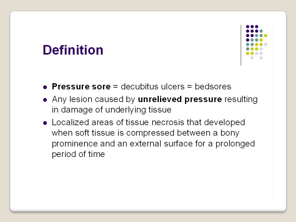
Pressure Sore

3
زخم بستر ( زخم فشاری ) واژه های متعددی جهت زخمهای فشاری ( بستر ) به کار رفته است که معمول ترین آنها Decubitus ulcer و Bedsore است. واژه Decubitus از کلمه لاتین دکومبر Decumber به معنای دراز کشیدن مشتق شده است و دلالت بر این دارد که این زخمها صرفا درنتیجه خوابیدن به مدت طولانی ایجاد می شوند. علت نامگذاری bedsore بروز مکرر این زخمها در بیماران بستری در تخت است. با توجه به تعاریف از آن جایی که فشار عامل اصلی ایجاد زخم است واژه pressure ulcer یا زخم فشاری صحیح ترین و مناسب ترین واژه برای توصیف این زخم ها است.

4
تعریف زخم فشاری باتوجه به اهمیت موضوع تعاریف متفاوتی در منابع مختلف عنوان گردیده است که در زیر به مهمترین آنها اشاره می کنیم. - زخم فشاری به زخمی گفته می شود که به علت وارد آوردن فشاری بیش از فشار طبیعی مویرگها به مدت طولانی بر سطح پوست ایجاد می گردد که موجب نکروز ناحیه محدودی از بافتهای نرم می شود. " برونر - سودارث 1998" - یک زخم فشاری ناحیه متمرکزی از نکروز بافتی است که هنگامیکه بافت نرم بین یک برجستگی استخوانی و یک سطح خارجی به مدت طولانی تحت فشار قرار می گیرد ایجاد می شود." پوتروپری وفیس براساس NPUAP " زخم فشاری عبارت از ناحیه نکروزه ای است که در نتیجه فقدان جریان خون کافی به آن ناحیه در اثر فشار ایجاد می شود."HARKNESS& DIBCLTER" - Margo lis زخم فشاری را این طور تعریف کنید : از بین رفتن ساختمان آناتومیک و عملکرد نرمال پوست که در نتیجه فشار خارجی وارد بر برجستگی های استخوانی ایجاد می شود و طبق قاعده ای منظم و در یک زمان معین بهبود نمی یابد.(( پوتروپری ))
).")
8
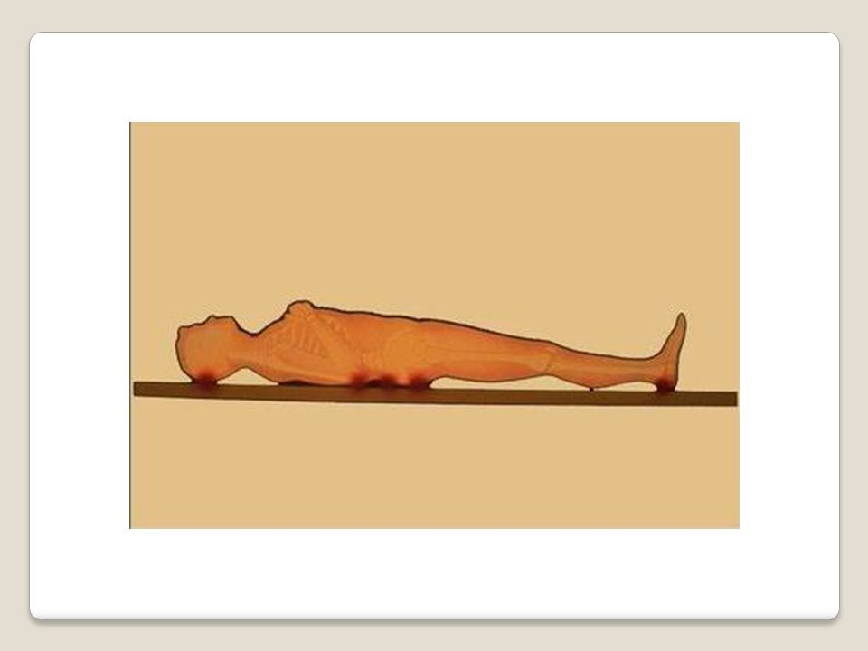
Etiology of pressure ulcers Pressure ulcers are due to localized ischemia, a deficiency in the blood supply to the tissue. The tissue is compressed between two hard surfaces, usually the surface between the bed and the skeleton, when the blood cannot reach the tissue, the cells are deprived of oxygen and nutrients, waste products of metabolism accumulate in the cells, and the tissue consequently dies. Prolonged, unrelieved pressure also damages the small blood vessels.

9
After the skin has been compressed, it appears pale, as if the blood had been squeezed out of it. When pressure is relieved, the skin takes on a bright red flush called reactive hyperthermia. The flush is due to vasodilatation, a process in which extra blood supply to compensate for the preceding period of impeded blood flow.

10
Risk factors Friction and Shearing Two other factors frequently act in conjunction with pressure to produce pressure ulcers: Friction: is a force acting parallel to the skin surface, such as sheets rubbing against skin create friction. Friction can abrade the skin, that is, remove the superficial layers, making it more prone to breakdown.

11
Pressure Ulcer Causes of Pressure Ulcer Development Pressure

12
Shearing force: combination of friction and pressure. It occurs commonly when the a client assumes a Fowler’s position. In this position, the body tends to slide downward toward the foot of the bed. This downward movement is transmitted to the sacral bone and the deep tissues. At the same time, the skin over the sacrum tends not to move because of the adherence between the skin and the bed linens. The skin and superficial tissues are thus relatively unmoving in relation to the bed surface, whereas the deeper tissues are firmly attached to the skeleton and move downward. This causes a shearing force in the area where the deeper tissues and the superficial tissues meet. and the superficial tissues meet. The force damages the blood vessels and tissues in this area.

13
Shearing forces can occur when a patient is moved carelessly or slides down in bed.

14
Immobility Refers to a reduction in the amount and control of movement a person has. Such as paralysis, extreme weakness, pain. Inadequate nutrition It causes weight loss, muscle atrophy, and loss of subcutaneous tissue. These three reduce the padding between the skin and the bones. More specifically, inadequate intake of protein, carbohydrates, fluids, and vitamin C.

15
Diminished sensation Paralysis, stroke, loss of consciousness may cause loss of sensation in a body area. loss of sensations reduce person’s ability to respond to trauma, to injuries heat and cold, and to the tingling (pins and needles) that signals loss of circulation. Sensory loss also impairs the body’s ability to recognize and provide healing mechanisms for a wound. Excessive body heat Increased body temperature increase metabolism, increase cell need for oxygen.
 that signals loss of circulation. Sensory loss also impairs the body’s ability to recognize and provide healing mechanisms for a wound. Excessive body heat Increased body temperature increase metabolism, increase cell need for oxygen..")
16
Advanced age Due to changes in body mechanisms such as loss of lean body mass, decreased strength and elasticity, diminished pain perception, increased dryness due to a decrease in the amount of oil produced by the sebaceous.

17
Chronic medical conditions D/M, cardio vascular diseases are risk for skin breakdown and delayed healing. These conditions compromise oxygen delivery to tissues by poor perfusion and thus cause poor and delayed healing and increase risk of pressure sores. Other factors Poor lifting techniques, incorrect positioning, repeating injection at the same area, incorrect application of pressure relieving devices.

18
Stages of pressure ulcers Stage 1:- red color and the skin don’t return to normal color even the pressure is released. Stage 2 :- redness accompanied by blisters or shallow break in the skin Stage 3 :- break in the skin extending to the subcutaneous tissue Stage 4:- ulcer involves loss of all skin layers exposing muscle and bone.

19
Pressure Ulcer Grades of Ulcers Grade 1 Epidermis Dermis Subcutaneous Fat Muscle Bone Redness that does not disappear when pressure is applied

20
Pressure Ulcer Grades of Ulcers Grade 2

21
Pressure Ulcer Grades of Ulcers Grade 3

22
Pressure Ulcer Grades of Ulcers Grade 4

23
1. Consider all bed- or chair-bound persons, or those whose ability to reposition is impaired, to be at risk for pressure ulcers. 2. Select and use a method of risk assessment, such as the Norton Scale or the Braden Scale, that ensures systematic evaluation of individual risk factors. 3. Assess all at-risk patients at the time of admission to health care facilities and at regular intervals thereafter. 4. Identify all individual risk factors (decreased mental status, moisture, incontinence, nutritional deficits) to direct specific preventive treatments. Modify care according to the individual factors. Risk Assessment Risk Assessment
 to direct specific preventive treatments. Modify care according to the individual factors. Risk Assessment Risk Assessment.")
25
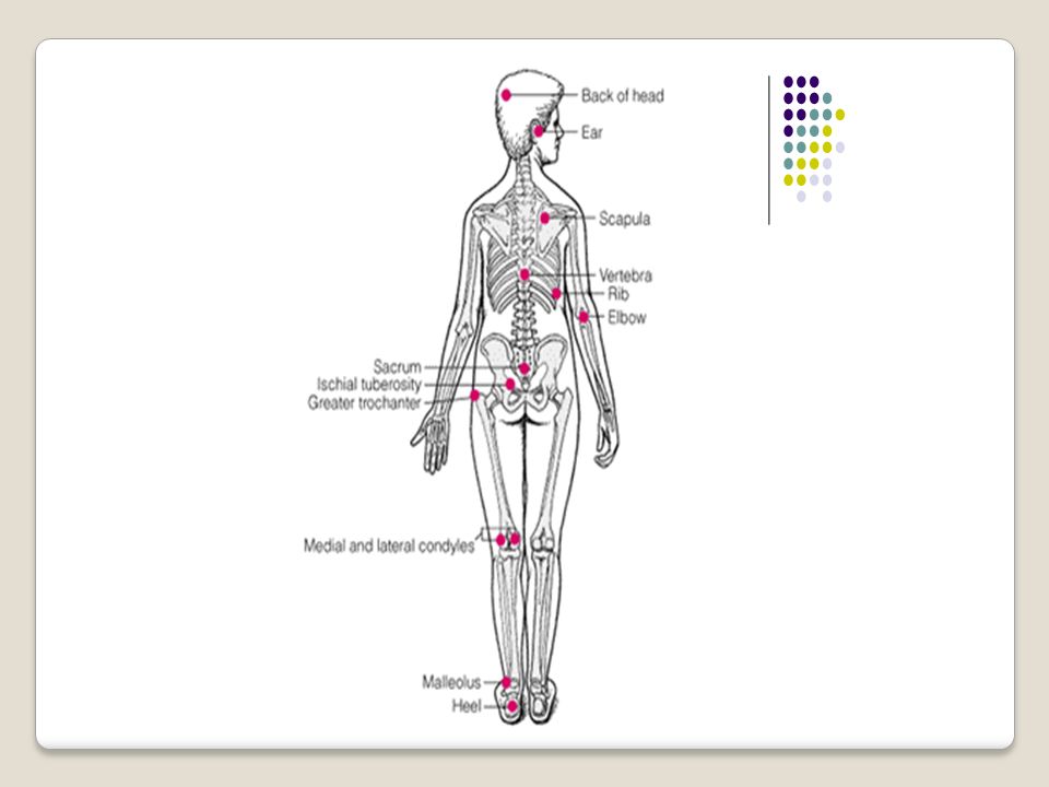
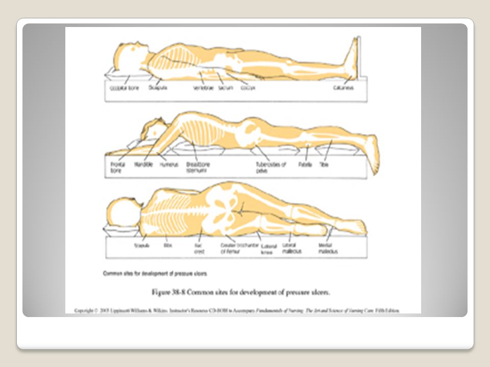
Pressure Ulcer Bony Prominences at Risk

26
Skin Care and Early Treatment Skin Care and Early Treatment 1. Inspect the skin at least daily, and document assessment results. 2. Individualize bathing frequency. Use a mild cleansing agent. Avoid hot water and excessive friction. 3. Assess and treat incontinence. When incontinence cannot be controlled, cleanse skin at time of soiling, use a topical moisture barrier, and select underpads or briefs that are absorbent and provide a quick drying surface to the skin. 4. Use moisturizers for dry skin. Minimize environmental factors leading to dry skin such as low humidity and cold air.

27
5. Avoid massage over bony prominences. 6. Use proper positioning, transferring, and turning techniques to minimize skin injury due to friction and shear forces. 7. Use dry lubricants or protective coverings to reduce friction injury. 8. Identify and correct factors compromising protein/ calorie intake and consider nutritional supplementation/support for nutritionally compromised persons. 9. Institute a rehabilitation program to maintain or improve mobility/activity status. 10. Monitor and document interventions and outcomes.

28
خسته نباشيد

Similar presentations


 A pressure ulcer is localized injury to the skin and/or underlying.>")


