Download presentation
Presentation is loading. Please wait.

1
Essentials of Human Anatomy & Physiology Copyright © 2003 Pearson Education, Inc. publishing as Benjamin Cummings Seventh Edition Elaine N. Marieb The Muscular System part 2a Muscle Physiology Modified by S. Mendoza 1/2015

2
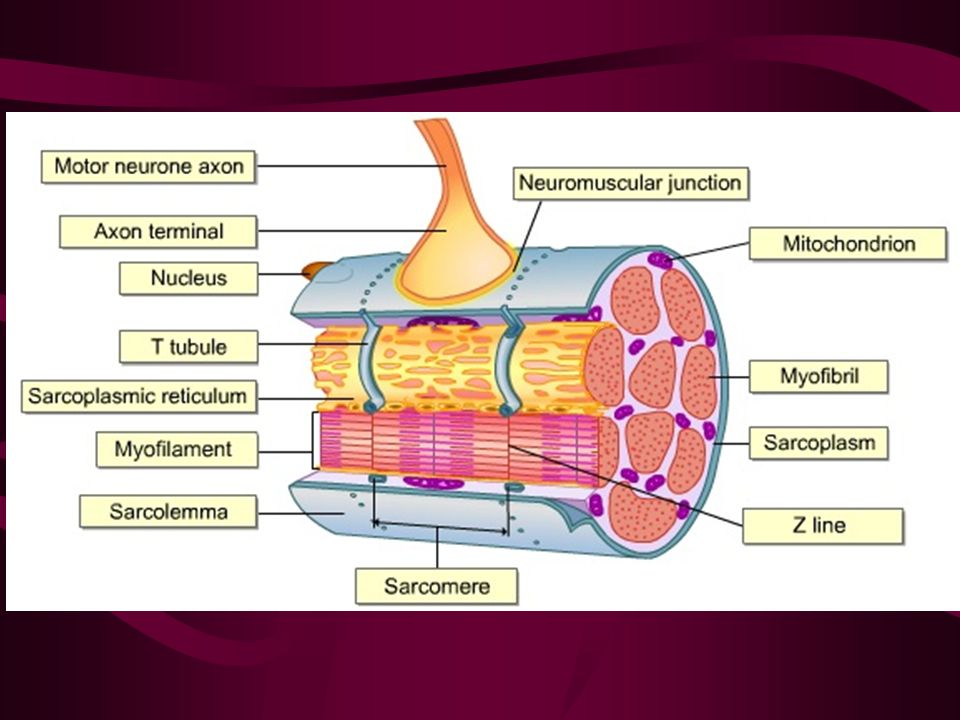
Microscopic Anatomy of Skeletal Muscle Slide 6.9b Copyright © 2003 Pearson Education, Inc. publishing as Benjamin Cummings Sarcolemma – specialized plasma membrane of muscle fiber Cells are multinucleate Nuclei are just beneath the sarcolemma Sarcoplasm – cytoplasm of a muscle fiber Figure 6.3a

3
Myoglobin Myoglobin is a common protein, which has the ability to store oxygen in muscle cells. The myoglobin has a high level of red pigment, so the more myoglobin the meat has, the redder it will be. The terms “red meat” and “white meat” are actually an indicator for the level of myoglobin.

4
Myoglobin Amounts

5
Myoglobin This protein is also the main reason that the red meat turns darker while you’re cooking it. During the heating process, iron atoms of the myoglobin lose electrons and they move up to a higher oxidation level. Thus, the meat turns from pinkish-red to brown.

6
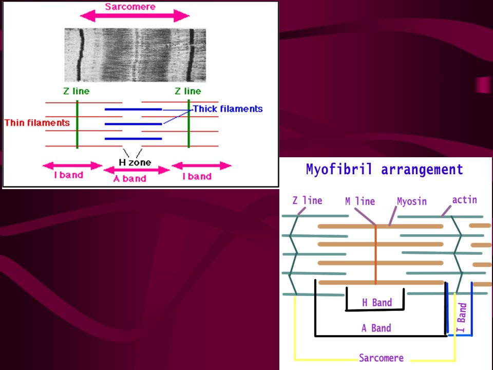
Microscopic Anatomy of Skeletal Muscle Slide 6.10a Copyright © 2003 Pearson Education, Inc. publishing as Benjamin Cummings Myofibril - Long rod like organelles comprising 80% of cell volume Running parallel the entire length of the cells the myofibrils are aligned to give distinct bands A band = dark band Contains lighter central H Zone visible only in relaxed fiber I band = light band Contains Z disc/line at midpoint

7
Micro anatomy Banding patterns/striations reveal the working structure of muscle fiber

9
Microscopic Anatomy of Skeletal Muscle Slide 6.10b Copyright © 2003 Pearson Education, Inc. publishing as Benjamin Cummings Sarcomere Region of myofibril Contractile unit of a muscle fiber Region between 2 successive Z discs Figure 6.3b

11
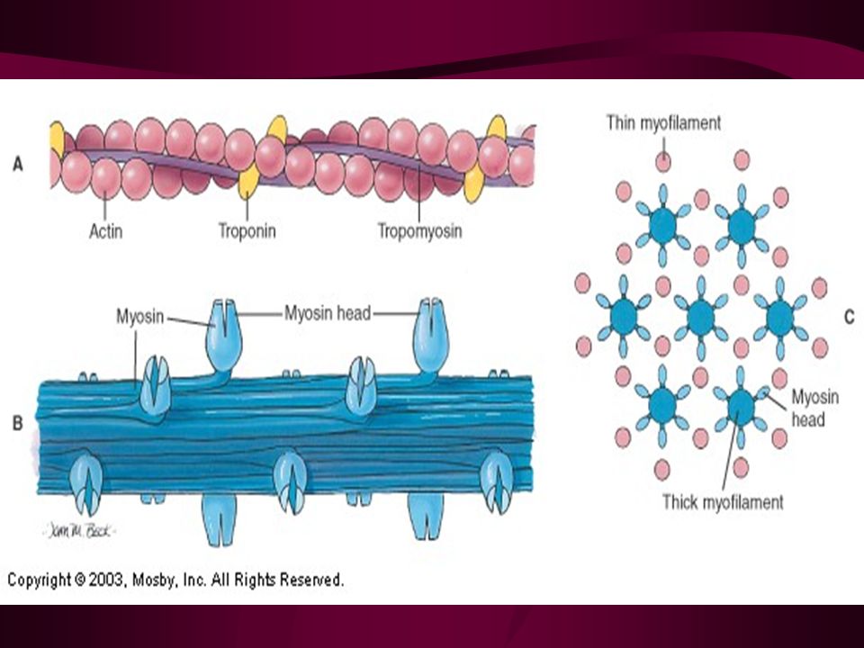
Microscopic Anatomy of Skeletal Muscle Slide 6.11b Copyright © 2003 Pearson Education, Inc. publishing as Benjamin Cummings Organization of the sarcomere Thin filaments = actin filaments Contain troponin & tropomyosin to regulate attachment of myofilaments to each other

12
Microscopic Anatomy of Skeletal Muscle Slide 6.11a Copyright © 2003 Pearson Education, Inc. publishing as Benjamin Cummings Thick filaments = myosin filaments Composed of the protein myosin with cross bridge heads Heads contain ATPase enzymes to split ATP & release energy for contraction Figure 6.3c

14
Sarcoplasmic Reticulum Sarcoplasmic reticulum – specialized smooth endoplasmic reticulum Function: Stores ionic calcium & releases it on demand

15
Sarcoplasmic Reticulum Surrounds myofibrils At junction of A band and I band, sarcolemma forms hollow T-tubule to conduct stimulus deep into every sarcomere

17
END OF QUIZ #1 MATERIAL Study all your info and diagrams!

18
How muscle knows WHEN to contract Mechanism of contraction on a cellular level

19
Motor Unit One motor neuron and ALL the muscle cells that it stimulates Spread throughout muscle

20
Explanation - then see next slide! Stimulation of one motor unit results in weak contraction of ENTIRE muscle –Since a motor unit is spread throughout the muscle & not clustered together, it stimulation will activate cells scattered throughout the entire muscle –This causes a weak contraction of the entire muscle –Muscles requiring fine control have small motor units that only activate a few cells at a time.

21
Slide 6.14 Copyright © 2003 Pearson Education, Inc. publishing as Benjamin Cummings Figure 6.4a

22
Nerve Stimulus to Muscles Slide 6.15a Copyright © 2003 Pearson Education, Inc. publishing as Benjamin Cummings Neuromuscular junctions – association site of nerve and muscle Figure 6.5b

23
Nerve Stimulus to Muscles Slide 6.15b Copyright © 2003 Pearson Education, Inc. publishing as Benjamin Cummings Each axon terminal forms junction with single muscle fiber Synaptic cleft – fluid filled gap between nerve and muscle Nerve and muscle do not make contact Importance: prevent continuous stimulation Figure 6.5b

25
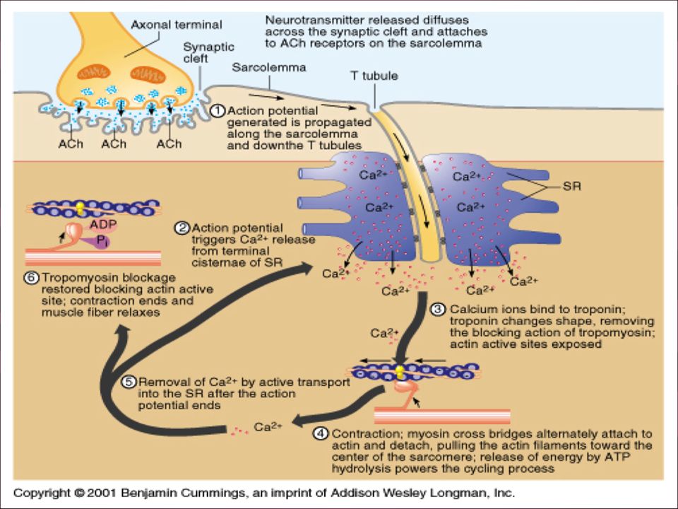
Transmission – know the steps Slide 6.16a Copyright © 2003 Pearson Education, Inc. publishing as Benjamin Cummings Vesicles in axon terminal filled with neurotransmitter – chemical released by nerve upon arrival of nerve impulse The neurotransmitter for skeletal muscle is acetylcholine (ACh) Neurotransmitter crosses synaptic cleft and attaches to receptors on the sarcolemma The Neuromuscular Junction The Neuromuscular Junction
 Neurotransmitter crosses synaptic cleft and attaches to receptors on the sarcolemma The Neuromuscular Junction The Neuromuscular Junction.")
26
Transmission – know the steps Sarcolemma becomes temporarily permeable to sodium (Na + ) Na+ ions rush into muscle cell which reverses electrical conditions Action potential is caused which moves along sarcolemma and down T tubules deep into muscle fiber Once initiated – action potential is unstoppable (all or none principle) resulting in full contraction of that particular muscle fiber (cell) Excitation-Contraction Coupling
 Na+ ions rush into muscle cell which reverses electrical conditions Action potential is caused which moves along sarcolemma and down T tubules deep into muscle fiber Once initiated – action potential is unstoppable (all or none principle) resulting in full contraction of that particular muscle fiber (cell) Excitation-Contraction Coupling")
27
Safeguard When nerve stimulation stops: –Ach is destroyed by acetylcholinesterase (AChE) to prevent continued contraction –Substances such as certain organophosphates found in pesticides and fertilizers destroy AChE causing convulsions
 to prevent continued contraction –Substances such as certain organophosphates found in pesticides and fertilizers destroy AChE causing convulsions")
28
End of stimulation K+ ions leaves cell rapidly to restore electrical balance Then Na-K pump restores ions to original positions for relaxation of muscle fiber

29
Sliding Filament Theory HOW a muscle contracts

30
Sliding Filament Theory The thin filaments slide past the thick filaments so the overlap increases This shortens the muscle fiber and thus the entire muscle

31
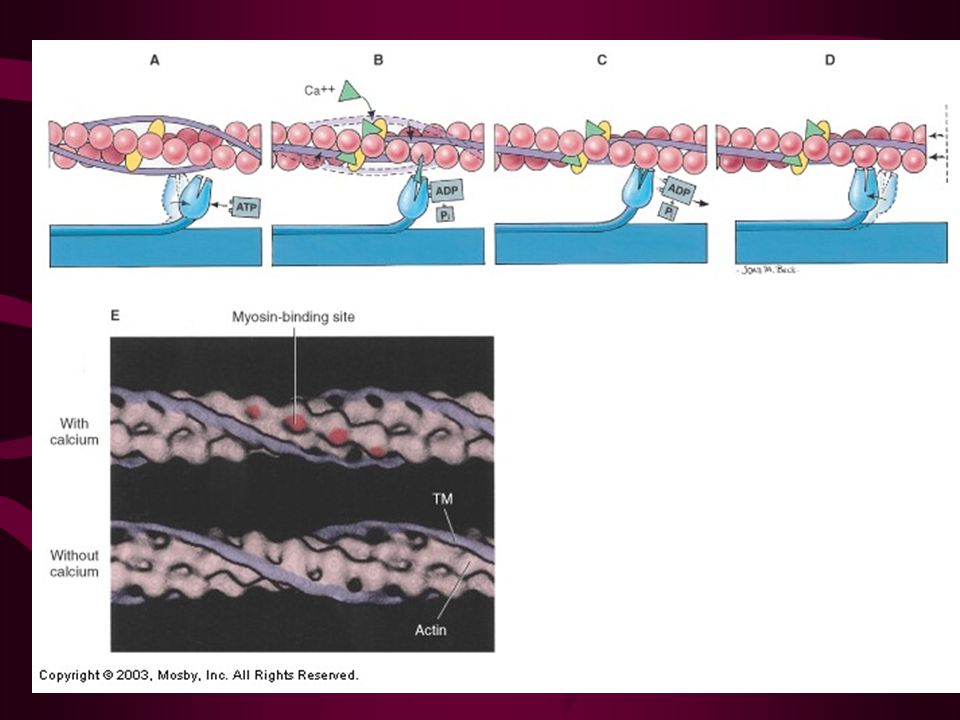
The Sliding Filament Theory Slide 6.17a Copyright © 2003 Pearson Education, Inc. publishing as Benjamin Cummings Activation by nerve causes myosin heads (crossbridges) to attach to binding sites on the thin filament Myosin heads then bind to the next site of the thin filament Figure 6.7
 to attach to binding sites on the thin filament Myosin heads then bind to the next site of the thin filament Figure 6.7.")
32
The Sliding Filament Theory of Muscle Contraction Slide 6.17b Copyright © 2003 Pearson Education, Inc. publishing as Benjamin Cummings This continued action causes a sliding of the myosin along the actin The result is that the muscle is shortened (contracted) Figure 6.7
 Figure 6.7.")
33
What causes the filaments to slide? Cross bridge attachment: in presence of Ca ions, high energy myosin cross bridge binds to actin binding site Power Stroke: energy from ATP is used to bend cross bridge and pull actin toward center of sarcomere 1% shortening for each power stroke

34
What causes the filaments to slide? Cross bridge detachment: ATP also provides energy to detach cross bridge rigor mortis on next slide “Cocking” of myosin head: energy returns myosin head to high energy configuration to prepare for next attachment

35
Cross Bridge Cycling

36
Rigor Mortis –stiffness of death Muscles begin to stiffen within 4 hours after death –Face, hand, feet, rest of body –Intensity depends on muscle mass Peak rigidity occurs 12 to 48 hours Gradually disappears over next 36-48 hours in cool climates 9-12 hours in hot Dead cells unable to exclude Ca ions so Ca ions come out of SR and promote cross bridge binding

37
Rigor Mortis ATP synthesis stops shortly after breathing stops –Exhaustion before death accelerates rigor since ATP has been depleted Cross bridge detachment impossible Actin & myosin become irreversible cross linked producing stiffening of dead muscle Gradual disappearance results from breakdown of biological molecules

38
The Role of Ionic Calcium Tropomyosin (regulatory protein) normally block binding sites on actin molecule so myosin cannot attach to actin When ionic Ca becomes available (released from SR), Ca binds to troponin (another regulatory protein Troponin “pulls” tropomysosin off of the binding site by causing it to change shape This allows attachment of cross bridges
 normally block binding sites on actin molecule so myosin cannot attach to actin When ionic Ca becomes available (released from SR), Ca binds to troponin (another regulatory protein Troponin pulls tropomysosin off of the binding site by causing it to change shape This allows attachment of cross bridges")
40
Excitation Contraction Coupling

41
Excitation Contraction coupling Study diagram & YouTube animations provided Action potential "wave" travels along sarcolemma and deep into T tubules. This triggers Ca 2+ release from terminal cisternae of SR Ca 2+ binds to troponin — binding sites exposed. Contraction: see sliding filament theory Action potential ends, Ca 2+ is taken back up into the SR Tropomyosin blockage restored, contraction ends, muscle fiber relaxes.

42
Contraction of a Skeletal Muscle Slide 6.19 Copyright © 2003 Pearson Education, Inc. publishing as Benjamin Cummings “All or none” principle applies only to each muscle fiber, NOT the whole muscle Within a skeletal muscle, not all fibers may be stimulated during the same interval Different combinations of muscle fiber contractions may give differing responses

43
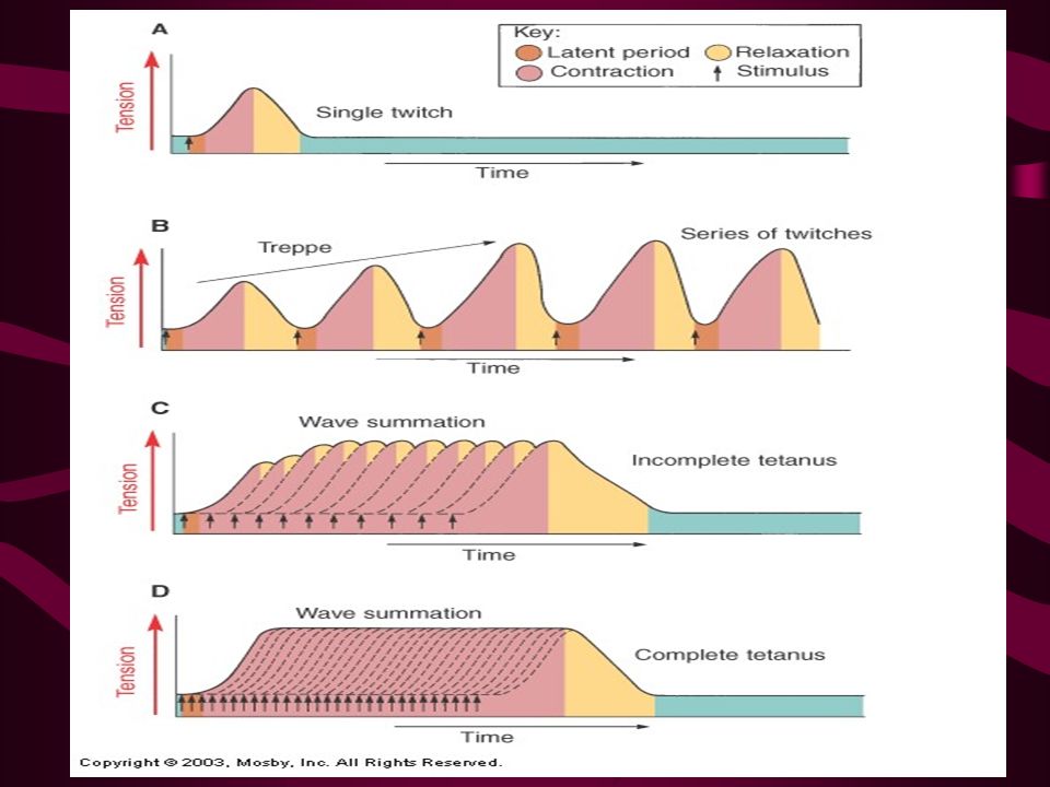
Graded Muscle Responses Graded responses – different degrees of skeletal muscle shortening, Produced in 2 ways By changing the frequency of muscle stimulation (Be able to recognize the diagrams & basic facts) By changing the # of muscle cells being stimulated
 By changing the # of muscle cells being stimulated")
44
Changing Frequency of Muscle Stimulation - recognize diagrams & underlined info

45
Types of Graded Responses Slide 6.20a Copyright © 2003 Pearson Education, Inc. publishing as Benjamin Cummings Twitch Response of muscle to single action potential Not a normal muscle function Metabolic properties & enzymes present in muscles cause different twitch responses Useful for diagnostics

46
Types of Graded Responses Treppe –Response that occurs when frequency & strength of stimulus held constant. –Staircase pattern on myogram recording –Each successive contraction is stronger than the previous one –Reflects increased availability of Ca 2+ ions –Heat also increases efficiency of enzymes in muscle –Another good reason to warm up !!!!! (along with thinning out your synovial fluid)
.")
47
Treppe

48
Types of Graded Responses Slide 6.20b Copyright © 2003 Pearson Education, Inc. publishing as Benjamin Cummings Wave Summation Impulses in rapid succession (increasing the frequency) cause contractions to build on one another The muscle does not completely return to a resting state The effects are added and show as unfused tetanus until the frequency is such that it becomes fused tetanus Figure 6.9a, b
 cause contractions to build on one another The muscle does not completely return to a resting state The effects are added and show as unfused tetanus until the frequency is such that it becomes fused tetanus Figure 6.9a, b.")
49
Types of Graded Responses Slide 6.21b Copyright © 2003 Pearson Education, Inc. publishing as Benjamin Cummings Fused Tetanus No evidence of relaxation before the following contractions The result is a sustained muscle contraction – usual manner of muscle contraction Figure 6.9a, b Figure 6.9c,d

51
CHANGING NUMBER OF MUSCLE CELLS BEING STIMULATED - RECOGNIZE DIAGRAMS & UNDERLINED INFO

52
Multiple Motor Unit Summation – aka. Recruitment Slide 6.22 Copyright © 2003 Pearson Education, Inc. publishing as Benjamin Cummings increasing the strength of the stimulus therefore increasing the number of motor neurons firing to gain more muscle force. Muscle force depends upon the number of fibers stimulated

53
Multiple Motor Unit Summation – aka. Recruitment Slide 6.22 Copyright © 2003 Pearson Education, Inc. publishing as Benjamin Cummings Larger # of Motor units recruited = larger force of contraction Weak or precise movements = few motor units Recruitment of fiber types: Type I first, then type IIa, then type IIb (will learn about later)
.")
54
Multiple Motor Unit Summation

55
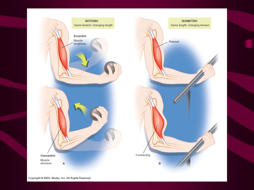
Types of Muscle Contractions Slide 6.28 Copyright © 2003 Pearson Education, Inc. publishing as Benjamin Cummings Isotonic (same tension) contractions Myofilaments are able to slide past each other during contractions The muscle shortens & movement occurs Isometric (same length) contractions Tension in the muscles increases The muscle does not shorten & no movement occurs Most movements involve both types of activity
 contractions Myofilaments are able to slide past each other during contractions The muscle shortens & movement occurs Isometric (same length) contractions Tension in the muscles increases The muscle does not shorten & no movement occurs Most movements involve both types of activity.")
Similar presentations