Download presentation
Presentation is loading. Please wait.

1
KCP-760 ( 토 의 자 ) 원 자 력 병 원 전공의 허일영
 원 자 력 병 원 전공의 허일영")
2
Male / 75 Chief complaint: Chest pain on exertion for 6 months Chest CT: Pleural effusion with diffuse pleural thickening Debridement and TB medication Pleural effusion and hemoptysis Past history: TB pleurisy (35 years ago)
")
10
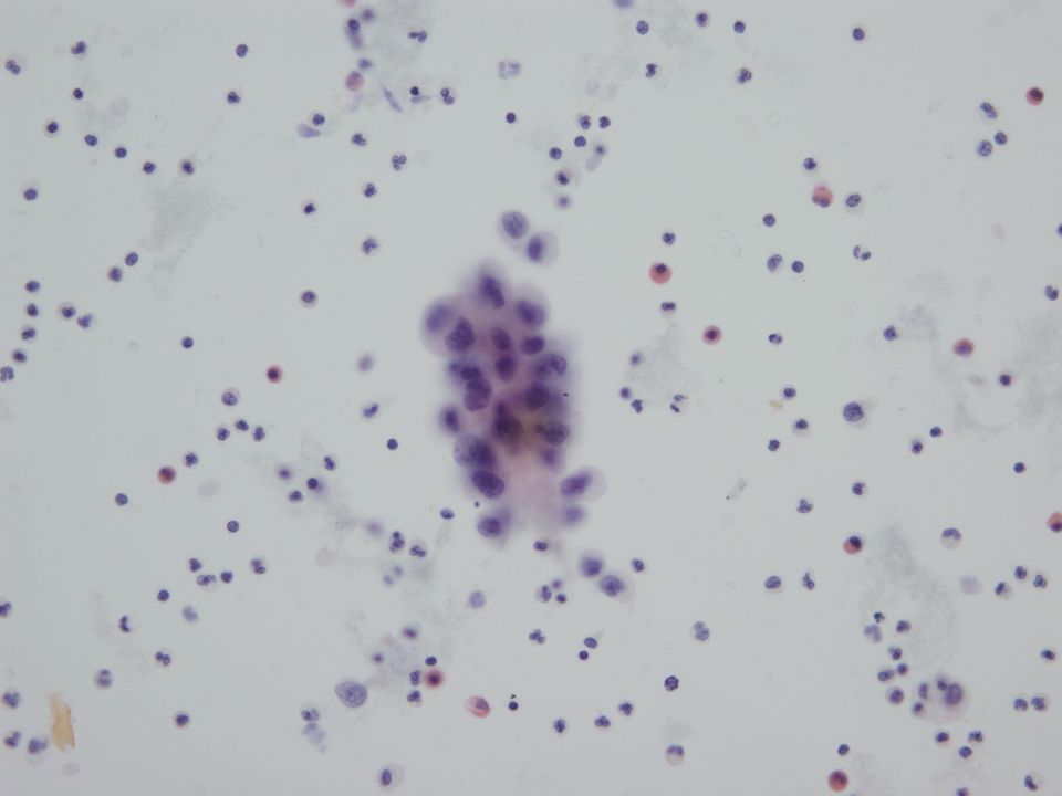
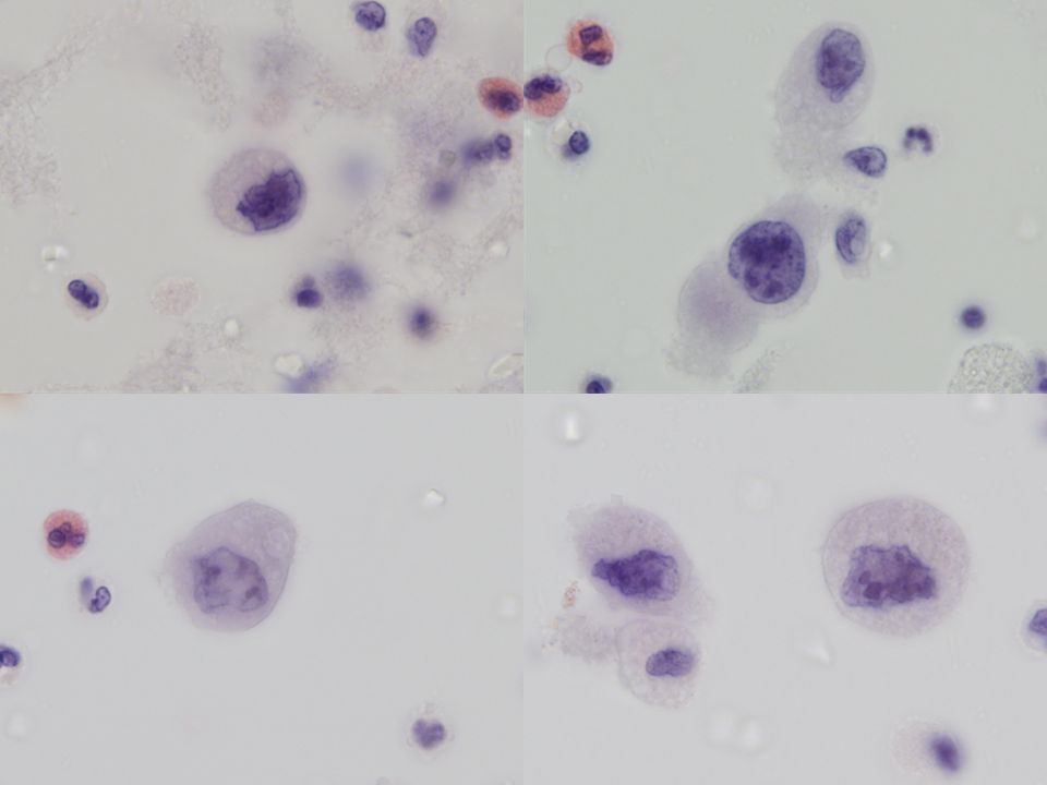
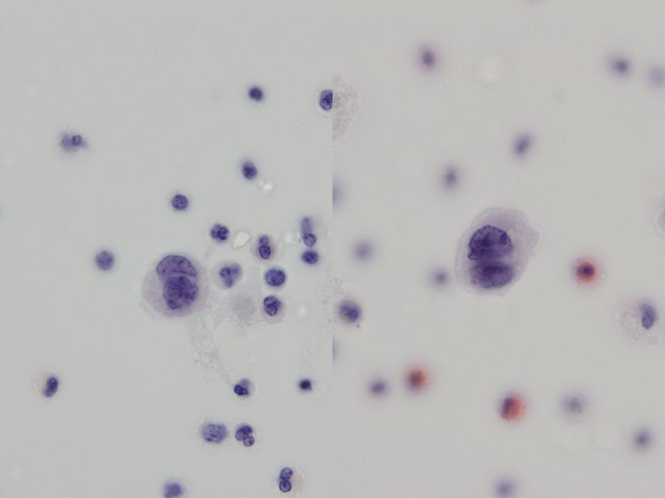
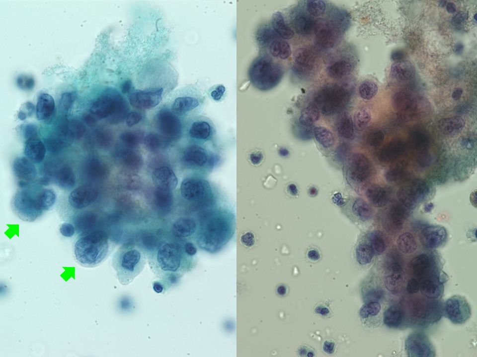
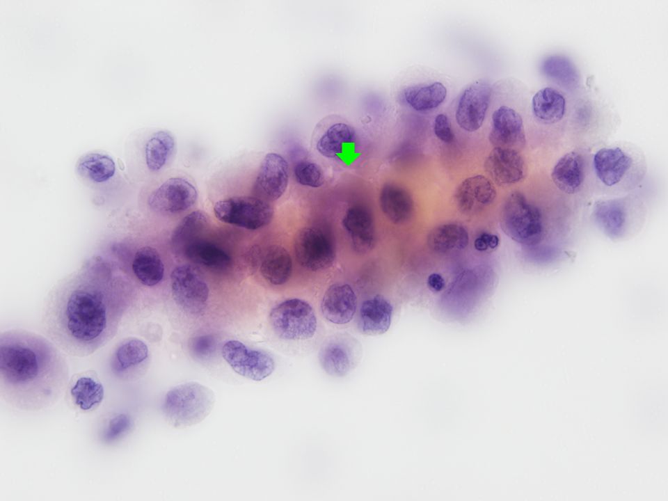
Single cells –Well-defined or fuzzy cell border –Ampler cytoplasm with peripheral vacuolization or small vacuoles –Sharp irregularities of nuclear contour –Prominent and multiple nucleoli –Bi-nucleated atypical cells Cells in aggregates –Less cohesive, single cell population, knobby outline and intercellular “window” –Fuzzy border, microvilli, “two-tone” cytoplasm, degenerative vacuole and peripheral vacuolization Neutrophils, eosinophils and lymphocytes in the background

11
Differential diagnosis Malignant mesothelioma Reactive mesothelial hyperplasia Metastatic adenocarcinoma

12
Malignant mesotheliomaReactive Mesothelial hyperplasiaMetastatic adenocarcinoma Mostly “one-cell” population Single cells and tissue fragments (three-dimensional balls, papillary- like branching fragments) Usually large cells with prominent nucleoli Malignant features such as sharp irregularities of nuclear contour, irregular chromatin distribution Cytoplasmic rim Hypercellular smears Nuclei usually round and centrally located Cell aggregates and small tissue fragments, mostly doublets and rarely in fragments with 2-6 cells Hypercellular specimen Predominantly single cells Single cells or cells in aggregates may vary in size and N/C ratio Chromatin texture varies but is usually evenly distributed Nucleoli, single or multiple, can become prominent Few papillary tissue fragments Predominantly tissue fragments Acinus formation Papillary architecture Secretory vacuoles Material in vacuoles Occupying most of the cytoplasm Well-defined border S.Z. Ali, E.S. Cibas. Serous Cavity Fluid and Cerebrospinal Fluid Cytopathology. Springer, 2011. Winifred Gray, MB BS FRCPath. DIAGNOSTIC CYTOPATHOLOGY. Elsevier, 2010.

13
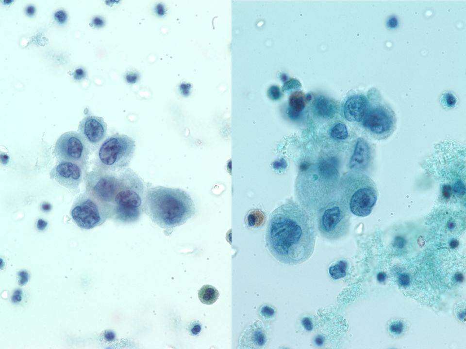
Long chain-like arrangement (hand-in-hand appearance) One cell population Intercellular window Relatively consistent nuclear-cytoplasmic ratio despite variable nuclear size Lacy border with microvilli Jin Ho Paik et al. Korean J Pathol 2009; 43: 458-66 Malignant mesothelioma

14
“Most often serous cavity fluids in patients with malignant mesothelioma will contain a large amount of malignant cells with well-developed cytomorphologic characteristics.” “Diagnostic issues arise when the amount of lesional cells is low” “It is not recommended to make a diagnosis of mesothelioma based on cytology alone because of the high risk of diagnostic error” “It is recommended that a cytological suspicion of mesothelioma is followed by tissue confirmation” Eur Respir J 2010; 35: 479–495. S.Z. Ali, E.S. Cibas. Serous Cavity Fluid and Cerebrospinal Fluid Cytopathology. Springer, 2011.

15
Pleural fluid, cytology: Atypical mesothelial cells, Suspicious for malignant mesothelioma

Similar presentations


 Reactive endocervical cells B) HSIL C) LSIL D) Squamous metaplasia E) ASCUS 1.>")


 Chapter: Neoplasia Definitions Nomenclature Characteristics of benign and malignant neoplasms Epidemiology.>")


