Download presentation
Presentation is loading. Please wait.

1
Shabnam tehrani, MD Assistant Professor of Shahid Beheshti University of Medical science Amebiasis

2
Definition Amebiasis is infection with the parasitic intestinal protozoan Entamoeba histolytica (the "tissue-lysing ameba"). Most infections are probably asymptomatic, but E. histolytica can cause disease ranging from dysentery to extraintestinal infections, including liver abscesses.

3
Life Cycle and Transmission E. histolytica exists in two stages: - a hardy multinucleate cyst form -the motile trophozoite stage. Infection is acquired by ingestion of cysts contained in fecally contaminated food or water. Trophozoites can live within the large-bowel lumen without causing disease or can invade the intestinal mucosa, causing amebic colitis. In some cases, E. histolytica trophozoites invade through the mucosa and into the bloodstream, traveling through the portal circulation to reach the liver and causing amebic liver abscesses.

5
Epidemiology It was a staple of most textbooks that 10% of the world's population was infected with E. histolytica. We now know that most asymptomatic individuals harboring amebic trophozoites or cysts in their stools are infected with a noninvasive species: Entamoeba dispar E. histolytica infections are most common in areas of the world where poor sanitation and crowding compromise the barriers to contamination of food and drinking water with human feces. Endemic areas include parts of Mexico, India, and nations in the tropical regions of Africa, South and Central America, and Asia.

6
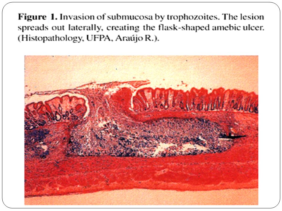
Pathogenesis and Pathology E. histolytica trophozoites possess a potent repertoire of adhesins, proteinases, pore-forming proteins Disease begins when trophozoites adhere to colonic mucosal epithelial cells,then disruption of the colonic mucin barrier secreting proteolytic enzymes( histolysine ) and cytotoxic substances. contact-dependent cell killing cytophagocytosis
 and cytotoxic substances. contact-dependent cell killing cytophagocytosis.")
9
Clinical Syndromes A. Intestinal Amebiasis: Most patients are asymptomatic, but individuals with E. histolytica infection can develop disease. Symptoms of amebic colitis generally appear 2–6 weeks after ingestion of the cyst form of the parasite. Diarrhea (classically heme-positive) and lower abdominal pain are the most common symptoms. Malaise and weight loss may be noted as disease progresses. Severe dysentery, with 10–12 small-volume, blood- and mucus-containing stools daily, may develop, but only 40% of patients are febrile.
 and lower abdominal pain are the most common symptoms. Malaise and weight loss may be noted as disease progresses. Severe dysentery, with 10–12 small-volume, blood- and mucus-containing stools daily, may develop, but only 40% of patients are febrile..")
10
… Fulminant amebic colitis, with even more profuse diarrhea, severe abdominal pain (including peritoneal signs), fever, and pronounced leukocytosis are rare, disproportionately affecting young children, pregnant women, individuals being treated with glucocorticoids, and possibly individuals with diabetes or alcoholism. Mortality rates from fulminant amebic colitis: 40% Recognized complications of amebic colitis also include - toxic megacolon (documented in 0.5% of patients with colitis), with severe bowel dilation and intramural air, -ameboma, which presents as an abdominal mass that may be confused with colon cancer.
, with severe bowel dilation and intramural air, -ameboma, which presents as an abdominal mass that may be confused with colon cancer..")
11
… B.Amebic Liver Abscess: Most individuals with amebic liver abscess do not have concurrent signs or symptoms of colitis, and most do not have E. histolytica trophozoites in their stools. The exceptions are individuals with fulminant amebic colitis, in which concurrent amebic liver abscess is not uncommon. Disease can arise from months to years after travel to or residence in an endemic area; therefore, a careful travel history is key in making the diagnosis. The classic presentations of amebic liver abscess are RUQ pain, fever, and hepatic tenderness. The pace of disease is usually acute, with symptoms lasting <10 days. Jaundice is unusual, but dullness and rales at the right lung base (secondary to pleural effusion) are common.
 are common..")
12
This is an amebic abscess of liver. Abscesses may arise in liver when there is seeding of infection from the bowel, because the infectious agents are carried to the liver from the portal venous circulation.

13
Diagnostic Tests The diagnosis of amebic colitis has traditionally been based on the demonstration of E. histolytica trophozoites or cysts in the stool or colonic mucosa of patients with diarrhea

14
… However, the inability of microscopy to differentiate between E. histolytica and other Entamoeba species, such as E. dispar limits its effectiveness as a sole diagnostic method. Examination of 3 stool samples improves sensitivity for the detection of Entamoeba species, and it has been argued that the presence of amebic trophozoites containing red blood cells in a diarrheal stool is highly suggestive of E. histolytica infection. Despite these inherent limitations, microscopy, often combined with serologic testing, remains the standard diagnostic approach. Culture of stools for E. histolytica trophozoites serves as a research tool but is generally not available for clinical use.

15
… PCR assay for DNA in stool samples is currently the most sensitive and specific method for identifying E. histolytica infection and has become a valuable epidemiologic and research tool Commercially available tests that use enzyme-linked immunosorbent assays (ELISAs) or immunochromatographic techniques to detect Entamoeba antigens are less expensive and more easily performed and are being used with increasing frequency. At this point, antigen detection–based ELISAs that can specifically identify E. histolytica in stool probably represent the best choice in endemic areas In instances in which amebiasis is suspected on clinical grounds in a patient with acute colitis but initial stool samples are negative, colonoscopy with examination of brushings or mucosal biopsies for E. histolytica trophozoites may be helpful in making the diagnosis or in identifying other diseases, such as inflammatory bowel disease or pseudomembranous colitis.
 or immunochromatographic techniques to detect Entamoeba antigens are less expensive and more easily performed and are being used with increasing frequency. At this point, antigen detection–based ELISAs that can specifically identify E. histolytica in stool probably represent the best choice in endemic areas In instances in which amebiasis is suspected on clinical grounds in a patient with acute colitis but initial stool samples are negative, colonoscopy with examination of brushings or mucosal biopsies for E. histolytica trophozoites may be helpful in making the diagnosis or in identifying other diseases, such as inflammatory bowel disease or pseudomembranous colitis..")
16
… The diagnosis of amebic liver abscess is based on the detection (generally by ultrasound or CT) of one or more space- occupying lesions in the liver and a positive serologic test for antibodies to E. histolytica antigens. Amebic liver abscesses are classically described as single, large, and located in the right lobe of the liver, but sensitive imaging techniques have shown that multiple abscesses are more common than previously suspected. When a patient has a space-occupying lesion of the liver, a positive amebic serology is highly sensitive (>94%) and highly specific (>95%) for the diagnosis of amebic liver abscess. False-negative serologic tests have been reported when serum samples were obtained very early in the course of abscess (within 7–10 days of onset), but repeat tests are almost always positive
 and highly specific (>95%) for the diagnosis of amebic liver abscess. False-negative serologic tests have been reported when serum samples were obtained very early in the course of abscess (within 7–10 days of onset), but repeat tests are almost always positive.")
17
Treatment Amebic Colitis or Liver Abscess: Tinidazole: Better tolerate & more effective for: colitis and liver abscess(2 gr/d. 3 d) Metronidazol: (750 mg tid po or IV 5-10 d) Entamoeba histolytica Luminal Infection: Paromomycin: 30mg/kg tid po 5-10 d Idoquinol: 650 mg tid po 20d
 Metronidazol: (750 mg tid po or IV 5-10 d) Entamoeba histolytica Luminal Infection: Paromomycin: 30mg/kg tid po 5-10 d Idoquinol: 650 mg tid po 20d.")
18
Giardia lamblia

19
… Giardiasis is one of the most common parasitic diseases in both developed and developing countries worldwide, causing both endemic and epidemic intestinal disease and diarrhea Infection follows the ingestion of environmentally hardy cysts, which excyst in the small intestine, releasing flagellated trophozoites Giardia remains a pathogen of the proximal small bowel and does not disseminate hematogenously World wide distribution Highest incidence in children, young adults in late summer.

20
Transmission 1-Person to person transmission 2- Water sports, surface contamination. Watershed contamination

21
Clinical Manifestations range from asymptomatic carriage to fulminant diarrhea and malabsorption. Most infected persons are asymptomatic, but in epidemics the proportion of symptomatic cases may be higher. Symptoms may develop suddenly or gradually In persons with acute giardiasis, symptoms develop after an IP that lasts at least 5–6 days and usually 1–3 weeks. Prominent early symptoms include diarrhea, abdominal pain, bloating, flatus, nausea, and vomiting. Although diarrhea is common, upper intestinal manifestations such as nausea, vomiting, bloating, and abdominal pain may predominate.

22
… The duration of acute giardiasis is usually >1 week, although diarrhea often subsides. Some persons who have relatively mild symptoms for long periods recognize the extent of their discomfort only in retrospect. Fever, the presence of blood and/or mucus in the stools, and other signs and symptoms of colitis are uncommon and suggest a different diagnosis or a concomitant illness. Because of the less severe illness and the propensity for chronic infections, patients may seek medical advice late in the course of the illness; however, disease can be severe, resulting in malabsorption, weight loss, growth retardation, and dehydration. Giardiasis can be severe in patients with hypogammaglobulinemia and can complicate other preexisting intestinal diseases, such as that occurring in cystic fibrosis. In patients with AIDS, Giardia can cause enteric illness that is refractory to treatment.

23
Diagnosis Giardia should be identified 50 to 70% of the time after one stool, and 90% identification after 3 stools Biopsy tissue/duodenal aspirate stained by trichrome or Giemsa stain.

24
Drugs Dose Metronidazole250mgtidX 5-7 d Nitazoxanide500mg bdX3d Paromomycin 25–30 mg/kg/d in 3 doses × 5–10 d Tinidazole 2 g × 1 dose

25
Treatment Drugs Dose Metronidazole 250mg tidX 5-7 d Nitazoxanide 500mg bdX3d Paromomycin 25–30 mg/kg/d in 3 doses × 5–10 d Tinidazole 2 g × 1 dose

26
Prevention The prevention of giardiasis requires proper handling and treatment of water Good personal hygiene on an individual basis Chlorination alone is sufficient to kill G. lamblia cysts, important variables, such as water temperature, clarity, pH, and contact time, alter the efficacy of chlorine, and higher chlorine levels (4 to 6 mg/liter) may be required. Bringing water to a boil is sufficient to kill all protozoal cysts; at high altitudes, boiling for longer periods may be necessary
 may be required. Bringing water to a boil is sufficient to kill all protozoal cysts; at high altitudes, boiling for longer periods may be necessary.")
Similar presentations


, Hepatovirus Picornavirus, enterovirus 72 27 nm 1 serotype only, although there are 4 genotypes.>")
, Department of Biological Sciences University of Alberta.>")


