Download presentation
Presentation is loading. Please wait.

2
Skeletal system includes bones of the skeleton, and the cartilage, ligaments, and other connective tissues that stabilize and/or connect the bones

3
SUPPORT › Provides structural support and framework for entire body STORAGE OF MINERALS AND LIPIDS › Mainly calcium salts and fats (in yellow marrow) BLOOD CELL PRODUCTION › RBCs, WBCs, and other blood elements are made in red marrow
 BLOOD CELL PRODUCTION › RBCs, WBCs, and other blood elements are made in red marrow")
4
PROTECTION › Many soft tissues are surrounded by bone LEVERAGE › Bones function as levers that change the magnitude and direction of the forces generated by skeletal muscles

6
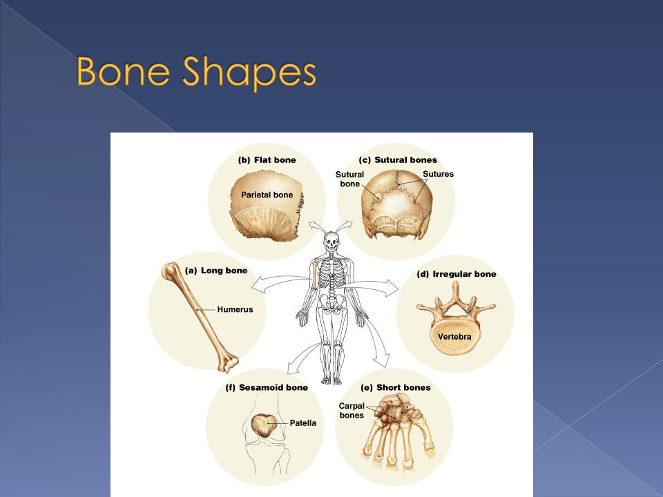
There are 206 bones in the human body, which are categorized into 6 shapes 1) Long bones › Long and slender › Located in arm, thigh, leg, palms, soles, fingers and toes. › The femur (thigh bone) is largest and heaviest bone in the body
 is largest and heaviest bone in the body.")
8
2) Flat bones › Thin, relatively parallel surfaces › Found in roof of skull, sternum, ribs, scapula › Provide protection of underlying tissue › Large surface area for attachment of muscles 3) Sutural bones (Wormian bones) › Small, flat, irregularly shaped › Found between flat bones of the skull
 Flat bones › Thin, relatively parallel surfaces › Found in roof of skull, sternum, ribs, scapula › Provide protection of underlying tissue › Large surface area for attachment of muscles 3) Sutural bones (Wormian bones) › Small, flat, irregularly shaped › Found between flat bones of the skull")
10
4) Irregular bones › Complex shapes with short, flat, notched or ridged surfaces › Vertebrae, pelvis, some skull bones 5) Short bones › Small and boxy › Wrist bones and ankle bones 6) Sesamoid bones › Small, flat, shaped like sesame seed › Kneecaps, some bones in hands and feet
 Irregular bones › Complex shapes with short, flat, notched or ridged surfaces › Vertebrae, pelvis, some skull bones 5) Short bones › Small and boxy › Wrist bones and ankle bones 6) Sesamoid bones › Small, flat, shaped like sesame seed › Kneecaps, some bones in hands and feet")
12
Bones have external and internal features

15
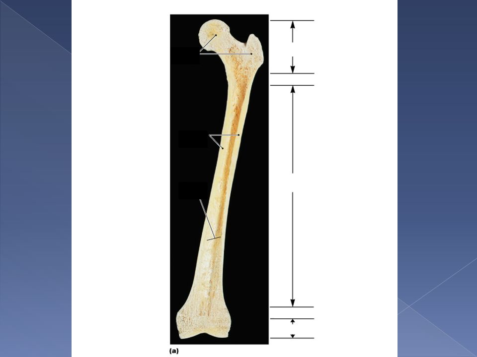
There are different parts to bone › Diaphysis – tubular shaft › Epiphysys – expanded area at each end of the diaphysis › Metaphysis – thin area that connects the epiphysis to the diaphysis

17
There are different parts to bone › Compact bone – outer portion of diaphysis Solid, sturdy layer that surrounds the marrow cavity › Spongy bone (cancellous) – mainly found in the epiphysis Open network of struts and plates with a thin covering of compact bone Marrow is present here, but no marrow cavity
 – mainly found in the epiphysis Open network of struts and plates with a thin covering of compact bone Marrow is present here, but no marrow cavity")
20
Bone tissue is a supporting connective tissue Contains specialized cells and matrix › Matrix is solid, due to calcium salts around protein fibers

21
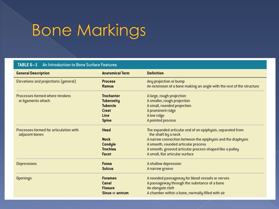
4 characteristics of bone 1. Matrix dense and contains calcium salts 2. Matrix contains bone cells (osteocytes), within pockets (lacunae), which are organized around blood vessels 3. Canaliculi, canals from lacunae to the blood vessels, allow for the transfer of materials 4. Except at joints, the outer surface of bone is covered by a periosteum
, within pockets (lacunae), which are organized around blood vessels 3. Canaliculi, canals from lacunae to the blood vessels, allow for the transfer of materials 4. Except at joints, the outer surface of bone is covered by a periosteum.")
22
Calcium phosphate (Ca 3 (PO 4 ) 2 ) makes up about 2/3 of bone About 1/3 of bone consists of collagen fibers About 2% of bone mass is cells
 2 ) makes up about 2/3 of bone About 1/3 of bone consists of collagen fibers About 2% of bone mass is cells")
23
Calcium phosphate is very hard, but inflexible and brittle › Can withstand compression, but will shatter when exposed to bending, twisting, or sudden impacts. Collagen fibers are strong and flexible › When subjected to tension, they are stronger than steel › Easily tolerate bending and twisting, but when compressed, they just bend out of the way

24
This mixture of calcium crystals and protein fibers give bone properties between the two › Strong, somewhat flexible, resistant to shattering › As good as steel reinforced concrete

25
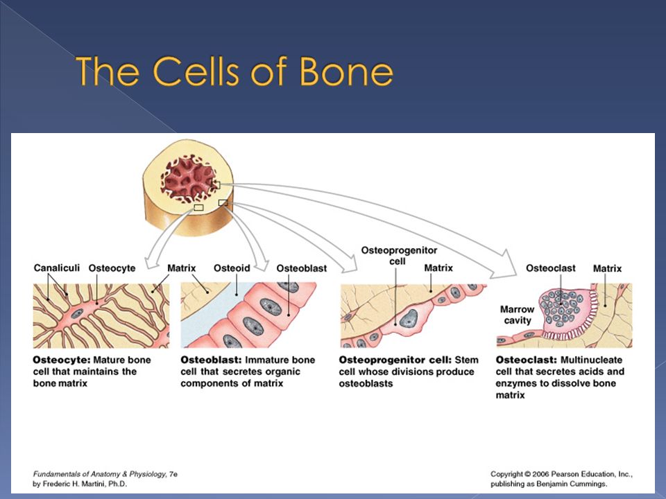
Bone tissue contains 4 types of cells 1) Osteocytes – mature bone cells Most bone cells are these Occupy a lacuna, between layers of matrix (lamella) Osteocytes can’t divide Canaliculi connect lacuna to each other and blood vessels
 Osteocytes – mature bone cells Most bone cells are these Occupy a lacuna, between layers of matrix (lamella) Osteocytes can’t divide Canaliculi connect lacuna to each other and blood vessels")
27
Bone tissue contains 4 types of cells Osteocytes have two major functions 1)Maintain mineral and protein content of surrounding matrix 2)Participate in the repair of bone 2) Osteoblasts – make new bone matrix › Process is called osteogenesis › Eventually turn into osteocytes
Maintain mineral and protein content of surrounding matrix 2)Participate in the repair of bone 2) Osteoblasts – make new bone matrix › Process is called osteogenesis › Eventually turn into osteocytes")
29
Bone tissue contains 4 types of cells 3) Osteoprogenitor cells – stem cells that give rise to osteoblasts 4) Osteoclasts – remove and recycle bone matrix › Process is called osteolysis
 Osteoprogenitor cells – stem cells that give rise to osteoblasts 4) Osteoclasts – remove and recycle bone matrix › Process is called osteolysis")
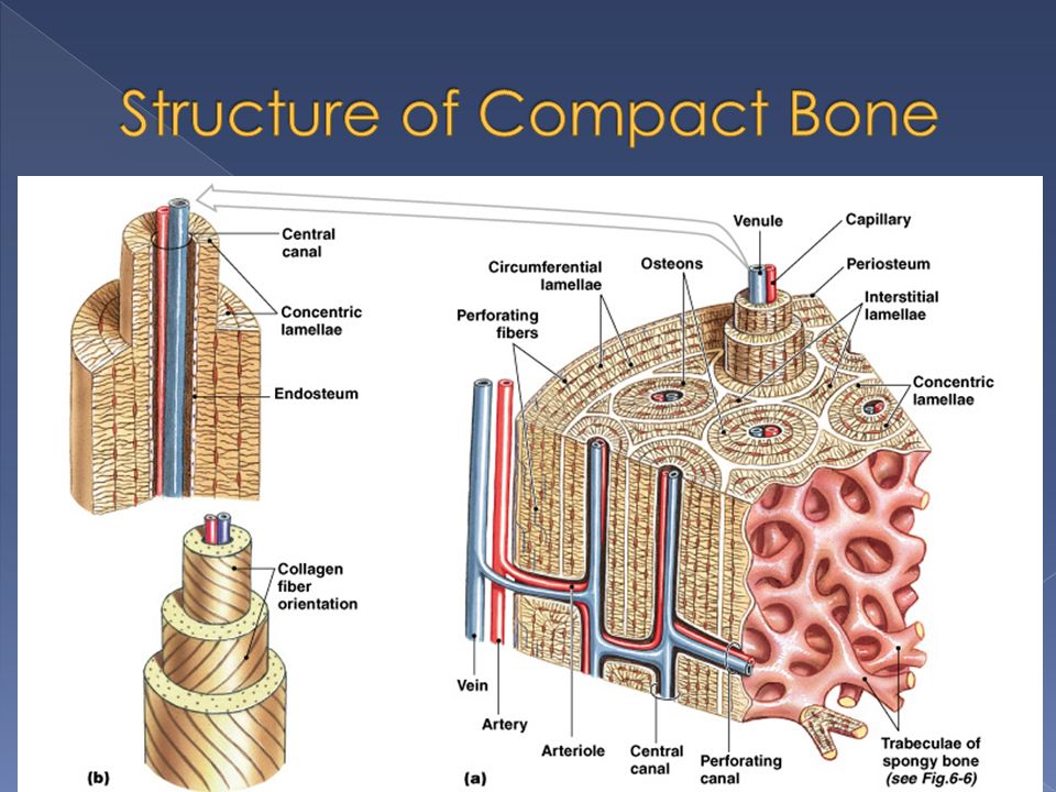
31
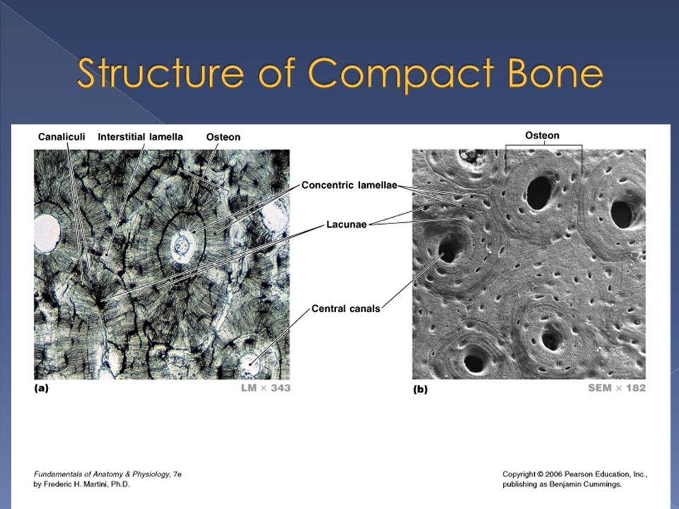
Osteon (Haversion System) – basic functional unit of mature compact bone › Osteocytes are arranged in layers around a central canal (Haversian canal) Canal contains blood vessels Canals run parallel to surface of bone › Perforating canals run perpindicular to surface of bone Contain blood vessels that take blood to deeper osteons and to marrow cavity
 – basic functional unit of mature compact bone › Osteocytes are arranged in layers around a central canal (Haversian canal) Canal contains blood vessels Canals run parallel to surface of bone › Perforating canals run perpindicular to surface of bone Contain blood vessels that take blood to deeper osteons and to marrow cavity")
33
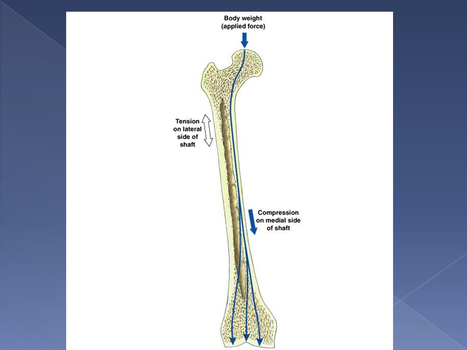
Compact bone is thickest where stresses arrive from limited directions Osteons in diaphysis are parallel to long axis › Shaft doesn’t bend. Your femur can withstand 15 times your body weight through long axis Much less force from the side will fracture your femur

36
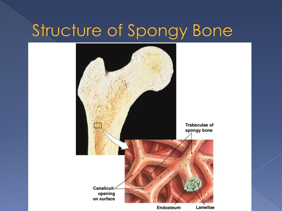
Lamellae are not arranged like in osteons Here, they form struts and plates called trabeculae This creates an open network No blood supply in spongy bone › Nutrients come from diffusion through canaliculi

38
Bone marrow is found in spongy bone › Red bone marrow makes blood cells › Yellow bone marrow is comprised of adipose tissue Fat storage for energy reserve

39
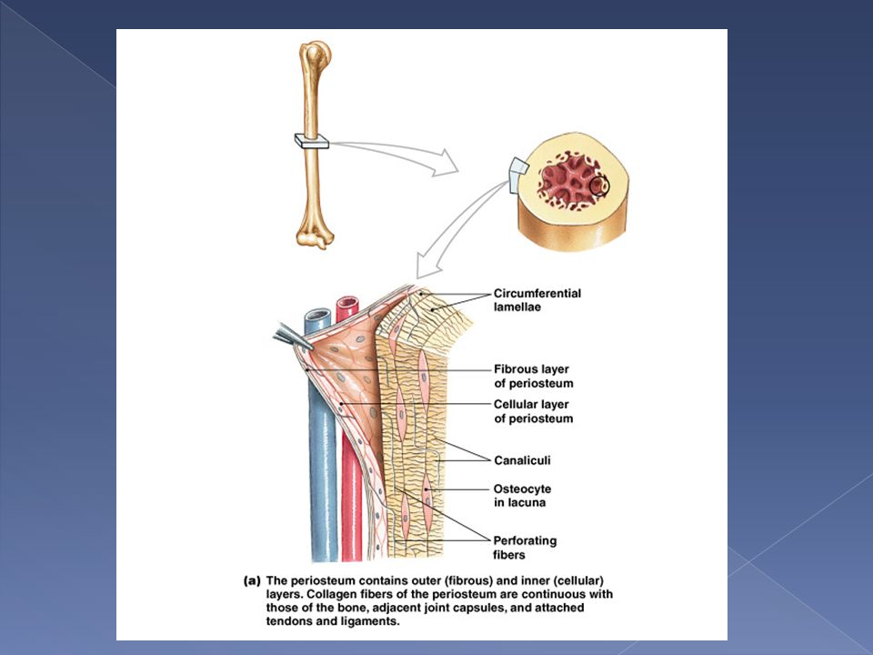
Periosteum covers superficial layer of compact bone, except at joints › 3 functions 1.Isolates bone from surrounding tissue 2.Provides route for circulatory and nervous supply 3.Actively participates in bone growth and repair Periosteum is interwoven with the synovial capsules and with tendons

41
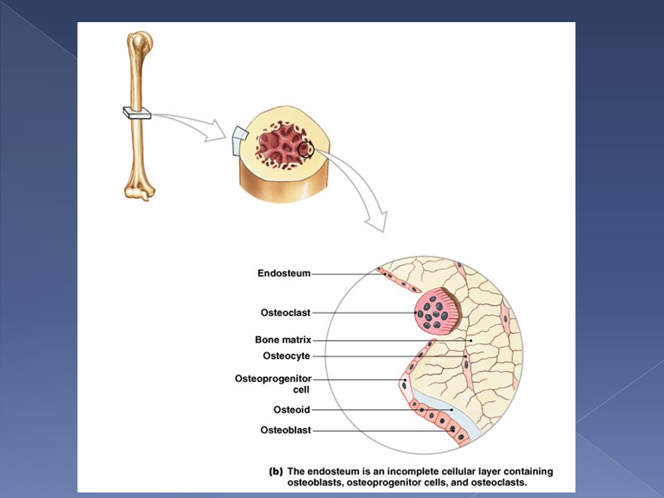
Endosteum › Lines marrow cavity › Active during bone growth, repair and remodeling › Single, incomplete layer of osteoprogenitor cells Where layer is incomplete, osteoblasts and osteoclasts can remodel bone tissue

44
Skeleton begins to form about 6 weeks after fertilization › Skeleton is cartilage Portions of the skeleton don’t stop growing until the age of 25

45
Ossification - process of replacing other tissues with bone › 2 types Endochondral ossification Intramembranous ossification Calcification – deposition of calcium, occurs during ossification

46
Bones are made of hyaline cartilage in the embryo Cartilage gradually converted to bone through endochondral ossification

47
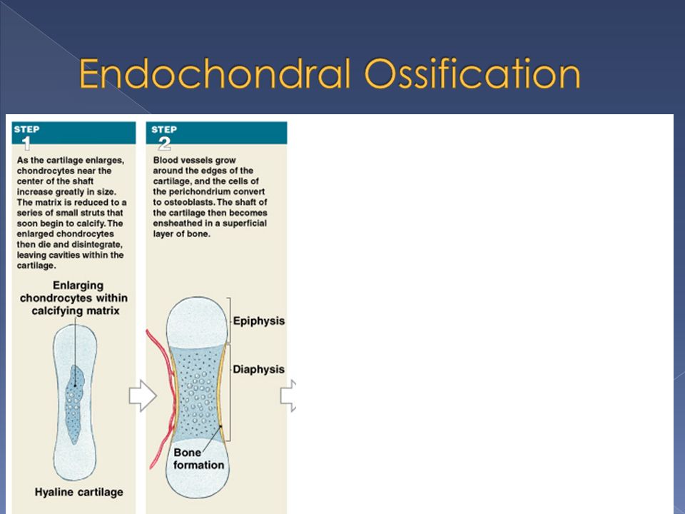
6 steps to endochondral ossification 1) Chondrocytes increase in size, cartilage is reduced to struts that calcify. Chondrocytes die and disintegrate 2) Blood vessels grow into perichondrium. Cells in inner layer become osteoblasts and they produce a thin layer of bone along shaft (perichondrium become periosteum
 Blood vessels grow into perichondrium. Cells in inner layer become osteoblasts and they produce a thin layer of bone along shaft (perichondrium become periosteum.")
49
6 steps to endochondral ossification 3) Blood vessels make their way into the center of the shaft. Fibroblasts become osteoblasts and spongy bone is created. Bone production begins at this site (primary ossification center) and spreads toward both ends of the bone 4) Osteoclasts appear and disintegrate the spongy bone in the diaphysis and create a marrow cavity. Diameter of bone enlarges
 and spreads toward both ends of the bone 4) Osteoclasts appear and disintegrate the spongy bone in the diaphysis and create a marrow cavity. Diameter of bone enlarges.")
51
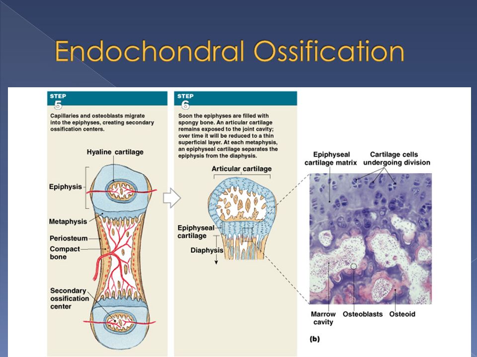
6 steps to endochondral ossification 5) Centers of the epiphyses begin to calcify. Capillaries and osteoblasts migrate in to create secondary ossification centers 6) Epiphyses fill in with spongy bone. Caps of cartilage (articular cartilage) remain on ends of bone exposed to the joint cavity. Narrow region of cartilage remains (epiphyseal cartilage) between epiphysis and diaphysis. This is your ‘growth plate’.
 Epiphyses fill in with spongy bone. Caps of cartilage (articular cartilage) remain on ends of bone exposed to the joint cavity. Narrow region of cartilage remains (epiphyseal cartilage) between epiphysis and diaphysis. This is your ‘growth plate’..")
53
Endochondral Ossification AnimationEndochondral Ossification Animation#2 Endochondral Ossification Animation #1

54
As long as cartilage is being produced on the epiphyseal side, and bone is replacing cartilage on the shaft side, the bone will continue to get longer. At puberty, sex hormones cause dramatic bone growth. Epiphyseal cartilage starts to disappear and become an epiphyseal line

56
Appositional Growth › Diameter of bone increases › Osteoblasts in inner layer of perisoteum create matrix along outer surface of shaft. › Osteoclasts slowly remove bone matrix along marrow cavity

57
Osteoblasts differentiate from mesenchymal stem cells in fibrous connective tissue › Result is dermal bones Ex – flat bones of skull, jaw, calvicle 3 steps to this process 1) Mesnechymal cells cluster together and secrete components of matrix, then turn into osteoblasts (called ossification center)
 Mesnechymal cells cluster together and secrete components of matrix, then turn into osteoblasts (called ossification center)")
58
2) Blood vessels grow into the initial spongy bone 3) Bone growth and remodeling produces compact bone
 Blood vessels grow into the initial spongy bone 3) Bone growth and remodeling produces compact bone")
59
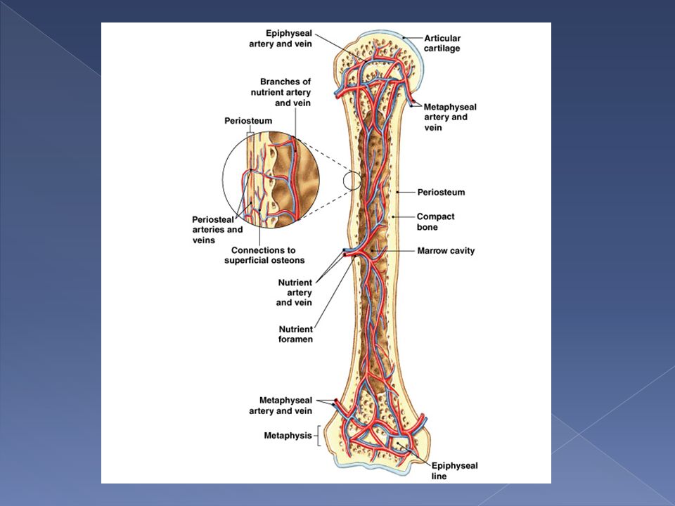
Bone is HIGHLY vascular 3 sets of blood vessels › Nutrient Artery and Vein Supply diaphysis › Metaphyseal vessels Supply epiphyses › Periosteal vessels Supply superficial osteons of shaft

62
The matrix is constantly being recycled › Process is called remodeling › Occurs throughout life › Osteoblasts constantly make matrix › Osteoclasts constantly dissolve matrix

63
Osteoblasts are attracted to minute electrical fields, which are created when bone is stressed. › More matrix is produced where stress is high Electrical stimulation is used in fracture healing Bone that is not stressed will lose matrix IF YOU DON’T USE IT, YOU WILL LOSE IT!!!!!!!

64
YOU NEED CALCIUM AND PHOSPHATE SALTS!!!!!!! Vitamin D - helps make calcitrol, which helps body absorb calcium Vitamin C – helps with collagen synthesis and with osteoblast differentiation Vitamin A – osteoblast activity Vitamins K, B12 – protein fiber production

65
Growth hormone (in pituitary gland) – stimulates bone growth Sex hormones – stimulate osteoblasts › Estrogen causes faster epiphyseal closure than testosterone, which is why women are shorter than men
 – stimulates bone growth Sex hormones – stimulate osteoblasts › Estrogen causes faster epiphyseal closure than testosterone, which is why women are shorter than men")
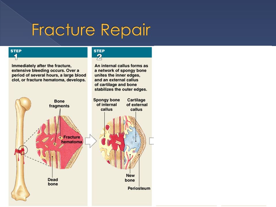
66
Damage to bone tissue is known as a fracture 4 steps to repair 1) Extensive bleeding occurs and a fracture hematoma forms 2) An external callus forms › Enlarged collar of cartilage and bone on surface of bone › An internal callus forms in the marrow cavity
 Extensive bleeding occurs and a fracture hematoma forms 2) An external callus forms › Enlarged collar of cartilage and bone on surface of bone › An internal callus forms in the marrow cavity")
68
3) Osteoblasts replace the cartilage with spongy bone in external and internal callus › Broken ends of bone are united 4) Continued remodeling of bone by osteoblasts and osteoclasts. › Lasts from 4 months to over a year › Bone is thicker and stronger than original

69
Fracture Repair Animation

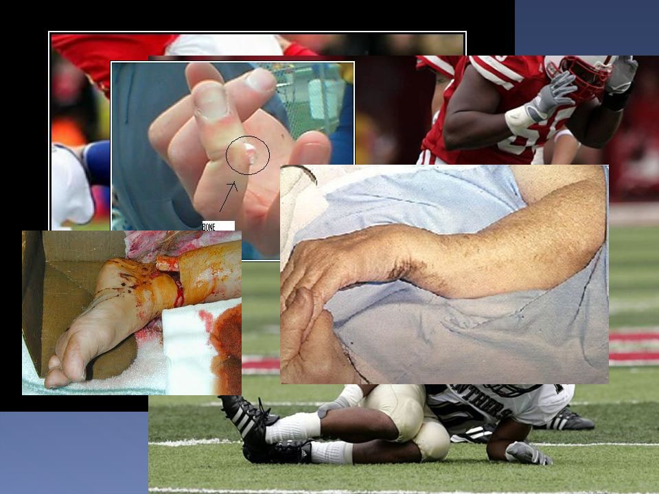
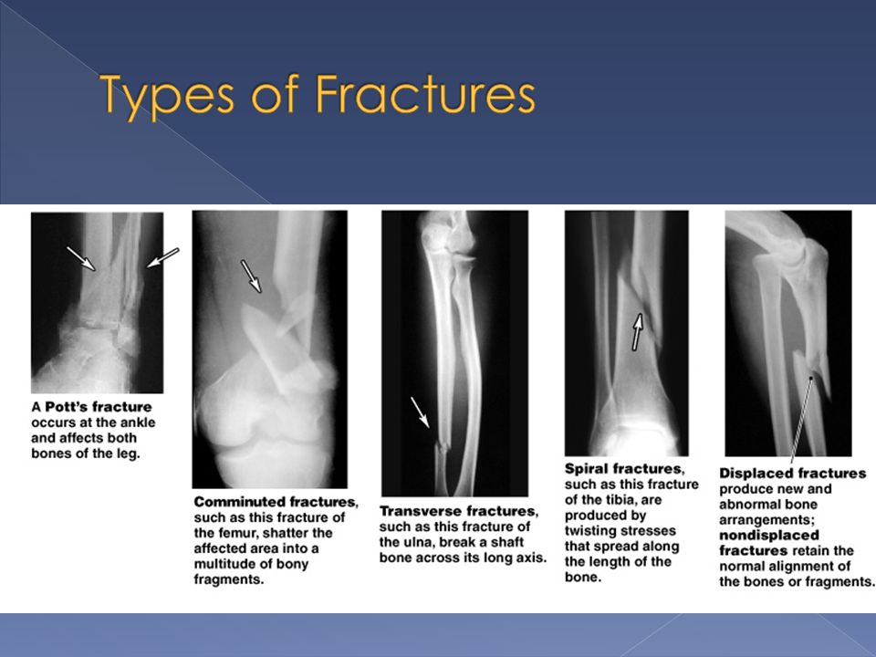
70
Fractures can be open (compound) or closed (simple)
 or closed (simple)")
Similar presentations


 Movement (Passive) Protection of Vital Organs Mineral Storage Blood Cell Formation.>")


 or an organ –Bone referred to as a connective tissue consists of: cells extracellular.>")


. BONE FUNCTION: Support and Protection bones shape and form body structures bones support and protect softer,>")
