Download presentation
Presentation is loading. Please wait.

1
KAU-Faculty of Science- Biochemistry department Clinical biochemistry lab (Bioc 416) 2013
 2013")
2
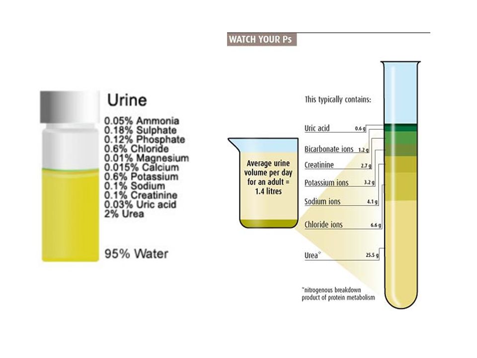
(96%) water Inorganic: Cl -, Na, K. trace amounts of: sulfate, HCO 3 etc.) Urine: Is an ultrafiltrate of plasma from which glucose, amino acids, water and other substances essential to body metabolism have been reabsorbed. Urine carries waste products and excess water out of the body. (4%) dissolved solids: (2%) Urea: ( half) (2%) Other compounds Organic: creatinine uric acid
 Urine: Is an ultrafiltrate of plasma from which glucose, amino acids, water and other substances essential to body metabolism have been reabsorbed. Urine carries waste products and excess water out of the body. (4%) dissolved solids: (2%) Urea: ( half) (2%) Other compounds Organic: creatinine uric acid.")
3
A urine test checks different components of urine, a waste product made by the kidneys. A regular urine test may be done to help find the cause of symptoms. The test can give information about your health and problems you may have. The kidneys take out waste material, minerals, fluids, and other substances from the blood to be passed in the urine. Urine has hundreds of different body wastes. What you eat, drink, how much you exercise, and how well your kidneys work can affect what is in your urine.

6
Factors affect on urine constituents: dietary intake, physical activity, body metabolism, endocrine function others.

7
Urine Analysis: Routine Urinalysis (Routine-UA): It consists of a group of tests performed as part of physical examination. It involves macroscopic and microscopic analysis. Type of analysis: macroscopic analysis: microscopic examination: urine sediment is examined under microscope to identify the components of the urinary sediments. physical characteristics chemical analysis

8
Steps in basic urine analysis Three steps analysis: First: physical characteristics of urine are noted and recorded. Second: series of chemical tests is run. Third: urine sediment is examined under microscope to identify the components of sediments.

9
Urine Collection: Types of urine specimens: type of specimen and collection procedure are determined by physician and depend on the tests to be performed. There are basically four types of urine specimens: 1.First morning specimen 2.Random urine specimen 3.Fractional collection 4.Timed collection Composition and concentration of urine changes during 24hr Urine conc. vary according: to water intake and physical activities.

10
Truly representative sample: it is necessary to regulate: time of collection, length of collection period, patient's dietary, medical intake and method of collection. Initial morning sample is preferred (particularly for protein analysis) because they are more concentrated from overnight retention in bladder. Time of analysis: - must analyzed within 1h at room temp. or within 8hr at 2 o C- 8 o C - If not assayed within these time limits, several changes will occur. sample should collected in a clean container. urine container must be sterile if the urine is to be cultured. For microscopic examination, the urine must be fresh.
 because they are more concentrated from overnight retention in bladder. Time of analysis: - must analyzed within 1h at room temp. or within 8hr at 2 o C- 8 o C - If not assayed within these time limits, several changes will occur. sample should collected in a clean container. urine container must be sterile if the urine is to be cultured. For microscopic examination, the urine must be fresh..")
11
direct visual observation. Normal fresh urine: Color: pale or dark yellow-amber, clear. Vol:750 - 2000 ml/24hr. Physical examination involves: 1.Color 2.Transparency 3.Odour 4.Volume 5.pH 6.Specific gravity

12
1- Color: Many things affect urine color, including fluid balance, diet, medicines, and diseases. Color intensity of urine correlates to concentration. Darker color means more concentrated sample. Urochrome Amber yellow Urochrome (derivative of urobilin, produce from bilirubin degradation, is pigment found in normal urine). Colorless due to reduced concentration. Silver or milky appearance Pus, bacteria or epithelial cells Reddish brown Blood (Hemoglobin). Yellow foam Bile or medications. Orange, green, blue or red medications. Vitamin B supplements can turn urine bright yellow.
. Colorless due to reduced concentration. Silver or milky appearance Pus, bacteria or epithelial cells Reddish brown Blood (Hemoglobin). Yellow foam Bile or medications. Orange, green, blue or red medications. Vitamin B supplements can turn urine bright yellow..")
13
2- Transparency: Urine is normally clear. Bacteria, blood, sperm, crystals, or mucus can make urine look cloudy. Is classified as clear or turbid. In normal urine: the main cause of cloudiness is crystals and epithelial cells. In pathological urine: it is due to pus, blood and bacteria. Degree of cloudiness depends on: pH and dissolved solids Turbidity: may be due to gross bacteriuria, Smoky appearance: is seen in hematouria. Thread-like cloudiness: is seen in sample full of mucus.

14
3- Odour: Odour has little diagnostic significance. 1.Aromatic odour------> Normal urine due to aromatic acids. 2.Ammonia odour------> On standing due to decomposition of urea. 3.Fruity odour--------> Diabetes due to the presence of ketones. Urine does not smell very strong, but has a slightly "nutty" odor. Some diseases cause a change in the odor of urine. For example, an infection with E. coli bacteria can cause a bad odor, while diabetes or starvation can cause a sweet, fruity odor.diabetes 4- Volume: Is important part of assessment for fluid balance and kidney functions. Adults produce from 750ml-2500ml / 24h, with the average of about 1.5L per person. For RUA, a 10ml-12ml of sample is optimal for accurate of analysis

15
5- pH: pH measure acidicity or alkalinity (basic) of urine Normal urine pH: 4.5-8. Increased acidity in urine: due to diabetes or medications. Urine sample must be fresh (why?) ( on standing urine become alkaline as a result of ammonia liberation due to urea decomposition). A urine pH of 4 is strongly acidic, 7 is neutral (neither acidic nor alkaline), and 9 is strongly alkaline. Sometimes the pH of urine is affected by certain treatments. For example, your doctor may instruct you how to keep your urine either acidic or alkaline to prevent some types of kidney stones from forming.kidney stones
 ( on standing urine become alkaline as a result of ammonia liberation due to urea decomposition). A urine pH of 4 is strongly acidic, 7 is neutral (neither acidic nor alkaline), and 9 is strongly alkaline. Sometimes the pH of urine is affected by certain treatments. For example, your doctor may instruct you how to keep your urine either acidic or alkaline to prevent some types of kidney stones from forming.kidney stones.")
16
6. Specific Gravity (SG): measures the amount of substances dissolved in urine. also indicates how well kidneys are able to adjust amount of water in urine. higher SG: more solid material is dissolved in urine When you drink a lot of fluid, your kidneys make urine with a high amount of water in it which has a low specific gravity. When you do not drink fluids, your kidneys make urine with a small amount of water in it which has a high specific gravity.

17
Organic: urea, uric acid, creatinine Inorganic: Cl -, PO 4 -3, HBO 3, NH 4, SO 4 -2 1- Urea: 1ml urine + 3ml NaOCL (sodium hypochlorite) ==>Evolution of N2 gas. 2- Uric acid UA: 1ml urine + 0.5 ml 10% NaOH +1ml Folins reagent ===> Blue color. 3- Creatinine: - 1ml urine + drops Picric acid + drops NaOH ====> red color ppt. Note: if reaction is acidified with HCL, the color changes to yellow.

18
4- Chloride: -1ml urine + drops HNO 3 +1 ml AgNO 3 ===> white ppt of AgCL 5- Phosphate: 1ml urine + 1ml conc. HNO 3 + 1ml NH 4 -molybdate===>Yellow color. 6- Carbonate: 1ml urine + drops conc. HCL ==> Na 2 CO 3 + 2 HCL ==> H 2 O + 2NaCL + CO 2 7- Ammonia: - Make urine alkaline with NaOH. Close the tube with a cork containing another side tube dipped in Nessler's reagent. Heat the urine and then notice the evolving of NH3 in Nessler's reagent - Detect NH3 by its odour. - 1ml urine + 1ml phenol + 1ml NaBr =======> Blue color. 8- Sulphates: - 1ml urine + 2 drops conc. HCL + few drops BaCL2 ===> White ppt of BaSO4. SO4 + BaCL2 =====> BaSO4 + 2CL- (effervescence)
.")
19
Chemical characterstics Protein. Protein is normally not found in the urine. Fever, hard exercise, pregnancy, and some diseases, especially kidney disease, may cause protein to be in the urine. Glucose. Glucose is the type of sugar found in blood. Normally there is very little or no glucose in urine. When the blood sugar level is very high, as in uncontrolled diabetes, the sugar spills over into the urine. Glucose can also be found in urine when the kidneys are damaged or diseased. Nitrites. Bacteria that cause a urinary tract infection (UTI) make an enzyme that changes urinary nitrates to nitrites. Leukocyte esterase (WBC esterase). Leukocyte esterase shows leukocytes (white blood cells [WBCs]) in the urine. Ketones. When fat is broken down for energy, the body makes substances called ketones (or ketone bodies). These are passed in the urine. Large amounts of ketones in the urine may mean a very serious condition, diabetic ketoacidosis, is present. A diet low in sugars and starches (carbohydrates), starvation, or severe vomiting may also cause ketones to be in the urine.
 make an enzyme that changes urinary nitrates to nitrites. Leukocyte esterase (WBC esterase). Leukocyte esterase shows leukocytes (white blood cells [WBCs]) in the urine. Ketones. When fat is broken down for energy, the body makes substances called ketones (or ketone bodies). These are passed in the urine. Large amounts of ketones in the urine may mean a very serious condition, diabetic ketoacidosis, is present. A diet low in sugars and starches (carbohydrates), starvation, or severe vomiting may also cause ketones to be in the urine..")
20
Lab Practices: Collect urine in a clean container. Run routine UA on the sample by using both urine strip and the method described before for chemical analysis. Record the results in the lab report of UA.

22
http://human-physiology---ashley-vg.wikispaces.com/Urology http://nursingcrib.com/medical-laboratory-diagnostic-test/nursing-considerations-for-routine-urinalysis/ http://ahdc.vet.cornell.edu/clinpath/modules/ua-rout/ua-rout.htm

23
Identification of Pathological Physical and Chemical Urine Constituents Abnormal (Pathological) constituents of urine: 1- Macroscopic analysis: physical tests chemical tests 2- Microscopic analysis: Pathological urine constituents are substances which are not usually present in urine such as glucose, protein, ketones, RBCs, Hb, bilirubin…. etc.

24
How to detect abnormal constituents: Urine strip: Strip is filter paper or plastic which has chemical substance (reagent) coated on it on different pads. It gives color when react with substance in urine. The produced color is compared with chart color visually or mechanically assessed. Glucose Bilirubin Ketones Specific Gravity Blood pH Protein Urobilinogen Nitrite Leukocyte

25
Results are reported as: In concentration (mg/dl) As small, moderate, or large Using the plus system (1+, 2+, 3+, 4+) As positive, negative, or normal Automated Urine Testing Machine Urinalysis test strip
 As small, moderate, or large Using the plus system (1+, 2+, 3+, 4+) As positive, negative, or normal Automated Urine Testing Machine Urinalysis test strip")
26
This method is rapid, easy, give early indication and qualitative. Therefore, usually there are other confirmatory tests: (chemistry, microbiology and microscopic analysis). Reaction in strip is effected by time, to reduce timing errors and to limit variations in color interpretation; automated instrument is used to read the reaction color on each test pad.
. Reaction in strip is effected by time, to reduce timing errors and to limit variations in color interpretation; automated instrument is used to read the reaction color on each test pad..")
27
Strip include the tests: Glucose Bilirubin Ketone Specific Gravity Blood Protein Urobilinogen Nitrite Leukocyte pH

28
1- Proteinurea: is the presence of abnormal amount of protein in urine. Urine of healthy individual contains no protein due to: In normal physiology, small M.wt. proteinsis reabsorbed by kidney tubules (proximal tubule) large M.wt of protein so it can't pass through kidney tubule to urine. unless kidney tubule has damage. The main protein in urine is albumin therefore, proteinuria=albuminuria
 large M.wt of protein so it can t pass through kidney tubule to urine. unless kidney tubule has damage. The main protein in urine is albumin therefore, proteinuria=albuminuria.")
29
Microalbuminuria: Is the presence of small amounts of albumin in urine. It is very important in detection of early stage of nephronpathy and in diagnosis of DM complication (nephropathy). High protein in urine makes urine looks foamy. Associated with face or feet abnormal odema, due to disturbance of liquid balance in body due to protein loss.
. High protein in urine makes urine looks foamy. Associated with face or feet abnormal odema, due to disturbance of liquid balance in body due to protein loss..")
30
Detection: Qualitative test: using a reagent test strip. Quantitative test: depends on volume and time of urine (protein conc. in urine may vary with time and volume) Most assays are performed on urine sample of 12-24h. Reference value: Quantitative for 24-h urine: Male:1-4 mg/dl Female: 3-10 mg/dl Child: 1-10mg/dl Qualitative reference value: Normal = Negative
 Most assays are performed on urine sample of 12-24h. Reference value: Quantitative for 24-h urine: Male:1-4 mg/dl Female: 3-10 mg/dl Child: 1-10mg/dl Qualitative reference value: Normal = Negative.")
31
2- Glucoseurea: is the presence of abnormal conc. of glucose in urine. Normally, glucose is reabsorbed by active transport in proximal tubule and therefore doesn't appear in urine. If the blood glucose level exceeds the reabsorption capacity of kidney tubules (renal threshold), glucose will appear in urine. Renal threshold of glucose: is around 160 mg/100 ml.
, glucose will appear in urine. Renal threshold of glucose: is around 160 mg/100 ml..")
32
Glucosuria indicates that glucose concentration in blood exceeds this amount and the kidneys are unable to reabsorb it efficiently. Glucosuria occurs in DM, which characterized by: hyperglycemia, usually polyurea (increased volume of urine), high SG urine may be light in color.
, high SG urine may be light in color..")
33
3- ketourea: is the presence of abnormal amount of ketone bodies in urine. Body normally uses carbohydrates as source of energy. If carbohydrate source depleted or there is defect in carbohydrate metabolism, body use fat as a source of energy. Fat metabolism is occurred for certain time, at certain point, fatty acid utilization occurs incompletely results in production of intermediate substances (keton bodies). Three ketone bodies: acetone, acetoacetate, hydroxybutayric acid Oxidation Fat Fatty AcidsH 2 O+CO 2 +energy
. Three ketone bodies: acetone, acetoacetate, hydroxybutayric acid Oxidation Fat Fatty AcidsH 2 O+CO 2 +energy.")
34
Normally ketone bodies are removed by liver. elevated levels of keton bodies in blood and urine cause acidosis which leads to coma and death. Ketourea is common in uncontrolled DM (why?) because diabetic patient has high blood glucose but can't use by cells, so lipids are used as source of energy. Ketourea present in: Disease Nutrition Vomiting for long time Results effected by: diet and drugs Normal values: negative test result is normal. Small: 80 mg/dl
 because diabetic patient has high blood glucose but can t use by cells, so lipids are used as source of energy. Ketourea present in: Disease Nutrition Vomiting for long time Results effected by: diet and drugs Normal values: negative test result is normal. Small: 80 mg/dl.")
35
4- Haematourea: It is the presence of red blood cells (RBCs) in the urine. Can’t detected by the naked eye so detection by strip or microscope as anucleated cells Positive result may be: normally: no pathological cause abnormally: due to stones or tumers. Need other confirmatory test.

36
5- Hemoglobinuria: Presence of heamoglobin in urine due to rupturing of RBCs This may occur in malaria, typhoid, yellow fever, hemolytic jaundice and other diseases.

37
6- Bilirubin (Bile): Result from hemoglobin breakdown Elevated in hepatitis and jaundice (biliary duct obstruction).
: Result from hemoglobin breakdown Elevated in hepatitis and jaundice (biliary duct obstruction).")
38
7- Nitrite: used for screening for bacteria. Normal urine contain nitrate but not contain nitrites. In the presence of bacteria, the normally present nitrate in the urine is reduced to nitrite. Positive test indicates presence of more than 10 organisms/ml. reduction nitrate nitrite "pink"

39
8- Urine leucocytes: This test detects any microbial infection in the body. Depends on esterase method: +ve result: means more than 5 leucocytes/hpf. (high power field) If urine stand long time leucocytes lysis and more intense reaction occur. False positives: occurs with vaginal contamination, presence of glucose, albumin, ascorbic acid False negative: large amounts of oxalic acid can inhibit the reaction. Esterase + Ester 3-0H-5-phenyl pyrrole diazonium salt pink -purple color neutrophils reagent strip
 If urine stand long time leucocytes lysis and more intense reaction occur. False positives: occurs with vaginal contamination, presence of glucose, albumin, ascorbic acid False negative: large amounts of oxalic acid can inhibit the reaction. Esterase + Ester 3-0H-5-phenyl pyrrole diazonium salt pink -purple color neutrophils reagent strip.")
40
9- pH: Phosphates will precipitate in an alkaline urine, and uric acid will precipitate in an acidic urine 10- SG: The specific gravity is a convenient index of urine concentration. It measures density and is only an approximate guide to true concentration. High SG is due to protein, glucose and other substances

Similar presentations


 Professor Austin Community College>")


 –Urea (from amino acids) –Creatinine (from muscle creatine.>")


