Download presentation
Presentation is loading. Please wait.

1
Nathan Lighthizer, O.D., F.A.A.O Assistant Professor, NSUOCO Chief of Specialty Care Clinics Chief of Electrodiagnostics Clinic COPE Approved: COPE # 41975-PD

2
What is electrodiagnostics testing? Visual Pathway – Basic Understanding VEP ERG Full field flash Pattern Clinical Cases

3
Upstream Photoreceptors Mid-retinal layers Ganglion cell layer NFL/Optic Nerve Optic Chiasm Optic Tract Downstream LGN Visual Cortex

4
A. Photoreceptors B. RPE layer C. Ganglion cell layer D. Nerve fiber layer & optic nerve E. Entire visual pathway

5
A. Glaucoma B. Traumatic brain injury C. Optic neuritis D. Amblyopia E. Unexplained vision loss F. VF defect G. All of the above

6
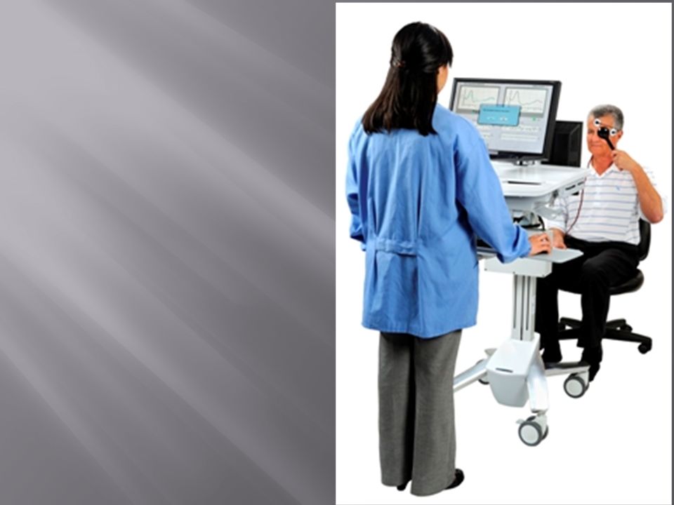
AKA Visually Evoked Response (VER) Flash vs. Pattern Measures the entire visual pathway From cornea to occipital lobe 3 electrodes Ground Reference Measuring -> occipital lobe 1” above inion

7
ReferenceGroundActive VEP Electrodes

8
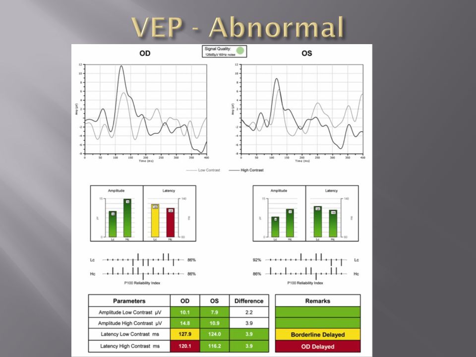
LATENCY (ms) AMPLITUDE (µv) Amplitude usually translates to the amount of axons conducting along the visual pathway Latency usually translates to the myelin status of the visual pathway VEP
 AMPLITUDE (µv) Amplitude usually translates to the amount of axons conducting along the visual pathway Latency usually translates to the myelin status of the visual pathway VEP")
9
Many optic nerve diseases are asymptomatic because central vision is not affected until late in the disease 1 Diagnosis and management of optic nerve disorders are often based on structural or subjective visual field tests 2 1 Glaucoma. American Optometric Association. www.aoa.orgwww.aoa.org 2 Prata, Tiago MD, G. De Moraes MD, J. Liebmann MD, R. Ritch, C. Tello MD. (2009). Diagnostic Ability of Fast Transient Visual Evoked Potential for Glaucoma Assessment [Poster & Abstract] American Academy of Ophthalmology. 128 VEP is an objective, functional test that can help discriminate between healthy and glaucomatous eyes 2
. Diagnostic Ability of Fast Transient Visual Evoked Potential for Glaucoma Assessment [Poster & Abstract] American Academy of Ophthalmology. 128 VEP is an objective, functional test that can help discriminate between healthy and glaucomatous eyes 2.")
11
“Increased pattern VEP latency was significantly correlated with both the severity and location of visual field defects and the degree of cupping and pallor of the optic disc.” The authors of this paper are world recognized electrophysiology specialist form New England Medical Center and University of Chicago

12
“The finding that is of clinical importance is the presence of abnormally long VEP latencies in some patients with ocular hypertension. The abnormal prolongation of VEP latency in these eyes may reflect subclinical optic nerve lesions that have not been uncovered with other techniques.”

13
Repeatability of short-duration transient visual evoked potentials in normal subjects. Tello C, De Moraes CG, Prata TS, Derr P, Patel J, Siegfried J, Liebmann JM, Ritch R. Doc Ophthalmol. 2010 Jun;120(3):219-28. Epub 2010 Jan 29. Short Duration Transient Visual Evoked Potentials in Glaucomatous Eyes. Prata TS, Lima VC, De Moraes CG, Trubnik V, Derr P, Liebmann JM, Ritch R, Tello C. J Glaucoma. 2011 May 10. [Epub ahead of print] Short-duration transient visual evoked potential for objective measurement of refractive errors. Anand A, De Moraes CG, Teng CC, Liebmann JM, Ritch R, Tello C. Doc Ophthalmol. 2011 Dec;123(3):141-7. Epub 2011 Sep 20.
: Epub 2010 Jan 29. Short Duration Transient Visual Evoked Potentials in Glaucomatous Eyes. Prata TS, Lima VC, De Moraes CG, Trubnik V, Derr P, Liebmann JM, Ritch R, Tello C. J Glaucoma May 10. [Epub ahead of print] Short-duration transient visual evoked potential for objective measurement of refractive errors. Anand A, De Moraes CG, Teng CC, Liebmann JM, Ritch R, Tello C. Doc Ophthalmol Dec;123(3): Epub 2011 Sep 20..")
14
dead Suffering Alive Glaucoma VEP OCT HRT GDX Effect of epigallocatechin-gallate on inner retinal function in ocular hypertension and glaucoma: a short-term study by pattern electroretinogram. Graefes Arch Clin Exp Ophthalmol. 2009 Sep;247(9):1223-33. Epub 2009 Mar 17.
: Epub 2009 Mar 17..")
15
Alive Glaucoma VEP OCT HRT GDX Alive dead Effect of epigallocatechin-gallate on inner retinal function in ocular hypertension and glaucoma: a short-term study by pattern electroretinogram. Graefes Arch Clin Exp Ophthalmol. 2009 Sep;247(9):1223-33. Epub 2009 Mar 17.
: Epub 2009 Mar 17..")
16
Many optic nerve diseases are asymptomatic because central vision is not affected until late in the disease 1 Diagnosis and management of optic nerve disorders are often based on structural or subjective visual field tests 2 1 Glaucoma. American Optometric Association. www.aoa.orgwww.aoa.org 2 Prata, Tiago MD, G. De Moraes MD, J. Liebmann MD, R. Ritch, C. Tello MD. (2009). Diagnostic Ability of Fast Transient Visual Evoked Potential for Glaucoma Assessment [Poster & Abstract] American Academy of Ophthalmology. 128 VEP is an objective, functional test that can help discriminate between healthy and glaucomatous eyes 2
. Diagnostic Ability of Fast Transient Visual Evoked Potential for Glaucoma Assessment [Poster & Abstract] American Academy of Ophthalmology. 128 VEP is an objective, functional test that can help discriminate between healthy and glaucomatous eyes 2.")
17
Low contrast testing demonstrates degradation of magnocellular pathways An early indication of glaucoma High contrast testing demonstrates degradation of parvocellular pathways An early indicator of central vision loss and issues caused by problems before signal reaches optic nerve **patient should be tested with best corrected vision**

18
Main Indications Glaucoma ***Glaucoma suspects*** Multiple Sclerosis Ischemic Optic Neuropathy Traumatic Brain Injury Amblyopia Other Neuropathies Unexplained vision loss VF defect FDT

20
Diopsys ® VEP Report

32
ERG’s are electrical signals that are a measure of the electrophysiological activity at the retina ***Mid-retinal layers, ganglion cell layer, and nerve fiber layer*** Objectively measures retinal function** ERG’s can help improve sensitivity and specificity in diagnosing optic neuropathies and maculopathies like glaucoma and macular degeneration when used in conjunction with other tests Can also help the clinician differentiate between retinal and optic nerve disorders when used in conjunction with Visual Evoked Potential (VEP).
.")
33
1. Concentric Stimulus Fields Drug toxicity Diabetic macular edema AMD 2. Contrast Sensitivity Glaucoma Diabetic retinopathy

34
1. Concentric Stimulus Fields Stimulus delivered at 15 flips/second BCVA Pt should be properly refracted for 24” 24” testing distance 100% contrast Right eye (OD) then Left Eye (OS) 25 seconds at 24 degrees 25 seconds at 16 degrees
 then Left Eye (OS) 25 seconds at 24 degrees 25 seconds at 16 degrees.")
35
2. Contrast Sensitivity Stimulus delivered at 15 flips/second BCVA Pt should be properly refracted for 24” 24” testing distance 85% and 15% Right eye (OD) then Left Eye (OS) 25 seconds at High Contrast (Hc) 25 seconds at Low Contrast (Lc)
 then Left Eye (OS) 25 seconds at High Contrast (Hc) 25 seconds at Low Contrast (Lc).")
36
“In patients who are glaucoma suspects, pERG signal anticipates an equivalent loss of OCT signal by several years (as many as 8 years). Invest Ophthalmol Vis Sci. 2013;54:2346-2352) DOI:10.1167/iovs.12-11026
 DOI: /iovs")
37
dead Suffering Alive Glaucoma VEP OCT HRT GDX Effect of epigallocatechin-gallate on inner retinal function in ocular hypertension and glaucoma: a short-term study by pattern electroretinogram. Graefes Arch Clin Exp Ophthalmol. 2009 Sep;247(9):1223-33. Epub 2009 Mar 17.
: Epub 2009 Mar 17..")
38
Alive Glaucoma VEP OCT HRT GDX Alive dead Effect of epigallocatechin-gallate on inner retinal function in ocular hypertension and glaucoma: a short-term study by pattern electroretinogram. Graefes Arch Clin Exp Ophthalmol. 2009 Sep;247(9):1223-33. Epub 2009 Mar 17.
: Epub 2009 Mar 17..")
39
“In patients who are glaucoma suspects, pERG signal anticipates an equivalent loss of OCT signal by several years (as many as 8 years). Invest Ophthalmol Vis Sci. 2013;54:2346-2352) DOI:10.1167/iovs.12-11026
 DOI: /iovs")
40
Glaucoma Optic Neuropathies Maculopathies AMD Diabetic retinopathy Diabetic macular edema Macular toxicity

45
VEP FLASH ERG 1. Glaucoma & glaucoma suspects 2. Unexplained vision loss 3. Transient vision loss 4. Unexplained VF defects 5. Unreliable VF 6. Optic neuropathies 7. Optic neuritis/MS 8. Amblyopia 9. TBI 1. RP & its variants 2. Cone dystrophies & Rod monochromat 3. Symptoms: “Night blindness” Restricted peripheral fields Color vision deficits PERG 1. Glaucoma & glaucoma suspects 2. Unexplained VF defects 3. Unreliable VF 4. Optic neuropathies 5. Maculopathies 1. AMD 2. Diabetic macular edema 3. High risk med use (Plaquenil) 4. Generalized DR
 4. Generalized DR.")
Similar presentations


 Inner retinal cells (bipolar.>")
 Waxman MD PhD>")

>")


 HPI: A 74-y Caucasian.>")


>")


