Download presentation
Presentation is loading. Please wait.

1
SUFIA HUSAIN ASSISTANT PROFESSOR & CONSULTANT PATHOLOGY KKUH, RIYADH September 2014

2
Pathology is the study of disease by scientific methods. It is the study of changes which occur in cells and tissues as a result of any injury to the cell or tissue. Disease is defined as an abnormality in structure or function of any part of the body.

3
The following are the 5 major aspects studied as part of pathology of any disease. 1.Epidemiology 2.Etiology, 3.Pathogenesis, 4.Morphologic changes and 5.Clinical features (signs and symptoms)
.")
4
Study of the occurrence and distribution of diseases or events in specified populations and the application of this knowledge to control the health problem. It is the study of the patterns, causes, and effects of health and disease conditions in defined populations. Epidemiology studies provide information regarding the following: 1.Sex 2.Age 3.Race 4.Occupation: workers in asbestos industry can have asbetosis or mesotheliomas. Workers in aniline dye industry can have urinary bladder cancer, hardwood workers can have nasal cancer from inhalation of wood dust etc.

5
5.Geographic location: Underdeveloped countries has more malnutrition and infection like tuberculosis. Developed countries have more cardiac problems, obesity related diseases etc. 6.Socioeconomic strata: 7.Prevalence: is the total number of cases of a particular disease in a particular defined population in a particular period of time. 8.Incidence: is the number of new cases of a particular disease in a defined population in a defined period of time. It measures the rate of occurrence of new cases. Immunization programmes affect the incidence of a disease. 9.Sequalea: it is the complication or the consequence of a disease. 10.Prognosis: it is the expected outcome of the disease. It is the estimate of the severity and possible results of the disease. 11.Morbidity: is the presence and extent of illness 12.Mortality rate is a measure of the number of deaths (in general, or due to a specific cause) in a defined population, during a defined period of time.
 in a defined population, during a defined period of time..")
6
Purposes and importance of Epidemiology 1. To investigate the extent of a disease in a community. 2. To study natural history and prognosis of disease 3. To identify causes and risk factors 4. To provide good health care based on the findings. 5. To recommend and assist in various interventions done to prevent or treat disease (preventive and therapeutic measures), e.g. immunizations and screening programs for different disease etc. 6. To evaluate all health care facilities and programs. 7. Provide information on public health in order to help plan and execute health care and prevention programmes, and informs policy decisions.
, e.g. immunizations and screening programs for different disease etc. 6. To evaluate all health care facilities and programs. 7. Provide information on public health in order to help plan and execute health care and prevention programmes, and informs policy decisions..")
7
Etiology of a disease means the cause of the disease. If the cause of the disease is unknown it is called idiopathic/ cryptogenic/ essential etc. There are two major classes of etiologic factors: 1. Genetic e.g. Down’s syndrome(extra chromosome 21), it is a chromosomal abnormality 2. Acquired Infectious: bacterial, viral, fungal. Nutritional: e.g. protein energy malnutrition Chemical: e.g. alcohol cause liver disease/cirrhosis, paraquat poisoning damages the lungs and excessive smoking causes lung and cardiac problems. Radiation: e.g. thyroid cancer after radiation to neck. Post radiation skin cancer (squamous cell carcinoma) etc. Mechanical: e.g. road traffic accident
, it is a chromosomal abnormality 2. Acquired Infectious: bacterial, viral, fungal. Nutritional: e.g. protein energy malnutrition Chemical: e.g. alcohol cause liver disease/cirrhosis, paraquat poisoning damages the lungs and excessive smoking causes lung and cardiac problems. Radiation: e.g. thyroid cancer after radiation to neck. Post radiation skin cancer (squamous cell carcinoma) etc. Mechanical: e.g. road traffic accident.")
8
Pathogenesis is the mechanism by which the cause leads to tissue injury (pathological manifestations). The pathogenetic mechanisms could take place in the latent or incubation period. The four basic pathogenetic mechanisms are as follows: Inflammation Degenerative Carcinogenesis: transformation of normal cells to malignant. Immunological All these will be dealt with in later chapters Pathogenesis leads to morphologic changes.

9
The morphologic changes are the structural changes that take place in cells or tissues after any disease. These morphological changes can be seen grossly (called macroscopic findings) with the naked eye or sometimes they can only be seen under the light microscope (called microscopic/histologic findings). Often a particular gross or microscopic change is specific for a particular disease so it helps in the diagnosis of that disease.
 with the naked eye or sometimes they can only be seen under the light microscope (called microscopic/histologic findings). Often a particular gross or microscopic change is specific for a particular disease so it helps in the diagnosis of that disease..")
10
The normal function of the affected organ and this will lead to the development clinical features (signs & symptoms) of a disease. ◦ Symptoms: is something experienced and reported by the patient e.g. ‘I am feeling tired’, ‘I have a headache’ etc. ◦ Signs: are findings discovered by the physician during examination of the patient e.g. doctor finds a lymph node when he/she examines the neck of patient or doctor find a liver or spleen enlargement while palpating the abdomen etc.

11
Epidemiology Etiology Pathogenesis Morphological or chemical alteration Clinical features (signs and symptoms)
")
12
Definition Epidemiology of disease Clinical features/presentation: signs and symptoms Pathogenesis and pathophysiology Morphology: it is divided into Gross/ macroscopic- visible to the naked eye Microscopic- visible under a microscope Differential diagnosis: is there any other alternative diagnosis/diagnoses with similar findings Treatment and management Prognosis

13
1. CONGENITAL DISEASE: is a condition existing at birth and often before birth, or that develops during the first month of life, regardless of causation. They can be: ◦ Genetic/ chromosomal: e.g. hemophilia which is an x- chromosome linked disorder), Down syndrome, inborn error of metabolism etc ◦ Non-genetic: e.g. a birth defect like cleft lip/palate or spina bifida.
, Down syndrome, inborn error of metabolism etc ◦ Non-genetic: e.g. a birth defect like cleft lip/palate or spina bifida..")
14
2. ACQUIRED DISEASES: They can be: ◦ Inflammatory e.g. rhematoid arthiritis ◦ Infective ◦ Vascular or Immune mediated e.g. atherosclerosis (causes stroke and heart attack), vasculitis etc. ◦ Degenerative e.g. Alzheimer’s disease and Parkinson’s disease ◦ Neoplastic (growth disorder) e.g. cancer ◦ Drug related e.g. liver or kidney failure secondary to certain drugs, bone marrow suppression, skin rash ◦ Metabolic e.g. gout, diabetes mellitus etc. ◦ Nutritional deficiency diseasese.g. anemia, protein energy malnutrition etc.
, vasculitis etc. ◦ Degenerative e.g. Alzheimer’s disease and Parkinson’s disease ◦ Neoplastic (growth disorder) e.g. cancer ◦ Drug related e.g. liver or kidney failure secondary to certain drugs, bone marrow suppression, skin rash ◦ Metabolic e.g. gout, diabetes mellitus etc. ◦ Nutritional deficiency diseasese.g. anemia, protein energy malnutrition etc..")
15
The course of a disease is the natural history of the disease in the absence of any intervention. The different stages in the natural history of disease especially infectious are: a) Exposure to various risk factors (causative agents) b) Latent period between exposure and onset of disease. Incubation (induction) period refers to variable period of time from the time of exposure to the development of signs or symptoms. c) Onset of disease: the beginning of signs or symptoms. d) Outcome and consequences of disease: Following clinical onset, disease may follow any of the following trends: Resolution/recovery without complication or sequalae. The disease can settle down but with sequelae. Complications Death.
 Exposure to various risk factors (causative agents) b) Latent period between exposure and onset of disease. Incubation (induction) period refers to variable period of time from the time of exposure to the development of signs or symptoms. c) Onset of disease: the beginning of signs or symptoms. d) Outcome and consequences of disease: Following clinical onset, disease may follow any of the following trends: Resolution/recovery without complication or sequalae. The disease can settle down but with sequelae. Complications Death..")
16
Any patient presenting to a clinic is put through history taking, clinical, radiological and pathological examination in order to come to a diagnosis. Clinical examination includes looking for signs of disease on the patient’s body (e.g. rash, lump etc.). The pathological examinations the are commonly done are various blood, urine and stool tests. Sometimes the patient is also asked to undergo a cytological or histopathological tests or other special pathological tests in order to obtain an accurate diagnosis. This way pathology plays an essential role in the management of the patient and diagnosis of their disease and therefore their treatment.
. The pathological examinations the are commonly done are various blood, urine and stool tests. Sometimes the patient is also asked to undergo a cytological or histopathological tests or other special pathological tests in order to obtain an accurate diagnosis. This way pathology plays an essential role in the management of the patient and diagnosis of their disease and therefore their treatment..")
17
1) Histopathology 2) Cytopathology 3) Immunohistochemistry 4) Hematopathology 5) Chemical pathology/ clinical biochemistry 6) Microbiology 7) Immunology 8) Toxicology 9) Cytogenetics 10) Molecular techniques 11) Autopsy
 Histopathology 2) Cytopathology 3) Immunohistochemistry 4) Hematopathology 5) Chemical pathology/ clinical biochemistry 6) Microbiology 7) Immunology 8) Toxicology 9) Cytogenetics 10) Molecular techniques 11) Autopsy")
18
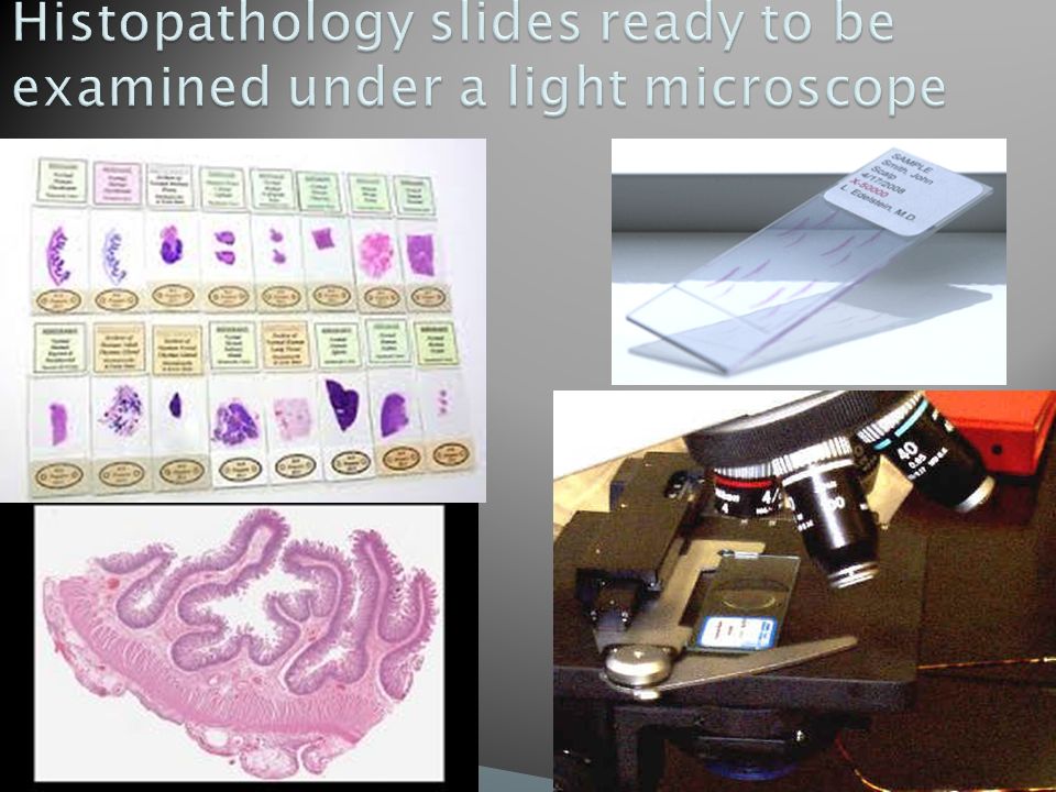
Histopathology is the study of tissues under the microscope. Tissues are obtained by biopsies and excision of organs etc. Once the tissue is removed from the patient, it has to be immediately fixed/preserved by putting it in 10% formaldehyde/formalin. The purpose of fixation is to prevent autolysis and bacterial decomposition.

19
Tissue is processed is special way resulting in very thin tissue slices (4 to 6 microns). The end result is thin slices of stained tissue on a slide. The Hematoxylin & Eosin stain is routinely used. It gives the nucleus a blue color & the cytoplasm a pinkish color. The pathologist will look at the slide under the microscope and give a diagnosis. Histopathology is usually the gold standard for pathologic diagnosis. NOTE: sometimes during surgery an urgent diagnosis is needed and tissue is processed rapidly to give results in 20 minutes. This is called frozen section.

21
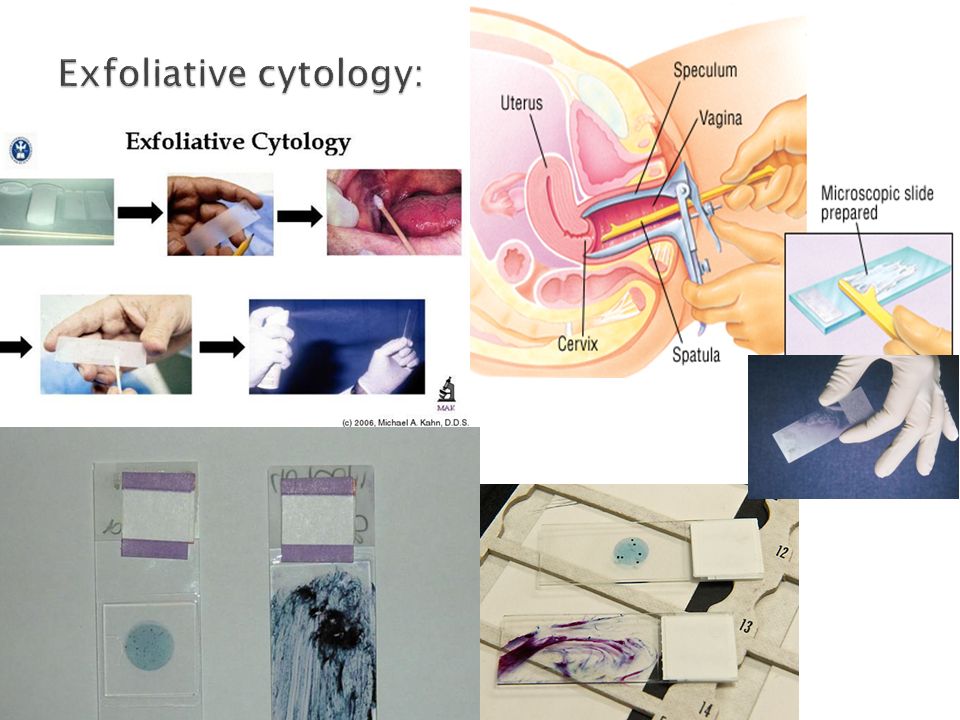
Cytopathology is the study of cells which are scraped (exfoliative cytology) or aspirated (fine-needle aspiration cytology) from various parts of body. The morphology of the cells are studied and a diagnosis made from it. It is used for the purpose of: Screening for cancer e.g. cervical cytology is used in the screening of carcinoma of cervix. Diagnosing cancer The advantage of cytologic technique when compared to histopathological techniques is that the procedure is cheap, takes less time and requires no anesthesia. Fine-needle aspiration cytology (FNAC): It is the suction of cells from diseased organ. In FNAC, cells are obtained by aspirating the diseased organ or mass using a needle. The cells obtained are stained and examined under a microscope. Exfoliative cytology: The cells are scraped of the mucosa using a spatula (e.g. cervix and oral cavity) or the cells exfoliate themselves and collect in a particular type of secretion (e.g. in urinary tract disease the cells which exfoliate collect in the urine). E.g. sputum, cerebrospinal fluid, urine, effusions in lung abdomen, wall of cervix etc.
: It is the suction of cells from diseased organ. In FNAC, cells are obtained by aspirating the diseased organ or mass using a needle. The cells obtained are stained and examined under a microscope. Exfoliative cytology: The cells are scraped of the mucosa using a spatula (e.g. cervix and oral cavity) or the cells exfoliate themselves and collect in a particular type of secretion (e.g. in urinary tract disease the cells which exfoliate collect in the urine). E.g. sputum, cerebrospinal fluid, urine, effusions in lung abdomen, wall of cervix etc..")
22
http://themedicalpost.files.wordpress.com/2010/05/fine-needle-biospy- 2_small.jpg

24
a) Histology b) Cytology b a
 Histology b) Cytology b a")
25
3. Hematology: a study of the constituents of blood and bone marrow, used in the diagnosis of anemias & leukemias. 4. Immunohistochemistry: is a specialized staining procedure is used to detect antigens in the tissue. 5. Biochemical tests/biochemistry: is the analysis of bodily fluids (blood, urine, etc) for diagnosis. 6. Microbiology: is the study of micro-organisms 7. Immunology: is the analysis of the immune system of the body. 8. Toxicology: study of various poisons and toxic substances. 9. Cytogenetics (clinical genetics): is a study of chromosomal abnormalities. 10. Molecular techniques: various molecular techniques such as fluorescent in situ hybridization, Southern blot are used to detect genetic diseases.
 for diagnosis. 6. Microbiology: is the study of micro-organisms 7. Immunology: is the analysis of the immune system of the body. 8. Toxicology: study of various poisons and toxic substances. 9. Cytogenetics (clinical genetics): is a study of chromosomal abnormalities. 10. Molecular techniques: various molecular techniques such as fluorescent in situ hybridization, Southern blot are used to detect genetic diseases..")
26
It is a sub-specialty of pathology which involves examining a dead body An autopsy is done to to determine the cause of death (this is the main reason why autopsy is done). It can be homicidal, suicidal, accidental, some disease (like cancer, cardiovascular disease) etc. provide useful information about various disease. do research. Also it can be used as a tool to educate students, surgeons etc Who does the autopsy? The pathologist.
 etc. provide useful information about various disease. do research. Also it can be used as a tool to educate students, surgeons etc Who does the autopsy. The pathologist..")
27
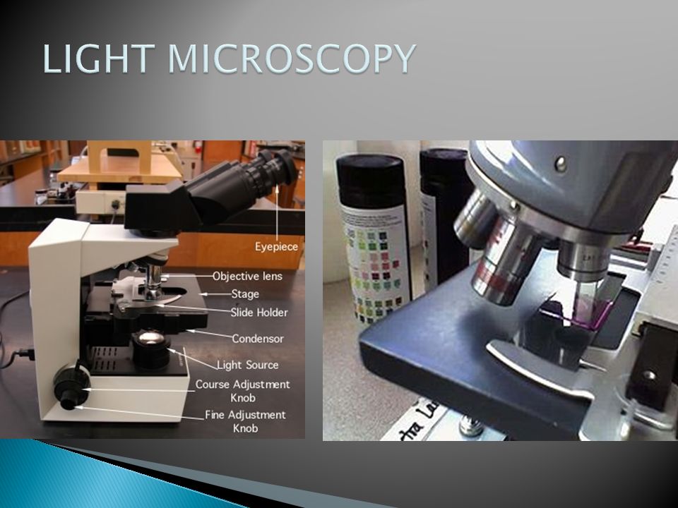
There are different diagnostic instruments used in pathology. Some of the instruments used in pathology are ◦ light microscope ◦ immunofluorescent microscope ◦ electron microscope.

29
Immunofluorescence micros copy uses a special blue filter to search for various antigens in a tissue with the help of a flourescent dye. It helps in diagnosing immunological diseases.

30
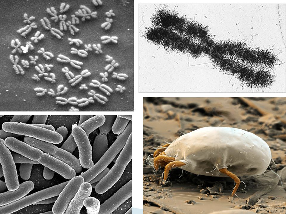
The electron microscope is a type of microscope that uses electrons to magnify objects up to two million times, which is much higher than a light microscope It enables us to see cell structure like mitochondria, endoplasmic reticulum, viral particles etc. It is also called as ultra structural studies. It is an expensive technique.

32
A tardigrade, or "water bear" is seen through an electron microscope. Less than 1 mm in length,

Similar presentations


 The following percentages (%) of the total grade will be assigned: In-Course Assessments..........................………………………..………60.>")

>")
