Download presentation
Presentation is loading. Please wait.

1
Transmediastinal Penetrating Injuries Tanya L. Zakrison Clinical Fellow Ryder Trauma Center University of Miami August 25 th, 2009

2
Thank you to Drs. Asensio, Stahl and Prichayudh for slide contributions

3
Objectives Anatomy & definitions History & Epidemiology Common Algorithms Operative Approach Injuries: Life threatening early: Cardiac, great vessels Life threatening late: Esophageal, tracheobronchial Unique Problems Foreign body embolization Azygous vein injury Spinal cord injury

4
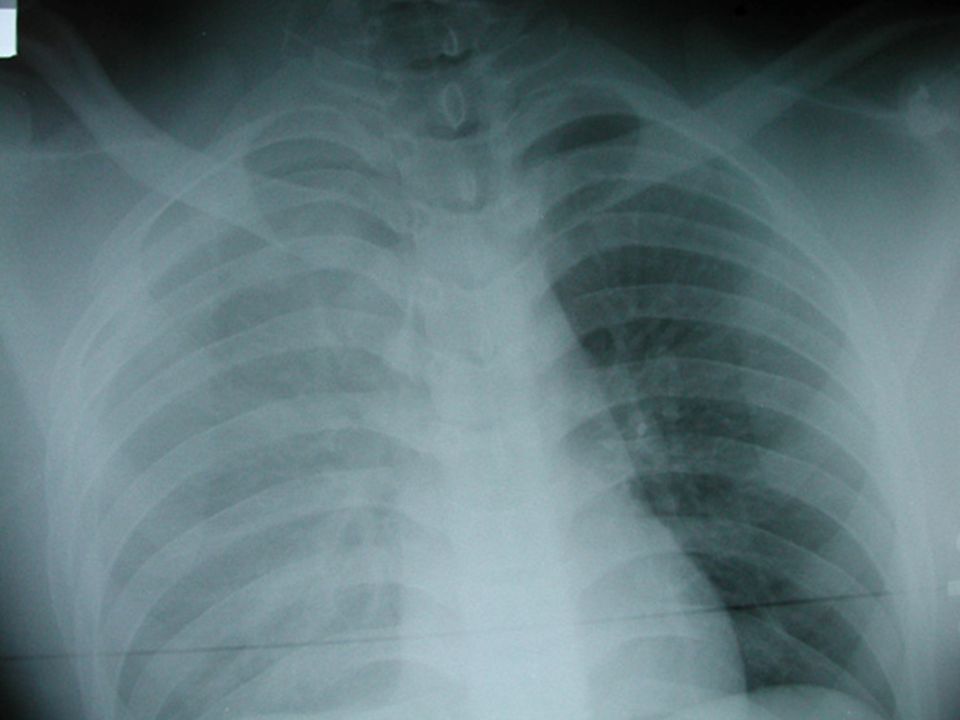
Case ID: 35M GSW X 1 chest One GSW R anterior axillary line 3 rd ICS One GSW L anterior axillary line 3 rd ICS Alert, confused GCS = 14 Diminished breath sounds bilaterally SaO2 = 92% RA BP = 70/30, faint bilateral radial pulses No gross neurologic deficits No other blunt or penetrating injuries What next?

5
ATLS initiated 2 400 cc crystalloid given CXR: Superior mediastinal hematoma Bilateral hemothoraces Bilateral chest tubes 400 cc blood out each side BP = 74/40 mmHg FAST negative What would you do you do next?

6
Definitions Mediastinum: (Dorland’s Medical Dictionary) 1. A median septum or partition 2. The mass of tissues and organs separating the two lungs, between the sternum in front and vertebral column behind Transmediastinal Penetrating Injury / GSW (Richardson et al. 1981) missile entry & exit wounds or missile entry wound & retained missile localized on radiography in opposite hemithoraces Traverse Mediastinal Gun Shot Wound Transverse Mediastinal Gun Shot Wound
 missile entry & exit wounds or missile entry wound & retained missile localized on radiography in opposite hemithoraces Traverse Mediastinal Gun Shot Wound Transverse Mediastinal Gun Shot Wound.")
7
A transmediastinal penetrating injury does not necessarily mean a mediastinal penetrating injury

8
Anatomy: Mediastinum “Interpleural Space”

9
Superior: the aortic arch; the innominate artery and the thoracic portions of the left common carotid and the left subclavian arteries; the innominate veins and the upper half of the superior vena cava; the left highest intercostal vein; the vagus, cardiac, phrenic, and left recurrent nerves; the trachea, esophagus, and thoracic duct; the remains of the thymus, and some lymph glands. Anterior: It contains a quantity of loose areolar tissue, some lymphatic vessels which ascend from the convex surface of the liver, two or three anterior mediastinal lymph glands, and the small mediastinal branches of the internal mammary artery. Middle: It contains the heart enclosed in the pericardium, the ascending aorta, the lower half of the superior vena cava with the azygos vein opening into it, the bifurcation of the trachea and the two bronchi, the pulmonary artery dividing into its two branches, the right and left pulmonary veins, the phrenic nerves, and some bronchial lymph glands. Posterior: It contains the thoracic part of the descending aorta, the azygos and the two hemiazygos veins, the vagus and splanchnic nerves, the esophagus, the thoracic duct, and some lymph glands. Mediastinal Contents

10
Anatomy: Mediastinum “Interpleural Space” Superior: the aortic arch; the innominate artery and the thoracic portions of the left common carotid and the left subclavian arteries; the innominate veins and the upper half of the superior vena cava; the left highest intercostal vein; the vagus, cardiac, phrenic, and left recurrent nerves; the trachea, esophagus, and thoracic duct; the remains of the thymus, and some lymph glands. Anterior: It contains a quantity of loose areolar tissue, some lymphatic vessels which ascend from the convex surface of the liver, two or three anterior mediastinal lymph glands, and the small mediastinal branches of the internal mammary artery. Middle: contains the heart enclosed in the pericardium, the ascending aorta, the lower half of the superior vena cava with the azygos vein opening into it, the bifurcation of the trachea and the two bronchi, the pulmonary artery dividing into its two branches, the right and left pulmonary veins, the phrenic nerves, and some bronchial lymph glands. Posterior: It contains the thoracic part of the descending aorta, the azygos and the two hemiazygos veins, the vagus and splanchnic nerves, the esophagus, the thoracic duct, and some lymph glands. Other includes: azygous vein, thoracic duct, spinal cord Great Vessels Nothing… Heart & Airways Esophagus & others Mediastinal Contents

11
Transmediastinal Penetrating Trauma What do we worry about as surgeons? Injuries that kill early: Heart Great vessels Injuries that kill ‘later’: Tracheobronchial tree Injuries that kill if missed: Esophagus Azygous vein Injuries that cause morbidity: Thoracic duct Spinal cord

12
Thoracoabdominal area Cardiac Box Mediastinum Additional Concerns: Associated Injuries: Thorax: Chest wall Lungs Abdomen: Any structure Timing of exploration important Asensio et al. World J Surgery, 2002

13
History of Penetrating Thoracic Trauma Described in the Edwin Smith Surgical Papyrus, dated 3000 BC, written by Imhotep Galen reported treatment of gladiators sustaining chest injuries with packing First recorded “operation” for thoracic trauma in North America Cabeza de Vaca, 1635, described removal of arrowhead from the chest wall of an Native American Contemporary management of transmediastinal penetrating injuries required mandatory exploration Bradley M. Transmediastinal wounds. Am. Surg. 1966;32:847–852

14
Epidemiology of Penetrating Thoracic Trauma 150 000 people die a year secondary to trauma 25% of deaths related to thoracic trauma Urgent operative intervention only required in 2.8% of penetrating thoracic injuries Mediastinal penetrating injuries have an increased need for operation Unstable: 43% - all to OR Stable: 57% - 35-60% to OR (Richardson et al., Surgery, 1981) Management dictated by hemodynamic status
 Management dictated by hemodynamic status")
15
Can we triage patients based on blood pressure? (OR vs. investigations) Prospective, N = 68 Group I = SBP > 100 mmHg Dx: CXR > PE > OR Group II = SBP 60 – 100 mmHg Dx: CXR > PE > OR Group III = SBP < 60 mmHg Dx: ED thoracotomy > death > OR Conclusions: CXR with PE can diagnose TM GSW in 90% of pts. SBP > 100 mmHg, investigate 60% do not need an operation SBP < 60 mmHg, act quickly 100% need an ‘operation’ SBP 60 – 100 mmHg, watch response 50% do not need an operation 19% of all pts. also had intraabdominal injuries Indication for immediate OR in stable patients: Massive hemothorax & hemopericaridum
 Prospective, N = 68 Group I = SBP > 100 mmHg Dx: CXR > PE > OR Group II = SBP 60 – 100 mmHg Dx: CXR > PE > OR Group III = SBP < 60 mmHg Dx: ED thoracotomy > death > OR Conclusions: CXR with PE can diagnose TM GSW in 90% of pts. SBP > 100 mmHg, investigate 60% do not need an operation SBP < 60 mmHg, act quickly 100% need an ‘operation’ SBP 60 – 100 mmHg, watch response 50% do not need an operation 19% of all pts. also had intraabdominal injuries Indication for immediate OR in stable patients: Massive hemothorax & hemopericaridum.")
16
Majority of stable patients with TM GSWs do not need an OR If they do, it’s bleeding from the heart or great vessels All unstable patients need surgery Those in between can be investigated if they respond to resuscitation

17
Algorithms for Stable Transmediastinal Injuries Overt Injuries: Heart, great vessels To OR Occult Injuries: Heart, great vessels Tracheobronchial tree Esophagus Azygous vein Thoracic duct Patients with negative helical CTAs can be safely observed

18
Algorithms for All Transmediastinal Injuries 1. In Extremis: ED thoracotomy 2. Hemodynamically unstable: CXR, FAST To the OR 3. Hemodynamically stable: Diagnosis (CTA) Trajectory & injuries Therapeusis Surgical vs. conservative
 Trajectory & injuries Therapeusis Surgical vs. conservative.")
19
1) Patient in Extremis = ED Thoracotomy
 Patient in Extremis = ED Thoracotomy")
20
2) Pt. Unstable = To the OR
 Pt. Unstable = To the OR")
21
Pre-Operative Approach in All ABCDEs, protocolized ATLS approach Tube thoracostomy both diagnostic & therapeutic Fluid restriction, lines above & below the diaphragm Bickell et al. N Engl J Med. 1994 Oct 27;331(17):1105-9 Document neurologic status Risk of paraplegia, stroke, brachial plexus injury high in reconstruction of great vessels Arrange for autotransfusion device in the OR Close communication with anesthesia
: Document neurologic status Risk of paraplegia, stroke, brachial plexus injury high in reconstruction of great vessels Arrange for autotransfusion device in the OR Close communication with anesthesia.")
22
Operative Approach What is injured? Great Vessels Massive hemothorax (unilateral or bilateral) Large mediastinal hematoma on CXR Heart FAST positive FAST indeterminate Ongoing blood loss from chest tube Both Both +/- other organs Other organs (massive tracheobronchial injury)
 Large mediastinal hematoma on CXR Heart FAST positive FAST indeterminate Ongoing blood loss from chest tube Both Both +/- other organs Other organs (massive tracheobronchial injury).")
25
Great Vessel Injury: Where to Cut?

26
Transmediastinal Injuries

27
Where to Cut? Median Sternotomy Principles: “Anywhere you need” (Dr. McKenney) Median Sternotomy +/- cervical extension (R or L): R subclavian artery, proximal R carotid, brachiocephalic artery, proximal L carotid artery NOT GOOD FOR L subclavian artery Too far posterior 3 rd ICS anterolateral thoracotomy for proximal control Supraclavicular incision with resection of medial 3 rd of clavicle (distal control) Median sternotomy (to join the incisions – book / trap door) Rarely used GOOD FOR concomitant cardiac injuries
 Median Sternotomy +/- cervical extension (R or L): R subclavian artery, proximal R carotid, brachiocephalic artery, proximal L carotid artery NOT GOOD FOR L subclavian artery Too far posterior 3 rd ICS anterolateral thoracotomy for proximal control Supraclavicular incision with resection of medial 3 rd of clavicle (distal control) Median sternotomy (to join the incisions – book / trap door) Rarely used GOOD FOR concomitant cardiac injuries.")
28
Where to Cut? Bilateral Anterolateral Thoracotomies Utility incision, access to heart and aorta for resuscitation Can access right lung hilum, ascending aorta, right subclavian vessels Also problematic for L subclavian artery injuries

29
What Can You Tie Off? ‘Any’ vessel Especially subclavian artery Brachiocephalic vein (gatekeeper) Do try to repair: Brachiocephalic artery, carotid, aorta Superior Vena Cava
 Do try to repair: Brachiocephalic artery, carotid, aorta Superior Vena Cava.")
30
Approach to the Mediastinal Hematoma – the Trail of Safety 1. Median sternotomy for superior mediastinum 2. Identify upper border of pericardium 1. Divide thymus if needed 3. Identify and ligate the left branch of the brachiocephalic vein 1. Access to superior aspect aortic arch 4. Orient self by opening pericardium 1. Blocks hematoma extension 5. Identify the bifurcation of the brachiocephalic artery 1. Protect vagus nerve 6. Proximal and distal control 1. Extend incision as needed

31
Great Vessels – Surgical Principles Great vessels are fragile, tear easily with dissection, therefore oversew proximal injury on aorta, sew graft into new location on aorta without tension Use prosthetic graft for vessels > 5 mm vs. saphenous vein (pseudoaneurysm vs. acute rupture) Dacron for fragile vessels – aorta, SCA
 Dacron for fragile vessels – aorta, SCA.")
32
Approach to the Subclavian Hematoma – the Trail of Safety 1. Is operation for ischemia or bleeding? 1. Are there alternative? 2. High 3 rd ICS incision on left side, median sternotomy for right SCA 3. Non-bleeding SCA injury: 1. Supraclavicular approach notch 10 cms distal 2. Divide platysma, clavicular head of SCM, omohyoid 4. Protect the internal jugular vein medially 5. Find subclavian vein first then artery 1. behind scalenus anterior 2. Watch phrenic nerve and thoracic duct (SCV & IJV) 6. Divide scalenus anterior sharply to find SCA 1. Ligate thyrocervical trunk to mobilize SCA 2. Watch internal mammary and vertebral arteries 7. Bleeding SCA injuries: 1. Perform claviculectomy
 6. Divide scalenus anterior sharply to find SCA 1. Ligate thyrocervical trunk to mobilize SCA 2. Watch internal mammary and vertebral arteries 7. Bleeding SCA injuries: 1. Perform claviculectomy.")
33
Retrospective review N = 79 pts. with penetrating subclavian or axillary artery injury Conclusion: Clavicular incision alone provides adequate exposure in 50% of pts. (R and L) In proximal injuries can use the addition of medial sternotomy More deaths seen with SCV injuries than SCA J Am Coll Surg 1999; 188:290-295
 In proximal injuries can use the addition of medial sternotomy More deaths seen with SCV injuries than SCA J Am Coll Surg 1999; 188:")
34
Exposure of Subclavian Artery Asensio et al.

35
Exposure of Subclavian Artery Asensio et al.

36
Subclavian Artery Injuries Pitfalls for SCA: Watch injury to phrenic nerve when dissecting out SCA Failure of proximal control with 3 rd ICS Failure to document brachial plexus status pre-op No tunica media, end to end anastomosis doomed to fail – Interposition graft Damage control: 1. Definitive repair of injuries with quick & simple techniques in one operation 2. Abbreviated thoracotomy to restore survivable physiology during a single operation

37
Cardiac Injuries Unstable pts. may present with tamponade or ongoing blood loss from chest tube Occult cardiac injuries may be present in 5% to 10% of patients after a TM-GSW Feliciano D, et al. J. Trauma 2000;48:416–422 TTE is the diagnostic test of choice in patients with wounds traversing the anterior mediastinum When ECHO is used to screen for pericardial fluid it is 97% sensitive, 100% specific, and 99% accurate Nagy K, et al. J. Trauma 1995;38:859 – 862

39
3) Pt. Stable = Investigate Carefully
 Pt. Stable = Investigate Carefully")
40
What is our local experience with investigating TM GSWs in stable pts.? Work up depends on trajectory Prospective, N = 50 pts. All pts. had a CXR followed by either: cardiac ultrasound, angiography, esophagoscopy, barium swallow and bronchoscopy 1. 8 pts. (16%) found to have a mediastinal injury (cardiac > vascular > tracheoesophageal) 2. 42 pts. (84%) had no mediastinal injury No difference between groups re: biochemical or clinical status (including chest tube outputs) Stable pts. may have life-threatening injuries Aggressive work-up needed in all to avoid missed injuries NO CTA
 found to have a mediastinal injury (cardiac > vascular > tracheoesophageal) pts. (84%) had no mediastinal injury No difference between groups re: biochemical or clinical status (including chest tube outputs) Stable pts. may have life-threatening injuries Aggressive work-up needed in all to avoid missed injuries NO CTA.")
41
Stable Patients: How Much to Investigate? After thoracic CT scan Esophagoscopy Esophageal swallow Bronchoscopy Angiography Mandatory pericardial window OR: Selective investigation depending on the trajectory of the bullet 83% of stable pts. had negative CTAs with no missed injuries

42
Can helical CT scan reduce the need for further investigations? N = 24 pts. mediastinal GSWs, HD stable All pts. received a helical contrast CT scan 12 pts. required further imaging Bullet tract close to mediastinum – to OR All other studies negative, no missed injuries Conclusion: 50% of pts. had a change in management based on CT scan Helical CT effective to evaluate missile trajectories to assess for mediastinal injuries and avoid unneccessary exams Tangential chest wounds excluded

43
Esophageal Injuries Incidence 0.7% after thoracic gun shot wounds Transmediastinal gun shot wounds close to spine may result in through and through injury Delay in repair disastrous Upper 2/3 rds of thoracic esophagus: Right posterolateral thoracotomy Distal 1/3: Left posterolateral approach (7 th ICS) Primary repair even if > 24 hrs Grillo pleural patch & decortication
 Primary repair even if > 24 hrs Grillo pleural patch & decortication")
44
N = 43 pts. with penetrating esophageal injury Conclusion: small sample size delay in esophageal repair likely increases mortality J Trauma 43(2), 1997
,")
45
How long is too long when investigating penetrating esophageal injuries? Retrospective, multicenter study, N = 45 pts. All pts. to OR, pre-operative evaluation vs. no evaluation 13 hrs vs. 1 hr Increased complications, LOS in group evaluated (OR > 3) Conclusion: Esophageal injuries carry high morbidity and mortality. Diagnostic testing, if done, should be expeditious with delays to definitive management reduced.
 Conclusion: Esophageal injuries carry high morbidity and mortality. Diagnostic testing, if done, should be expeditious with delays to definitive management reduced..")
46
Tracheobronchial Injuries Conservative: Small injuries (< 1/3 diameter of airway) Asymptomatic Controlled with tube thoracostomy No need for PPV Or place ETT beyond injury with cuff inflated below Operative: R posterolateral thoracotomy for injuries of: Intrathoracic tracheal, right bronchial, and proximal left mainstem bronchus or complex bilateral injuries L posterolateral thoracotomy for injuries of: Distal left bronchial injuries > 3 cm from carina Mobilize anterior and posterior trachea as needed Interrupted, absorbable sutures (4-0 Vicryl), sutures tied on the outside, end to end anastomosis Buttress repair with intercostal muscle Suture chin to chest for healing Follow up: Pre-op baseline flow-volume loops Beware later presentation of ‘adult asthma’ stricture
 Asymptomatic Controlled with tube thoracostomy No need for PPV Or place ETT beyond injury with cuff inflated below Operative: R posterolateral thoracotomy for injuries of: Intrathoracic tracheal, right bronchial, and proximal left mainstem bronchus or complex bilateral injuries L posterolateral thoracotomy for injuries of: Distal left bronchial injuries > 3 cm from carina Mobilize anterior and posterior trachea as needed Interrupted, absorbable sutures (4-0 Vicryl), sutures tied on the outside, end to end anastomosis Buttress repair with intercostal muscle Suture chin to chest for healing Follow up: Pre-op baseline flow-volume loops Beware later presentation of ‘adult asthma’ stricture")
47
Tracheobronchial Injuries

49
Special Problems Bullet embolization Thoracic duct injury Spinal cord Azygous vein

50
Bullet (Foreign Body) Embolization Bullet entry into large diameter vessels of chest Diagnosis frequently delayed as course of bullet not always apparent Usually lodges in femoral or iliac vessels Control site of entry for hemorrhage first Remove bullet emboli Surgery Endovascular methods
 Embolization Bullet entry into large diameter vessels of chest Diagnosis frequently delayed as course of bullet not always apparent Usually lodges in femoral or iliac vessels Control site of entry for hemorrhage first Remove bullet emboli Surgery Endovascular methods")
51
Special Problems Thoracic duct leaks: Chylothorax Refer to Dr. Nabri Spinal cord injury Always assess neurologic function pre-operatively Neurogenic shock may occur C > T > L spine

52
Azygous Vein Injury and Repair N = 22 patients over 40 years Mortality 36% Maintain an index of suspicion when there is continued dark venous ooze from behind the pulmonary hilum Posterolateral thoracotomy best exposure Ligate the vein unless no IVC

53
35M - Penetrating wound to chest? 1. Is it a transverse mediastinal wound? (CXR & PE) 2. Did this transverse mediastinal wound cause a mediastinal injury (especially heart and / or great vessels)? (superior hematoma CXR, chest tube outputs, FAST) 3. If FAST (+):If FAST (-): If FAST (?): Median sternotomyit depends window 4. If chest tube outputs are massive: Right: median sternotomy with clavicular extension Left: median sternotomy with clavicular extension 5. If superior mediastinal hematoma (+) on CXR: Median sternotomy…+/- laparotomy 6. If dying: Left anterolateral thoracotomy + / - clamshell
 2. Did this transverse mediastinal wound cause a mediastinal injury (especially heart and / or great vessels). (superior hematoma CXR, chest tube outputs, FAST) 3. If FAST (+):If FAST (-): If FAST ( ): Median sternotomyit depends window 4. If chest tube outputs are massive: Right: median sternotomy with clavicular extension Left: median sternotomy with clavicular extension 5. If superior mediastinal hematoma (+) on CXR: Median sternotomy…+/- laparotomy 6. If dying: Left anterolateral thoracotomy + / - clamshell.")
54
Thank you!

Similar presentations