Download presentation
Presentation is loading. Please wait.

1
The Cytoskeleton Functions
Structural scaffold creating and supporting cell shape Framework positioning organelles within cytoplasm Network of molecular “roads” for intracellular transport of materials Framework for whole cell movement Framework for cell division

2
The Cytoskeleton Three major structural components Microtubules
Major role: support, intracellular transport Intermediate filaments Major role: mechanical strength to resist physical stresses Microfilaments Major role: muscle contraction, motility

3
The Cytoskeleton Microtubules (MTs) Major role Intracellular transport
Motor proteins drag cargo along them Structural support Resist compression forces Resist shear (bending) forces Hollow, rigid 25nm diameter, 4nm wall thickness Radiate outward toward plasma membrane from near nucleus (MTOC)
 forces. Hollow, rigid. 25nm diameter, 4nm wall thickness. Radiate outward toward plasma membrane. from near nucleus (MTOC)")
4
The Cytoskeleton plus-end minus-end Microtubules (MTs)
Unit = alpha / beta tubulin heterodimer alpha subunit + beta subunit Heterodimer is asymmetric Beta end is called “plus” end Alpha end is called “minus” end Not referring to a charge difference plus-end minus-end

5
The Cytoskeleton Microtubules (MTs)
alpha / beta (a/b)-tubulin heterodimer Beta subunit is a GTPase Assembly Polymer grows by addition of units at the “plus” end GTP-bound tubulin can add GTP form hydrolyzes to GDP form GDP-bound tubulin cannot add GDP-bound tubulin can release only from “plus” end GDP-bound tubulin cannot release from “minus” end or from central region
-tubulin heterodimer. Beta subunit is a GTPase. Assembly. Polymer grows by addition of units at the plus end. GTP-bound tubulin can add. GTP form hydrolyzes to GDP form. GDP-bound tubulin cannot add. GDP-bound tubulin can release only from plus end. GDP-bound tubulin cannot release from minus end or from central region.")
6
The Cytoskeleton Dynamic instability
MTs can assemble/disassemble at different rates in different locations within a single cell Various proteins can bind and stabilize MTs

7
The Cytoskeleton Microtubule-associated proteins (MAPs)
Form bridges crosslinking adjacent MTs for parallel alignment Increase MT stability Promote assembly Regulated by phosphorylation state Anti-tubulin antibody stain

8
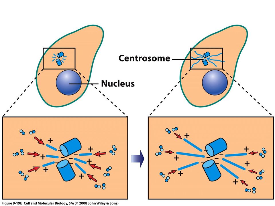
The Cytoskeleton Microtubule organizing centers (MTOCs)
GTP-bound a/b-tubulin spontaneously assembles into MTs very slowly GTP-bound a/b-tubulin add to an existing MT very rapidly MTOCs are the nucleation points for MT assembly Centrosome Basal body

9
The Cytoskeleton Microtubule organizing centers (MTOCs) Centrosome
2 centrioles at right angles to each other near nucleus Contain gamma-tubulin subunit Nucleate “minus” end of a/b-tubulin Plus-end is oriented outward toward plasma membrane

11
The Cytoskeleton Microtubule organizing centers (MTOCs) Basal body
Single centriole at the base of cilia and flagella

12
Eukaryotic cilia and flagella
Hair-like motile organelle projecting from cell surface Covered by plasma membrane

13
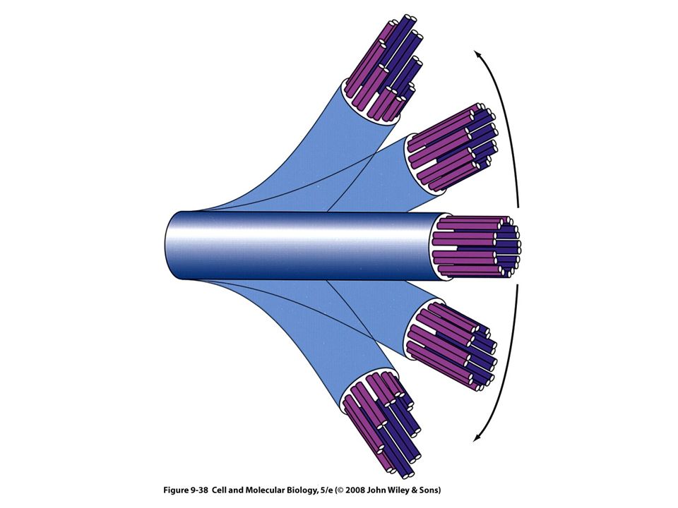
Eukaryotic cilia and flagella
Central protein core is called an “axoneme”

14
Eukaryotic cilia and flagella
Central protein core is called an “axoneme” Composed of 11 MTs arranged in a “9+2” array 9 outer MTs 2 central MTs Connected by various MAPs Locomotion caused by sliding outer tubules past each other Action of motor proteins (dynein)
")
16
The Cytoskeleton Motor proteins that “walk” on MTs Kinesin gene family
Plus-end directed Outward or “anterograde” transport Dynein gene family Minus-end directed Inward or “retrograde”

17
The Cytoskeleton Kinesins are composed of 2 heavy and 2 light polypeptides Cargo-interaction domain “tail” Different kinesins have different specificities ATPase “head” Binds to MT ATP hydrolysis propels heads forward Highly processive

18
The Cytoskeleton Kinesins are composed of 2 heavy and 2 light polypeptides ATPase “head” Binds to MT ATP hydrolysis propels heads forward Highly processive

19
The Cytoskeleton Motor proteins that “walk” on MTs Dynein gene family
Minus-end directed Inward or “retrograde” transport Very large (1.5MDa) Involved in cilia/flagella movement
 Involved in cilia/flagella movement.")
20
The Cytoskeleton Three major structural components
Intermediate filaments (~65 genes) Major role: mechanical strength to resist physical stresses Hemidesmosomes and desmosomes
 Major role: mechanical strength to resist physical stresses. Hemidesmosomes and desmosomes.")
21
Intermediate filaments (IFs)
Animal specific Strong, rope-like

22
Intermediate filaments (IFs) Animal specific Strong, rope-like
Bridged together with other cytoskeletal elements (e.g. plectin crosslinks MTs and IFs)
")
23
The Cytoskeleton Intermediate filaments Composition and assembly
Monomers form dimers Dimers form tetramers lacking polarity Tetramers form larger fibers Incorporation into existing filaments not limited to end regions

24
The Cytoskeleton Three major structural components
Microfilaments (MFs) Major role: muscle contraction, motility Solid, branched 8nm diameter Molecular unit= actin
 Major role: muscle contraction, motility. Solid, branched. 8nm diameter. Molecular unit= actin.")
25
The Cytoskeleton Microfilaments (MFs) Actin molecule is asymmetric
“plus”-end versus “minus”-end Actin is an ATPase ATP-bound actin can be incorporated into growing MFs plus-end of MFs grows 10x faster than minus-end Higher dissociation rate from minus-end leads to treadmilling

26
The Cytoskeleton Microfilaments (MFs) + cytochalasin D Drugs
Cytochalasin D blocks plus-end addition leading to complete MF depolymerization Phalloidin blocks turn-over locking MFs into polymerized state + cytochalasin D

27
The Cytoskeleton Microfilaments (MFs) + cytochalasin D Drugs
Cytochalasin D blocks plus-end addition leading to complete MF depolymerization Phalloidin blocks turn-over locking MFs into polymerized state + cytochalasin D

28
The Cytoskeleton Actin binding proteins + cytochalasin D

29
The Cytoskeleton Motors that walk on Microfilaments (MFs)
Myosin gene family ATPase “head” domain Cargo-interacting “tail” domain

30
The Cytoskeleton Motors that walk on Microfilaments (MFs)
Myosin gene family Type V can walk on actin filaments carrying a bound cargo Type II forms bipolar filaments via tail - tail interactions

31
The Cytoskeleton Myosin type II in muscle contraction Muscle fiber
Large cell, 100mm long, microns thick Contain >100 nuclei Derived from the fusion of many myoblast cells Myofibrils thin protein strands composed of repeating units called “sarcomeres” that give muscle its “striated” appearance Sarcomere Z, I, A, H and M regions

32
Myosin II

33
Sliding filament model of muscle contraction

34
Sliding filament model of muscle contraction

Similar presentations


 by W. H. Freeman and Company Chapter 18 Cell Motility and Shape I: Microfilaments.>")