Download presentation
Presentation is loading. Please wait.

2
The Circulatory System ROSELYN A. NARANJO www.roselynnaranjo.vze.com

3
Functions of the Circulatory System Brings blood containing oxygen, nutrients, and hormones to cells Transports CO 2 and other wastes away from cells

4
Functions Continued Fights infection Regulates body temperature Helps stabilize pH and ionic concentration of body fluids.

5
Circulatory System Components Heart Blood Vessels Arteries Veins Capillaries

6
The Heart A muscular pump Moves blood through the body Is suspended in the pericardial sac Composed of four chambers Divided into right and left halves Made up of cardiac muscle cells

7
Pericardium Protective sac of connective tissue Surrounds the heart Filled with fluid

8
Myocardium The muscle of the heart Strong and thick Composed of spontaneously contracting cardiac muscle fibers Can conduct electricity like nerves It’s blood supply comes from the coronary arteries Myocardium (heart muscle) shown in red Epicardium (Outer surface of myocardium) Endocardium (Inner surface of myocardium)
 shown in red Epicardium (Outer surface of myocardium) Endocardium (Inner surface of myocardium)")
9
Structures of the Heart Chambers Atria- (2) upper chambers Thin walled Receive blood from veins Send blood to ventricles Ventricles- (2) lower chambers Thick walled Receive blood from atria Pump blood out through arteries Septum Wall that divides heart into right and left halves Septum Pulmonary valve Right atrium Tricuspid valve Right ventricle Left atrium Aortic valve Mitral valve Left ventricle
 upper chambers Thin walled Receive blood from veins Send blood to ventricles Ventricles- (2) lower chambers Thick walled Receive blood from atria Pump blood out through arteries Septum Wall that divides heart into right and left halves Septum Pulmonary valve Right atrium Tricuspid valve Right ventricle Left atrium Aortic valve Mitral valve Left ventricle")
10
Structures of the Heart Valves PPrevent backflow of blood KKeep blood moving in one direction BBetween the chambers AAt junctions of artery and chamber Tricuspid valve Pulmonary veins Mitral valve Left atrium Pulmonary valve Aortic valve Right atrium Valves seen from above Chordea tendinea Pulmonary valve

11
Structures of the Heart Chordae tendinease “Heart strings” Cord-like tendons Connect papillary muscles to tricuspid and mitral valves Prevent inversion of valve Papillary muscles Small muscles that anchor the cords Papillary muscle

12
Cardiac Cycle Refers to all of the events from the beginning of one heart beat to the beginning of the next heart beat When cardiac muscle contracts it does so as a single unit, creating a heart beat One heartbeat - a cardiac cycle - consists of two parts called systole and diastole

13
Cardiac Cycle Diastole is the period of time when the heart relaxes after contraction Oxygenated blood from the lungs fills the left atrium Deoxygenated blood from other parts of the body fills the right atrium. At the end of the diastole, the atria contract, starting the Systole

14
Cardiac Cycle Atrial systole Atrial systole is the contraction of the heart muscle of the left and right atria. Both atria contract at the same time, sending blood into the corresponding ventricle Ventricular systole Ventricular systole is the contraction of the muscles of the left and right ventricles, which contract at the same time. systole The term systole is synonymous with contraction of a muscle.

15
Cardiac Cycle During systole the ventricles contract, forcing the blood into the pulmonary artery to be re-oxygenated in the lungs, and into the aorta for systemic distribution of oxygenated blood

16
Two normal heart sounds with each heart beat described as a….. Heart Sounds Lub “Lub”- sound- due to closure of the atrioventricular valves (mitral and tricuspid) Dub “Dub”- sound- due to closure of the aortic valve and pulmonary valve
 Dub Dub - sound- due to closure of the aortic valve and pulmonary valve.")
17
Cardiac Cycle Heart Rate - count of each heart beat On average, a heart beats 72 times a minute when at rest Usually it is calculated as number of contractions of heart (heart beats) in one minute and expressed as "beats per minute" (bpm). The pulse is the most straightforward way of measuring the heart rate Heart rate is controlled by nervous system Hearse on an emergency

18
Cardiac Cycle Resting heart rate can be significantly lower in athletes Sympathetic division increases heart rate Parasympathetic division decreases heart rate Heart rate increases when more food and oxygen are needed by the cells, or when under stress

19
Cardiac Cycle An electrocardiogram abbreviated as EKG or ECG is a test that measures the electrical activity of the heartbeat or one cardiac cycle.

20
Cardiac Conduction System Why don’t the atria and ventricles contract at the same time? Inefficient…. Blood would not be moved in one direction, some would flow backwards

21
Includes: SA node AV node Bundle of His Purkinje fibers Purkinje fibers

23
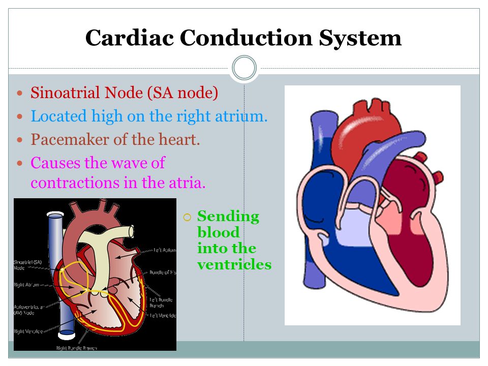
Cardiac Conduction System Sinoatrial Node (SA node) Located high on the right atrium. Pacemaker of the heart. Causes the wave of contractions in the atria. Sending blood into the ventricles

24
Cardiac Conduction System Atrioventricular Node (AV node) Located in the interatrial septum close to the tricuspid valve Carries the electrical impulse from the SA node to fiber bundles in the ventricles. This causes the ventricles to contract The location of nerve fiber bundles cause the ventricles to contract from the apex (bottom) up squeezing blood up and out
 up squeezing blood up and out.")
25
Pathway of Circulation Oxygen-poor blood draining from the body through veins into the superior and inferior vena cava flows to the right atrium, through the tricuspid valve, and into the right ventricle. As the right ventricle contracts, oxygen-poor blood passes through the pulmonary valve into the pulmonary arteries and on to the lungs to receive oxygen.

26
Oxygen-rich blood from the lungs enters the heart through the pulmonary veins, passing into the left atrium. Then through the mitral valve to the left ventricle. Contraction of the left ventricle forces blood through the aortic valve into the aorta. Various arteries branch off from the aorta to supply blood to all parts of the body.

27
Arteries branch into smaller and smaller vessels (arterioles) They eventually become capillaries, which supply blood to all body parts Capillaries merge into (venuoles) which join into veins and carry blood back to the heart. Nutrients pass into tissues Waste products filter back Blood pumped out of heart into arteries, which branch into smaller and smaller vessels until blood flows into capillaries Blood returns to the heart through the veins Heart Capillary network

28
Pathway of Circulation

29
14 9 11 10 8 6 13 12 6 5 4 3 2 1 1 16 15 14 7 And so on… It takes about 1 min. for blood to make 1 complete cycle

30
Get ¼ sheet of paper for a short quiz...

31
Questions Why don’t the atria and ventricles contract at the same time? How does a blood is distributed throughout the body?

Similar presentations


 Transport O 2, nutrients, hormones, cell wastes, etc…>")


