Download presentation
Presentation is loading. Please wait.

1
Anatomy and Physiology Chapter 13

2
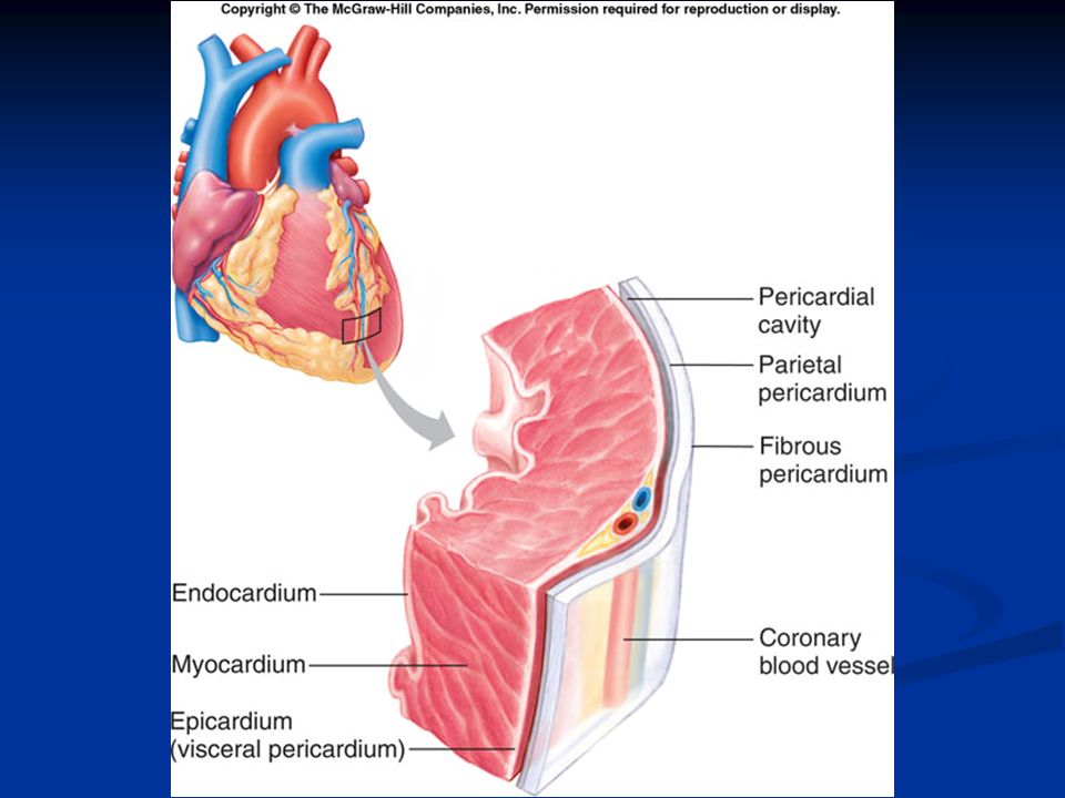
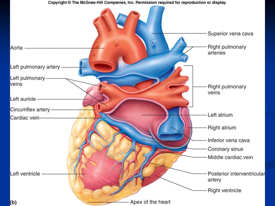
Introduction The cardiovascular system consists of the heart, and vessels, arteries, capillaries and veins. A functional cardiovascular system is vital for supplying oxygen and nutrients to tissues and removing wastes from them. The average adult heart is 14 cm long and 9 cm wide. The heart lies under the sternum The pericardium encloses the heart. The wall of the heart is composed of three distinct layers. The outermost layer, the epicardium, is the same as the visceral pericardium. The middle layer called myocardium consists of cardiac muscle and is the thickest layer of the heart wall. The inner endocardium is smooth and is made up of connective tissue and epithelium. The endocardium contains the Purkinje fibers.

5
Heart Chambers and Valves
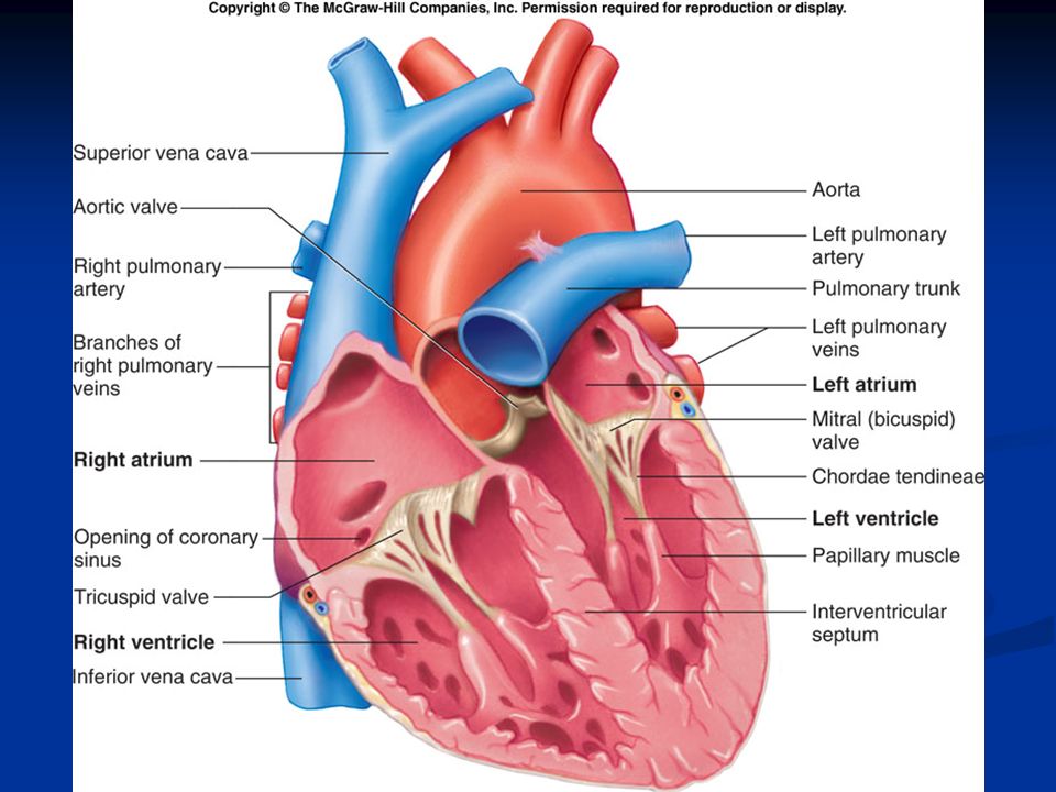
The heart has four internal chambers Two thin walled atria on top which receive the blood Two thick walled ventricles below which are more muscular and send the blood out to the body. A septum divides the atrium and ventricle on each side. Each also has an atrioventricular (A-V) valve to ensure one way flow of blood. The right A-V valve (tricuspid) and left A-V valve (bicuspid or mitral valve) have cusps to which chordae tendinae attach.
 valve to ensure one way flow of blood. The right A-V valve (tricuspid) and left A-V valve (bicuspid or mitral valve) have cusps to which chordae tendinae attach.")
7
The superior and inferior vena cavae bring de-oxygenated blood from the body to the right atrium.
At the base of the pulmonary trunk leading to the lungs is the pulmonary valve, which prevents a return flow of blood to the ventricle. The left atrium receives blood from four pulmonary veins. The left ventricle pumps blood into the entire body through the aorta. The aorta has an aortic valve that prevents backflow of blood into the ventricle.

9
Path of Blood through the Heart
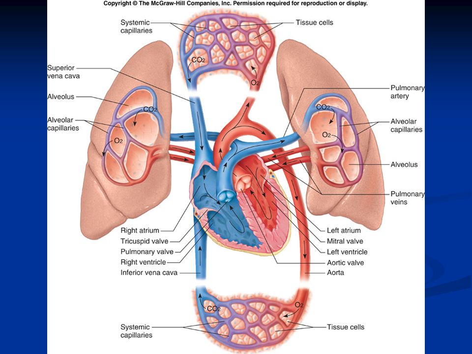
Blood low in oxygen returns to the right atrium via the vena cava and coronary sinus. The right atrium contracts, forcing blood through the tricuspid valve into the right ventricle. The right ventricle contracts, closing the tricuspid valve, and forcing blood through the pulmonary valve into the pulmonary trunk and arteries. The pulmonary arteries carry blood to the lungs where it can rid itself of excess carbon dioxide and pick up a new supply of oxygen. Freshly oxygenated blood is returned to the left atrium of the heart through the pulmonary veins. The left atrium contracts, forcing blood through the bicuspid valve into the left ventricle. The left ventricle contracts, closing the bicuspid valve and forcing open the aortic valve as blood enters the aorta for distribution to the body.

11
Pulmonary circulation is when the blood flows from the heart to the lungs and back.
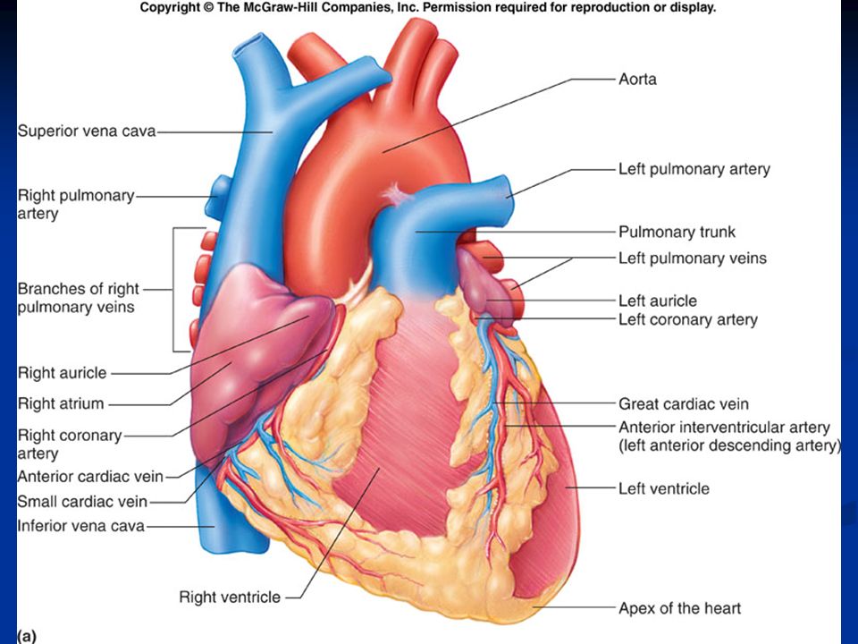
Systemic circulation occurs when the flow is from the heart to the body and back. To supply blood to the heart itself, coronary arteries and veins are used. The aorta has branches which lead to the coronary arteries The arteries diverge into arterioles and capillaries The blood then returns through coronary veins The coronary circulation ends when blood enters the right atrium through the coronary sinus. It is these vessels that become blocked during a heart attack.

15
Heart Actions The cardiac cycle consists of the atria beating in unison (atrial systole) followed by the contraction of both ventricles, (ventricular systole) then the entire heart relaxes for a brief moment (diastole). The pressure of the blood on each of the chambers causes the blood to flow from one to the next. Heart sounds are due to vibrations in heart tissues as blood rapidly changes velocity within the heart. Heart sounds can be described as a "lub-dub" sound. The first sound (lub) occurs as ventricles relax and aortic and pulmonary valves are closing. The second sound (dub) occurs as ventricles contract and A-V valves are closing. Heart Murmur
 followed by the contraction of both ventricles, (ventricular systole) then the entire heart relaxes for a brief moment (diastole). The pressure of the blood on each of the chambers causes the blood to flow from one to the next. Heart sounds are due to vibrations in heart tissues as blood rapidly changes velocity within the heart. Heart sounds can be described as a lub-dub sound. The first sound (lub) occurs as ventricles relax and aortic and pulmonary valves are closing. The second sound (dub) occurs as ventricles contract and A-V valves are closing. Heart Murmur.")
18
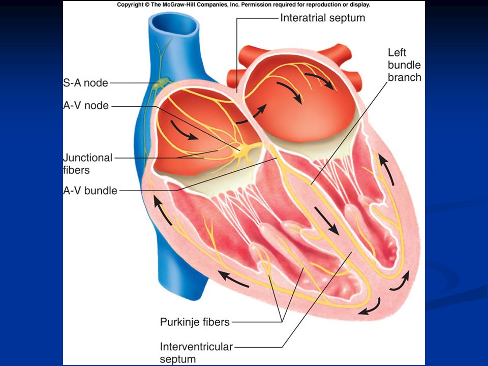
Contraction of the Heart
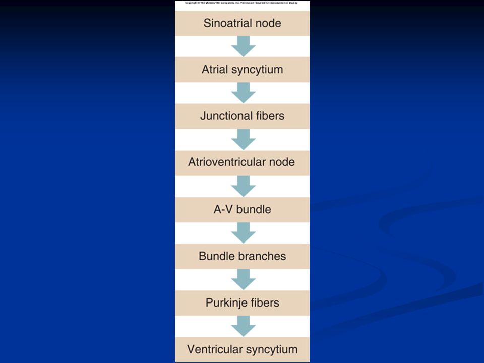
The heart contracts by means of electrical signals. The pacemaker of the heart is found in the right atrium and is known as the Sinoatrial (SA) Node. The impulse begins in the SA Node and causes the atria to contract together. It then travels to the atrial syncytium which cause the atria to contract. The impulse then travels to the Atrioventricular (AV) Node From the AV Node, it is passed down to the AV Bundle, to the bundle branches and into the Purkinje fibers. The signal is then sent to the ventricular syncytium. This causes the ventricles to contract.
 Node. The impulse begins in the SA Node and causes the atria to contract together. It then travels to the atrial syncytium which cause the atria to contract. The impulse then travels to the Atrioventricular (AV) Node. From the AV Node, it is passed down to the AV Bundle, to the bundle branches and into the Purkinje fibers. The signal is then sent to the ventricular syncytium. This causes the ventricles to contract.")
21
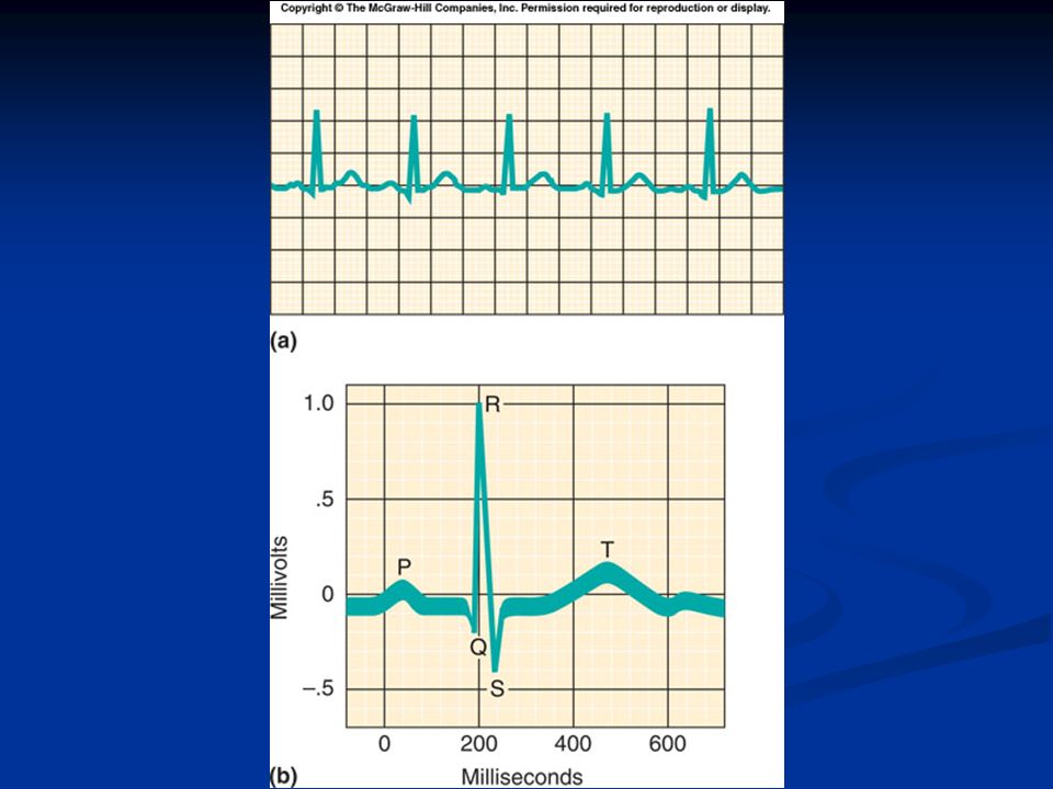
Electrocardiogram An electrocardiogram is a recording of the electrical changes that occur during a cardiac cycle. The first wave, the P wave, corresponds to the depolarization of the atria. The QRS complex corresponds to the depolarization of ventricles and hides the repolarization of atria. The T waves end the ECG pattern and corresponds to ventricular repolarization.

23
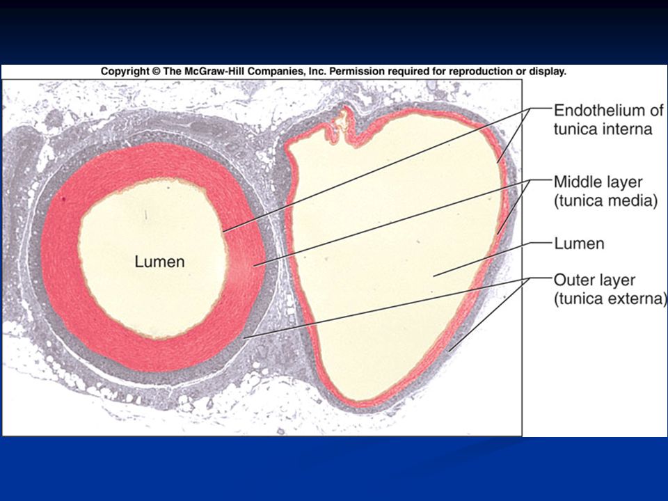
Blood Vessels The blood vessels (arteries, arterioles, capillaries, venules, and veins) form a closed tube that carries blood away from the heart, to the cells, and back again. Arteries are strong, elastic vessels adapted for carrying high-pressure blood Arteries become smaller as they divide and give rise to arterioles. The wall of an artery consists of an endothelium, tunica media (smooth muscle), and tunica externa (connective tissue). Arteries are capable of vasoconstriction and vasodilation. Capillaries are the smallest vessels, consisting only of a layer of endothelium through which substances are exchanged with tissue cells. Precapillary sphincters can regulate the amount of blood entering a capillary bed and are controlled by oxygen concentration in the area. If blood is needed elsewhere in the body, the capillary beds in less important areas are shut down.
 form a closed tube that carries blood away from the heart, to the cells, and back again. Arteries are strong, elastic vessels adapted for carrying high-pressure blood. Arteries become smaller as they divide and give rise to arterioles. The wall of an artery consists of an endothelium, tunica media (smooth muscle), and tunica externa (connective tissue). Arteries are capable of vasoconstriction and vasodilation. Capillaries are the smallest vessels, consisting only of a layer of endothelium through which substances are exchanged with tissue cells. Precapillary sphincters can regulate the amount of blood entering a capillary bed and are controlled by oxygen concentration in the area. If blood is needed elsewhere in the body, the capillary beds in less important areas are shut down.")
26
Transfer of materials at the capillary/cell junction

27
Venules leading from capillaries merge to form veins that return blood to the heart.
Veins have the same three layers as arteries have and have a flap-like valve inside to prevent backflow of blood. Veins are thinner and less muscular than arteries They do not carry high-pressure blood. Veins also function as blood reservoirs. Blood flow through the venous system depends on skeletal muscle contraction, breathing movements, and vasoconstriction of veins. Contractions of skeletal muscle squeeze blood back up veins one valve at a time.

29
Blood Pressure Blood pressure is the force of blood against the inner walls of blood vessels anywhere in the cardiovascular system Usually the term "blood pressure" refers to arterial pressure. Arterial blood pressure rises and falls following a pattern established by the cardiac cycle. During ventricular contraction, arterial pressure is at its highest (systolic pressure). When ventricles are relaxing, arterial pressure is at its lowest (diastolic pressure). The surge of blood that occurs with ventricular contraction can be felt at certain points in the body as a pulse. Blood pressure is normally directly proportional to the volume of blood within the cardiovascular system. Blood pressure varies with age, body size, and gender. Many things can affect BP including the diameter of blood vessels, distance from the heart, stress, and exercise.
. When ventricles are relaxing, arterial pressure is at its lowest (diastolic pressure). The surge of blood that occurs with ventricular contraction can be felt at certain points in the body as a pulse. Blood pressure is normally directly proportional to the volume of blood within the cardiovascular system. Blood pressure varies with age, body size, and gender. Many things can affect BP including the diameter of blood vessels, distance from the heart, stress, and exercise.")
31
Atherosclerosis refers to the buildup of fats, cholesterol and other substances in and on your artery walls (plaques), which can restrict blood flow Hypertension (HTN) or high blood pressure, sometimes called arterial hypertension, is a chronic medical condition in which the blood pressure in the arteries is elevated
 or high blood pressure, sometimes called arterial hypertension, is a chronic medical condition in which the blood pressure in the arteries is elevated.")
35
The End

Similar presentations


. Structure of the Heart About as big as your fist. Located within your thoracic cavity. –In the mediastinum –Sits.>")


 Transport O 2, nutrients, hormones, cell wastes, etc…>")


 and extensive system of tubes (blood vessels) B. Functions to transport oxygen, nutrients, and wastes. A. Size 1. Varies.>")

>")