Download presentation
Presentation is loading. Please wait.

1
SHOCK

2
Life is provided through a variety of mechanisms, however all of them depend on proper circulation. Circulation itself consists of 2 parts: work of heart (pump of the body) and vessels, through which blood is pumped to the most remote organs and tissues. During every systolic contraction heart pump 70-80 ml of blood 9so called stroke volume). Thus in case of heart rate 70 beats per minute heart pumps nearly 5 liters of blood, what makes more than 7 tones per day. From the left ventricle blood gets to the arterial system of the systemic circuit. Arteries contain 15% of the whole circulating blood volume; they carry blood from the heart to their distal departments – arterioles (vessels of resistance). Arterioles themselves are defining blood distribution: in condition of constriction (spasm) they make blood supply of the capillaries impossible (ischemia appears). On the contrary, in condition of dilatation they provide maximal oxygenation. When arterioles are blocked due to the spasm blood is flowing through the arterio-venous anastomosis directly to the venous system.
 and vessels, through which blood is pumped to the most remote organs and tissues. During every systolic contraction heart pump ml of blood 9so called stroke volume). Thus in case of heart rate 70 beats per minute heart pumps nearly 5 liters of blood, what makes more than 7 tones per day. From the left ventricle blood gets to the arterial system of the systemic circuit. Arteries contain 15% of the whole circulating blood volume; they carry blood from the heart to their distal departments – arterioles (vessels of resistance). Arterioles themselves are defining blood distribution: in condition of constriction (spasm) they make blood supply of the capillaries impossible (ischemia appears). On the contrary, in condition of dilatation they provide maximal oxygenation. When arterioles are blocked due to the spasm blood is flowing through the arterio-venous anastomosis directly to the venous system..")
3
Distribution of blood in the vascular bed (% of CBV). heart cavity 3% arteries 15% capillaries 12% venous system 70% Among the natural vasoconstrictors (agents, which cause constriction of the blood vessel) are epinephrine, norepinephrine, serotonin, angiotensin II. Stress enhances the secretion of cathecholamines, their blood concentration increases and arterioles constrict. Spasm of the arterioles is the basis of blood flow centralization: peripheral flow is disregarded in order to provide brain with the oxygenated blood as long as possible.
 are epinephrine, norepinephrine, serotonin, angiotensin II. Stress enhances the secretion of cathecholamines, their blood concentration increases and arterioles constrict. Spasm of the arterioles is the basis of blood flow centralization: peripheral flow is disregarded in order to provide brain with the oxygenated blood as long as possible..")
4
To the group of vasodilatators (agents, which provide dilatation of the vessels) belong “acid” metabolites (lactate, pyruvate, adenylic acid, inosinic acid), bradykinin, acetylcholine, different medicines (neuroleptics, α-adrenergic antagonists, peripheral vasodilatators, ganglionic blocking agents, etc.), some exogenous poisons. All of them cause blood flow decentralization: opening of arterioles and distribution of the blood from central vessels to the capillary bed. Capillaries are the interweaving network of the smallest body vessels with the general length of 90-100 thousands of kilometers. However simultaneously work only 20-25% of them. They provide metabolic exchange bringing oxygen and nutrients to the tissues and take back wastes of metabolism.

5
Periodically, every 30-40 seconds one of them get closed and others open (vasomotion effect). Capillaries contain 12% of the whole circulating blood volume, but different pathological conditions can increase this amount even 3 and more times. “Used” blood from the capillaries flows to the venous system. Veins are the blood reservoir, which contains 70% of the total circulating blood volume. Unlike arteries they are capable of volume control and thus they influence the amount of blood, which returns to the heart. The most important haemodynamic index of venous system is central venous pressure. CVP represents the pressure which blood causes to the walls of cava veins and right atrium. This parameter is an integral index of circulating blood volume, systemic vascular resistance and pump function of the heart. It can be measure with a special device called “phlebotonometer” (pic. 4.9) or with a usual infusion set and a ruler. Normally CVP measured from the sternum point is 0-14 cm H2O and from midaxillary line is 8-15 cm H2O.
 or with a usual infusion set and a ruler. Normally CVP measured from the sternum point is 0-14 cm H2O and from midaxillary line is 8-15 cm H2O..")
6
Central venous pressure decreases (sometimes even to negative) in case of: - blood loss - excessive water loss (dehydration) - distributive shock (decrease of peripheral resistance due to venous and arterial dilatation) In those conditions decreases volume of blood returning to the heart and thus suffers cardiac output. In case of negative CVP cardiac arrest is highly probable. Central venous pressure increases in case of: - heart failure (insufficiency of left or right ventricle) - hypervolemia (excessive blood infusion, improper infusion therapy) - obstructions to blood flow (pulmonary embolism, cardiac tamponade, etc.) When CVP over 15-16 cm H2O is combined with left ventricle insufficiency the risk of pulmonary edema is very high.
 - hypervolemia (excessive blood infusion, improper infusion therapy) - obstructions to blood flow (pulmonary embolism, cardiac tamponade, etc.) When CVP over cm H2O is combined with left ventricle insufficiency the risk of pulmonary edema is very high..")
7
Blood pressure is an integral index of arterial part of systemic haemodynamics. Talking about blood pressure we may refer to systolic, diastolic, pulse and mean arterial pressure. Systolic (Psyst) and diastolic (P diast) pressures are measured with the manometer (method with the usage of phonendoscope was invented by M. Korotkoff). Pulse pressure (PP) is a difference between systolic and diastolic blood pressure. Mean arterial pressure (MAP) is calculated according to the formula: MAP= P dias + 1/3 PP mm Hg
 and diastolic (P diast) pressures are measured with the manometer (method with the usage of phonendoscope was invented by M. Korotkoff). Pulse pressure (PP) is a difference between systolic and diastolic blood pressure. Mean arterial pressure (MAP) is calculated according to the formula: MAP= P dias + 1/3 PP mm Hg.")
9
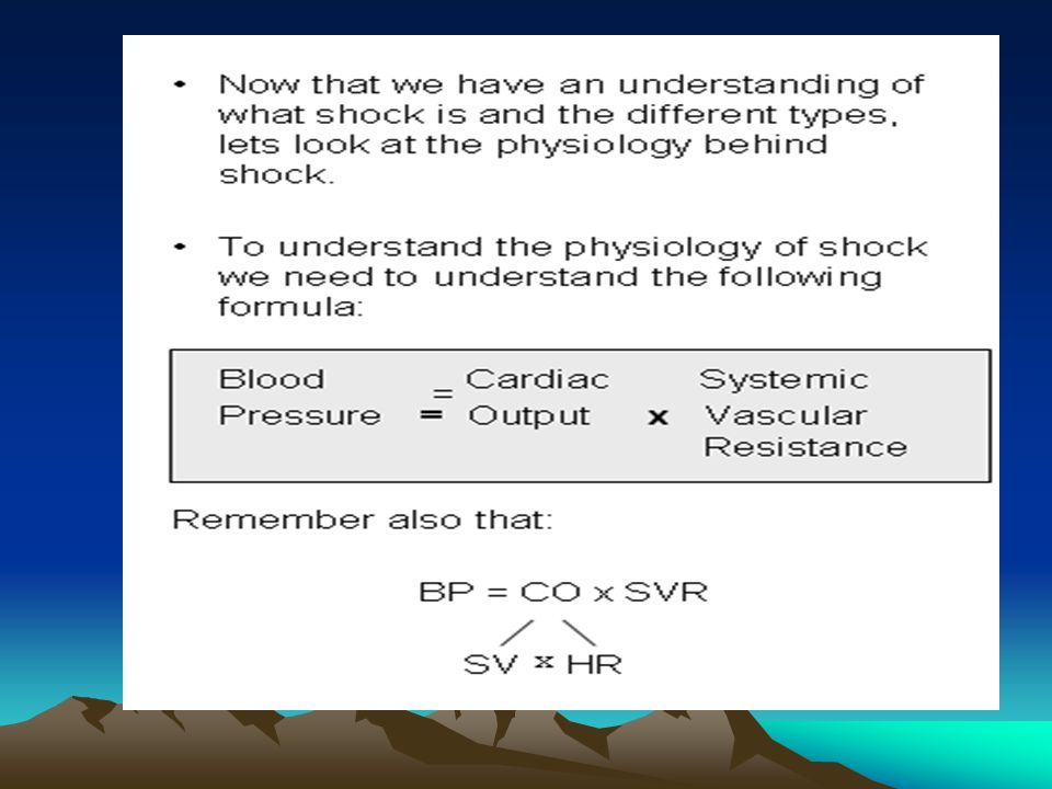
MAP defines the level of pressure necessary for the metabolic exchange in the tissues. Its measurement allows the evaluation of tissue perfusion level. Blood pressure depends on different factors, but the most important are cardiac output and vascular resistance (mostly arterioles). This dependence is direct, thus you can increase blood pressure using: - infusion of vasoconstrictors - solutions of epinephrine, phenylephrine (mesaton), etc. (they will increase the vascular resistance); - infusion of hydroxyethyl starch solutions or saline (they will increase circulating blood volume) - infusion of cardiac glycosides or other medicine which stimulate myocardium
. This dependence is direct, thus you can increase blood pressure using: - infusion of vasoconstrictors - solutions of epinephrine, phenylephrine (mesaton), etc. (they will increase the vascular resistance); - infusion of hydroxyethyl starch solutions or saline (they will increase circulating blood volume) - infusion of cardiac glycosides or other medicine which stimulate myocardium.")
10
General volume of blood in the body of a healthy adult is nearly 7% from the body weight: 70 ml per kilogram for male and 65 mil per kilogram of body weight for female. Of course circulating blood volume is lower, because part of blood is out of metabolic processes as a reserve. CBV can be measured with the infusion of coloring substance to the blood flow (Evans blue, polyglucin) and later evaluation of its dissolution degree. Therefore measurement of CVP, BP, cardiac output and circulating blood volume allow to evaluate condition of circulation system of the patients and to provide adequate correction.
 and later evaluation of its dissolution degree. Therefore measurement of CVP, BP, cardiac output and circulating blood volume allow to evaluate condition of circulation system of the patients and to provide adequate correction..")
11
Shock is a pathological state which can be described as a tissue hypoxia caused by hypoperfusion. Pathogenetic basis of shock depends on its reason (trauma, toxins, thermal injury) and at the same time on reactivity of the organism (level of defense mechanisms mobilization).
 and at the same time on reactivity of the organism (level of defense mechanisms mobilization)..")
12
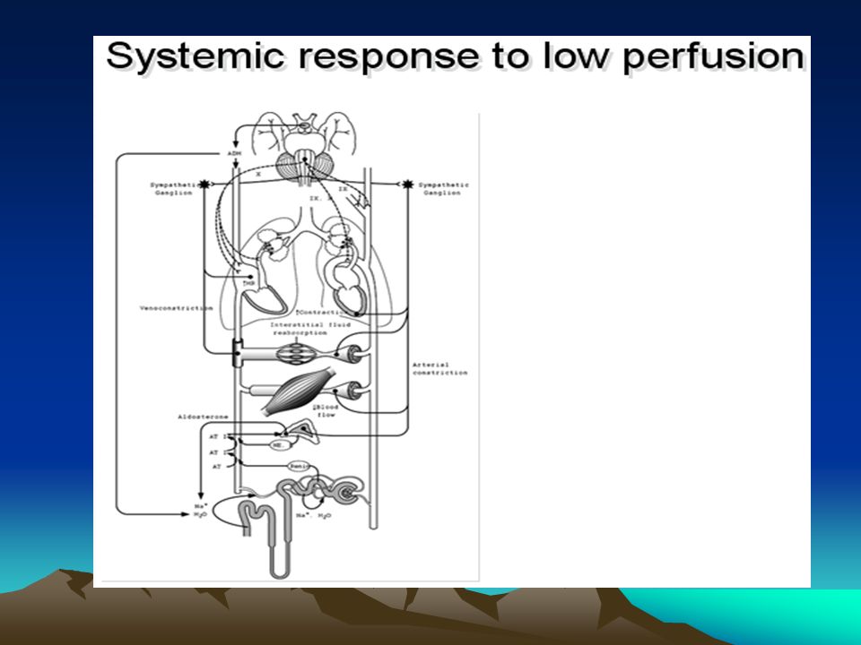
Stimulation of sympathetic nervous system - production of catecholamines and other vasoactive substances by hypothalamus and adrenal glands are the universal response of the body to the stress. Those mediators interact with the receptors of peripheral vessels causing their constriction and at the same time they dilatate the vascular bed of life-important organs. This is so called “centralization of the flow”: rational decrease of blood flow in less important tissues (skin, organs of abdominal cavity, kidneys) in case of aggressive external influence for protecting life itself (brain, heart, lungs).
 in case of aggressive external influence for protecting life itself (brain, heart, lungs)..")
13
However influence of shock agents (pain, hypovolemia, destroyed cells, toxic metabolites), extended microcirculation violations (vascular spasm, microthrombosis and sludge) and caused by them tissue ischemia lead to hypoxic affection and cellular death of the internal organs. Further it can bring multiple organ dysfunction syndrome.

14
Collapse is a vascular failure. It occurs when body is not able to provide blood flow according to the new level of its needs (either because reaction is not fast enough or because sympathetic activation fails).Vascular bed volume and circulating blood volume are disproportional: too much blood gets to the microcirculation vascular reserve and the amount, which returns to the heart is not enough for the systemic needs (so called “decentralization” of the blood flow). Cardiac output and blood pressure decrease, that causes hypoperfusion of the central nervous system and thus unconsciousness and life-threatening complications.
.Vascular bed volume and circulating blood volume are disproportional: too much blood gets to the microcirculation vascular reserve and the amount, which returns to the heart is not enough for the systemic needs (so called decentralization of the blood flow). Cardiac output and blood pressure decrease, that causes hypoperfusion of the central nervous system and thus unconsciousness and life-threatening complications..")
15
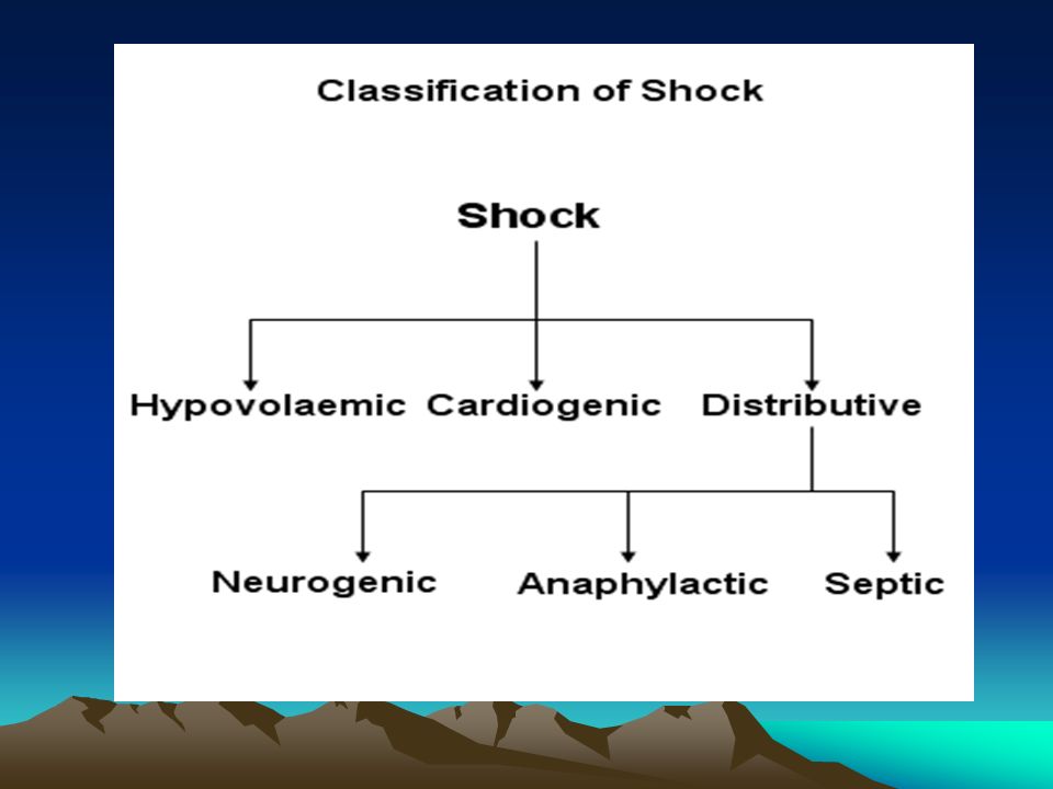
Pathogenetic classification of shock (according to P. Marino, 1998): - hypovolemic - cardiogenic - distributive - mixed (two and more factors). Clinical classification of shock: - traumatic shock; - haemorrhagic shock; - dehydration shock; - burn shock; - septic shock; - anaphylactic shock; - cardiogenic shock; - exotoxic shock.
: - hypovolemic - cardiogenic - distributive - mixed (two and more factors). Clinical classification of shock: - traumatic shock; - haemorrhagic shock; - dehydration shock; - burn shock; - septic shock; - anaphylactic shock; - cardiogenic shock; - exotoxic shock..")
17
Outline Definition Epidemiology Physiology Classes of Shock Clinical Presentation Management Controversies

40
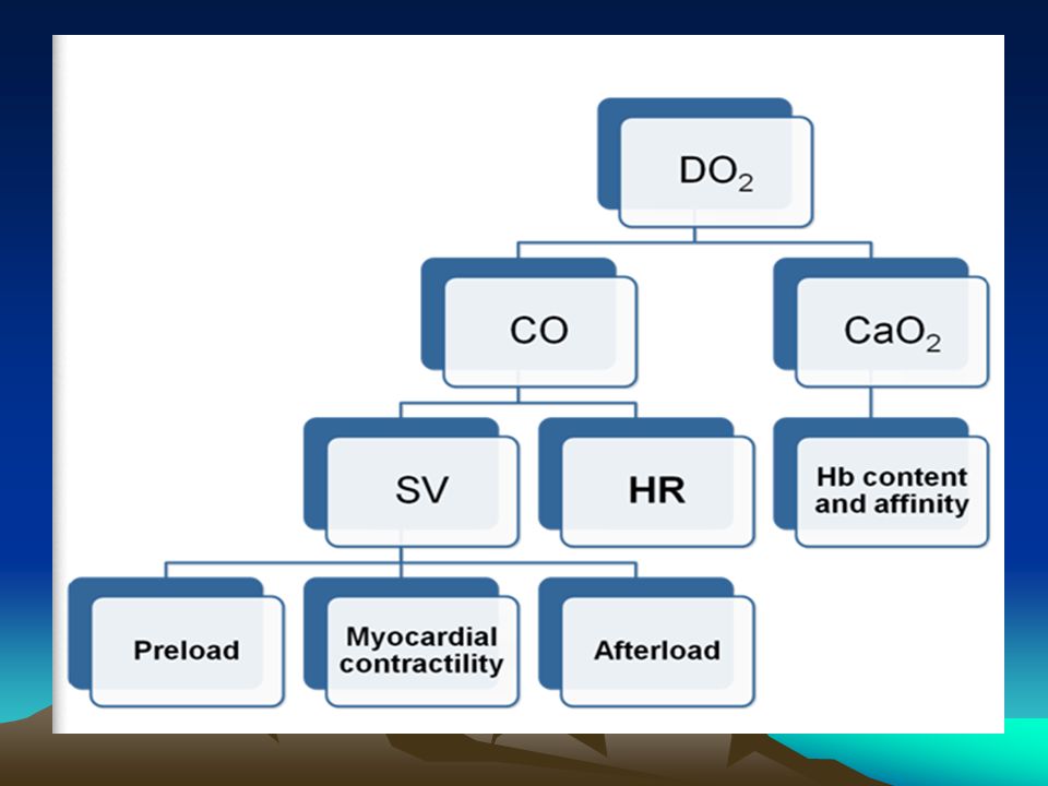
Definition A physiologic state characterized by –Inadequate tissue perfusion Clinically manifested by –Hemodynamic disturbances –Organ dysfunction

64
Epidemiology Mortality –Septic shock – 35-40% (1 month mortality) –Cardiogenic shock – 60-90% –Hypovolemic shock – variable/mechanism
 –Cardiogenic shock – 60-90% –Hypovolemic shock – variable/mechanism")
65
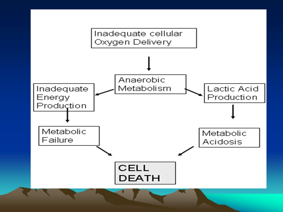
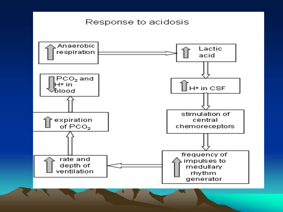
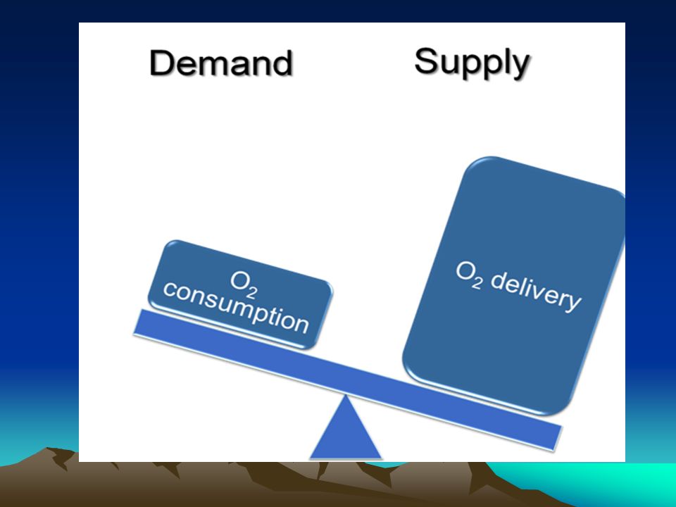
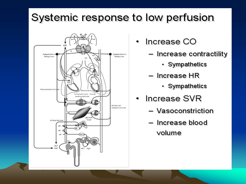
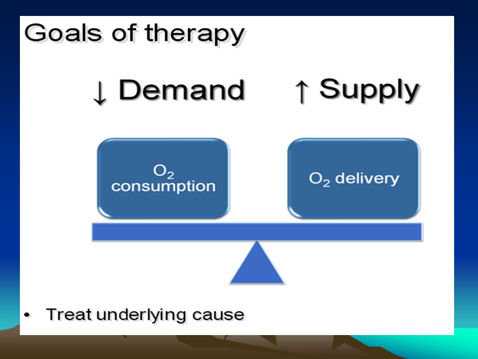
Pathophysiology Imbalance in oxygen supply and demand Conversion from aerobic to anaerobic metabolism Appropriate and inappropriate metabolic and physiologic responses

66
Pathophysiology Cellular physiology –Cell membrane ion pump dysfunction –Leakage of intracellular contents into the extracellular space –Intracellular pH dysregulation Resultant systemic physiology –Cell death and end organ dysfunction –MSOF and death

67
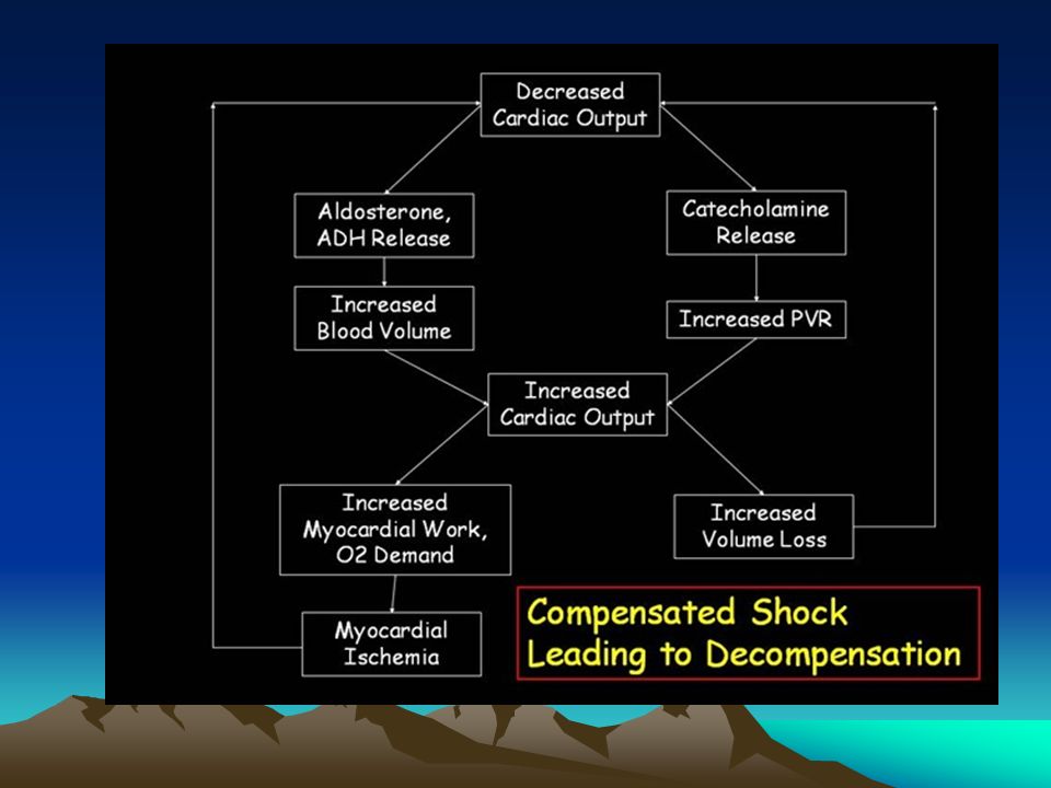
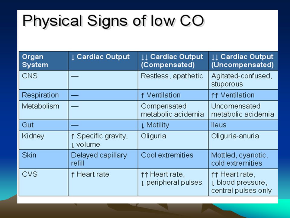
Physiology Characterized by three stages –Preshock (warm shock, compensated shock) –Shock –End organ dysfunction
 –Shock –End organ dysfunction")
68
Physiology Compensated shock –Low preload shock – tachycardia, vasoconstriction, mildly decreased BP –Low afterload (distributive) shock – peripheral vasodilation, hyperdynamic state
 shock – peripheral vasodilation, hyperdynamic state")
69
Pathophysiology Shock –Initial signs of end organ dysfunction –Tachycardia –Tachypnea –Metabolic acidosis –Oliguria –Cool and clammy skin

70
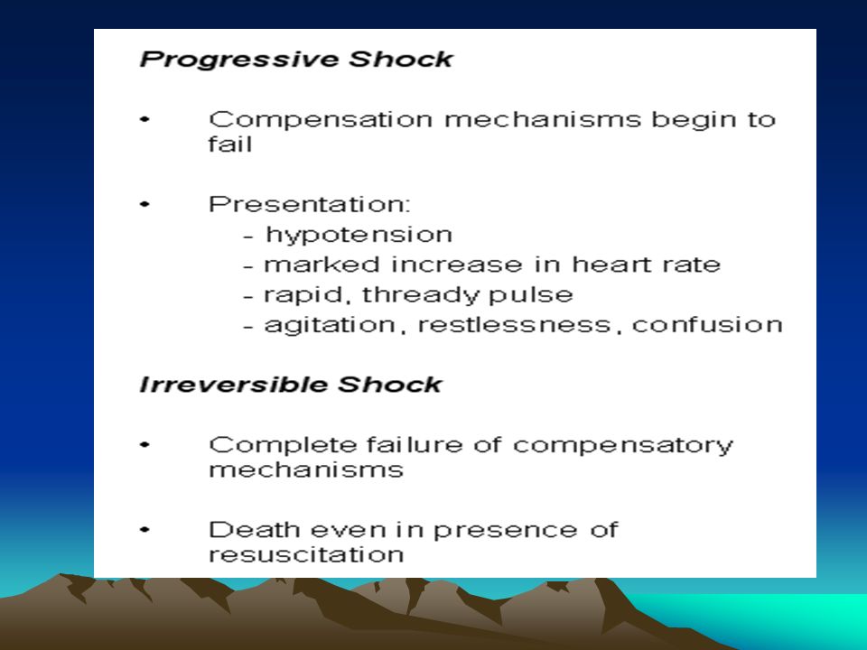
Physiology End Organ Dysfunction –Progressive irreversible dysfunction –Oliguria or anuria –Progressive acidosis and decreased CO –Agitation, obtundation, and coma –Patient death

71
Classification Schemes are designed to simplify complex physiology Major classes of shock –Hypovolemic –Cardiogenic –Distributive

72
Hypovolemic Shock Results from decreased preload Etiologic classes –Hemorrhage - e.g. trauma, GI bleed, ruptured aneurysm –Fluid loss - e.g. diarrhea, vomiting, burns, third spacing, iatrogenic

75
Hypovolemic Shock Hemorrhagic Shock ParameterIIIIIIIV Blood loss (ml)<750750–15001500–2000>2000 Blood loss (%)<15%15–30%30–40%>40% Pulse rate (beats/min)<100>100>120>140 Blood pressureNormalDecreased Respiratory rate (bpm)14–2020–3030–40>35 Urine output (ml/hour)>3020–305–15Negligible CNS symptomsNormalAnxiousConfusedLethargic Crit Care. 2004; 8(5): 373–381.
: 373–381..")
76
Cardiogenic Shock Results from pump failure –Decreased systolic function –Resultant decreased cardiac output Etiologic categories –Myopathic –Arrhythmic –Mechanical –Extracardiac (obstructive)
")
77
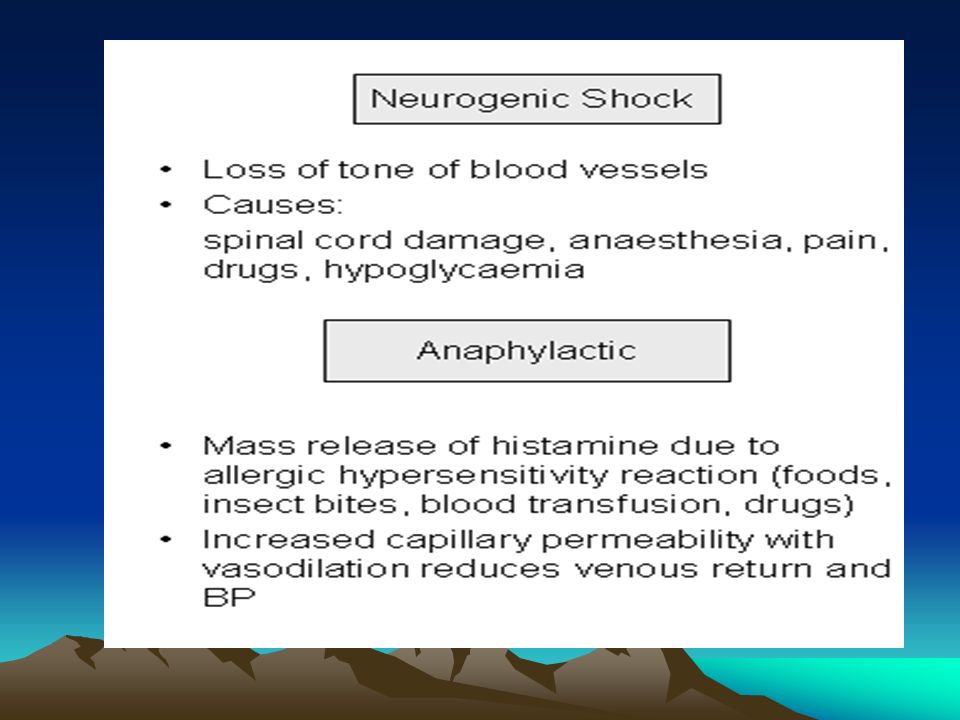
Distributive Shock Results from a severe decrease in SVR –Vasodilation reduces afterload –May be associated with increased CO Etiologic categories –Sepsis –Neurogenic / spinal –Other (next page)
")
79
Distributive Shock Other causes –Systemic inflammation – pancreatitis, burns –Toxic shock syndrome –Anaphylaxis and anaphylactoid reactions –Toxin reactions – drugs, transfusions –Addisonian crisis –Myxedema coma

80
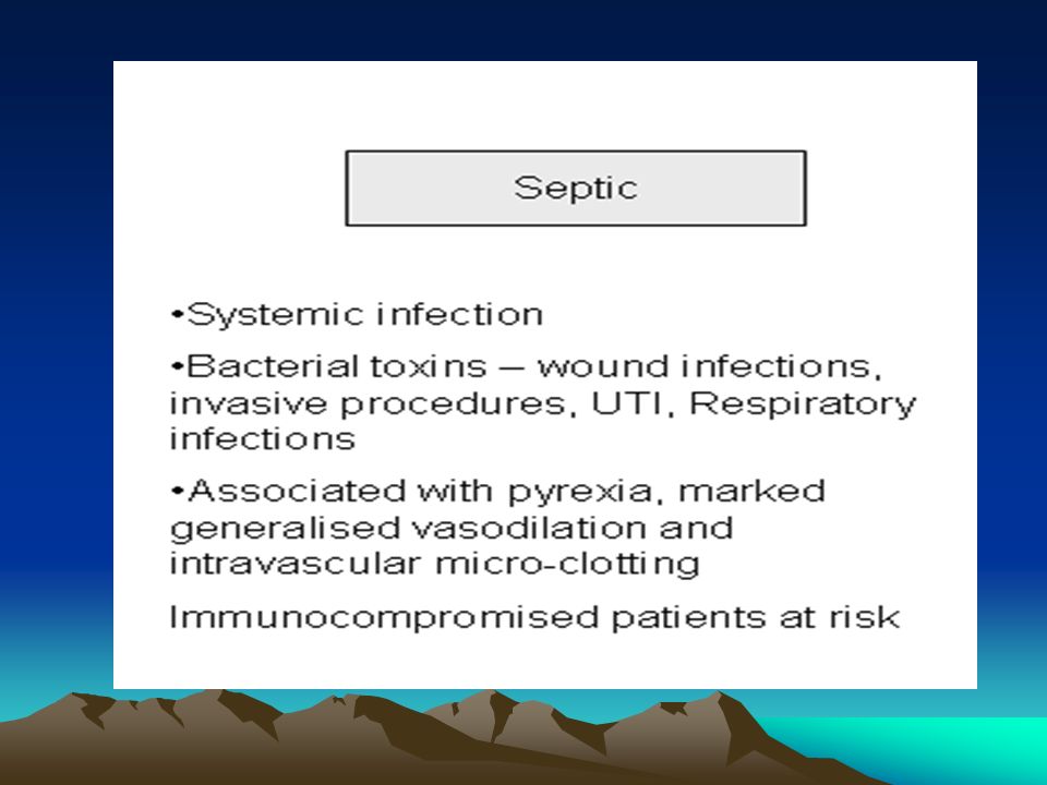
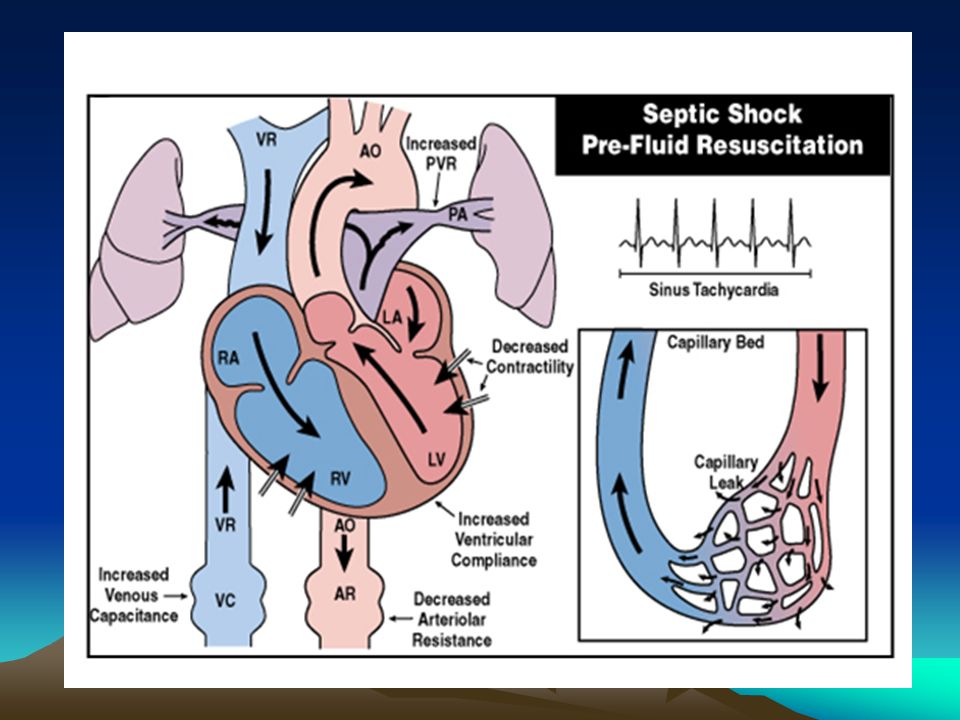
Distributive Shock Septic Shock

84
Clinical Presentation Clinical presentation varies with type and cause, but there are features in common Hypotension (SBP 40) Cool, clammy skin (exceptions – early distributive, terminal shock) Oliguria Change in mental status Metabolic acidosis
 Cool, clammy skin (exceptions – early distributive, terminal shock) Oliguria Change in mental status Metabolic acidosis")
85
Evaluation Done in parallel with treatment! H&P – helpful to distinguish type of shock Full laboratory evaluation (including H&H, cardiac enzymes, ABG) Basic studies – CxR, EKG, UA Basic monitoring – VS, UOP, CVP, A-line Imaging if appropriate – FAST, CT Echo vs. PA catheterization –CO, PAS/PAD/PAW, SVR, SvO2
 Basic studies – CxR, EKG, UA Basic monitoring – VS, UOP, CVP, A-line Imaging if appropriate – FAST, CT Echo vs. PA catheterization –CO, PAS/PAD/PAW, SVR, SvO2.")
86
Treatment Manage the emergency Determine the underlying cause Definitive management or support

87
Manage the Emergency Your patient is in extremis – tachycardic, hypotensive, obtunded How long do you have to manage this? Suggests that many things must be done at once Draw in ancillary staff for support! What must be done?

88
Manage the Emergency One person runs the code! Control airway and breathing Maximize oxygen delivery Place lines, tubes, and monitors Get and run IVF on a pressure bag Get and run blood (if appropriate) Get and hang pressors Call your senior/fellow/attending
 Get and hang pressors Call your senior/fellow/attending.")
89
Determine the Cause Often obvious based on history Trauma most often hypovolemic (hemorrhagic) Postoperative most often hypovolemic (hemorrhagic or third spacing) Debilitated hospitalized pts most often septic Must evaluate all pts for risk factors for MI and consider cardiogenic Consider distributive (spinal) shock in trauma
 Postoperative most often hypovolemic (hemorrhagic or third spacing) Debilitated hospitalized pts most often septic Must evaluate all pts for risk factors for MI and consider cardiogenic Consider distributive (spinal) shock in trauma")
90
Determine the Cause What if you’re wrong? 85 y/o M 4 hours postop S/P sigmoid resection for perforated diverticulitis is hypotensive on a monitored bed at 70/40 Likely causes Best actions for the first 5 minutes?

91
Definitive Management Hypovolemic – Fluid resuscitate (blood or crystalloid) and control ongoing loss Cardiogenic - Restore blood pressure (chemical and mechanical) and prevent ongoing cardiac death Distributive – Fluid resuscitate, pressors for maintenance, immediate abx/surgical control for infection, steroids for adrenocortical insufficiency
 and control ongoing loss Cardiogenic - Restore blood pressure (chemical and mechanical) and prevent ongoing cardiac death Distributive – Fluid resuscitate, pressors for maintenance, immediate abx/surgical control for infection, steroids for adrenocortical insufficiency")
92
Controversies IVF Resuscitation –Limited resuscitation in penetrating trauma –Use of hypertonic saline resuscitation in trauma –Endpoints for prolonged resuscitation Pressors –Best pressors for distributive shock Monitoring –Most appropriate timing and use for PA catheterization or intermittent echocardiogram

Similar presentations


 system is an important endocrine component of autoregulation. Renin is released by kidneys when.>")

 The McGraw-Hill Companies, Inc. Permission required for reproduction or display.>")
