Download presentation
Presentation is loading. Please wait.

1
Chapter 4 CELLS Accelerated Biology
Mrs. Schalles Notes from Holt Biology & other references as listed. Picture:

2
Each cell is an amazing world unto itself:
it can take in nutrients, convert these nutrients into energy, carry out specialized functions, and reproduce as necessary. Even more amazing is that each cell stores its own set of instructions for carrying out each of these activities.

3
Unicellular vs. multicellular
Some organisms, such as bacteria & some protists, are unicellular, consisting of a single cell. Other organisms, such as fungi, plants & animals are multicellular, or have many cells—humans have an estimated 100,000,000,000,000 cells! one celled body, Bacilli bacteria This is a picture of a newly-hatched C. elegans larva (a worm with 556 cells, the adult worms have about 1,000 cells in their bodies.

4
What is a cell? The smallest unit that can perform all of life’s processes All living things are made of cells It is often called “the building block of life”

5
2 general categories of cells: prokaryotes and eukaryotes
Prokaryotic cells: lack a nuclear membrane, they have no true nucleus. Examples: Bacteria & Archaea Eukaryotic cells: Have a true membrane bound nucleus & membrane bound organelles. They are larger cells- about 10 times or more larger than a prokaryotic cell, Examples: fungi, animals, plants as well as some unicellular organisms.

6
What is an Organelle? They are small structures within
cells that perform dedicated functions in eukaryotic cells. As the name implies, you can think of organelles as small organs.

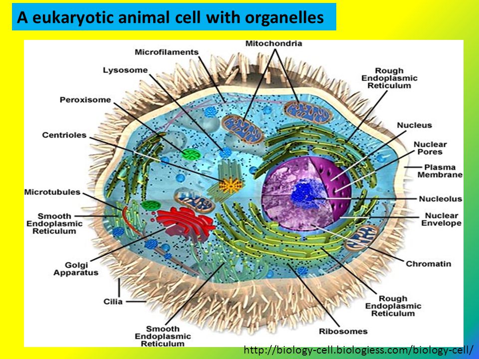
7
A eukaryotic animal cell with organelles
8
Notice that there are no organelles in the prokaryotic cell!

9
Summary – Differences- Prokaryotic & Eukaryotic Cells
Prokaryotic Cells Eukaryotic cells small cells (< 5 mm) larger cells (> 10 mm) always unicellular often multicellular no nucleus or any membrane-bound organelles always have nucleus and other membrane-bound organelles DNA is circular, without proteins DNA is linear and associated with proteins to form chromatin ribosomes are small (70S) ribosomes are large (80S) no cytoskeleton always has a cytoskeleton cell division is by binary fission cell division is by mitosis or meiosis reproduction is always asexual reproduction is asexual or sexual
 larger cells (> 10 mm) always unicellular. often multicellular. no nucleus or any membrane-bound organelles. always have nucleus and other membrane-bound organelles. DNA is circular, without proteins. DNA is linear and associated with proteins to form chromatin. ribosomes are small (70S) ribosomes are large (80S) no cytoskeleton. always has a cytoskeleton. cell division is by binary fission. cell division is by mitosis or meiosis. reproduction is always asexual. reproduction is asexual or sexual.")
10
History of cells: 1st person to see & name cells:
In 1665, Englishman, Robert Hooke, first saw & named "cells" while he was using a new instrument called a "microscope." He cut thin slices from cork, looked under a microscope, saw tiny box-like shapes. These tiny boxes reminded him of the small rooms that monks lived in called "cells".

11
Illustration of cork by R. Hooke

12
Anton Van Leeuwenhoek The cork cells Hooke saw were actually the remains of dead plant cells. Leeuwenhoek ( ) (Dutch) was actually the first man to observe live cells. Using microscopes he made, was the first to observe sperm, bacteria, & RBCs. Observations laid the foundations for bacteriology and microbiology.
 (Dutch) was actually the first man to observe live cells. Using microscopes he made, was the first to observe sperm, bacteria, & RBCs. Observations. laid the foundations for. bacteriology and microbiology.")
13
Together made “Cell Theory”
Other Scientists 1838 Matthias Schleiden –concluded that all plants were composed of cells. 1839 Theodor Schwann concluded all animals were made of cells. 1855 Rudolf Virchow reasoned that cells come from only other cells Together made “Cell Theory”

14
Cell Theory (be able to state these 3 parts!)
All living things are made of cells. May be unicellular or multicellular. Cells are the smallest basic unit of structure and function in an organism A cell is the smallest unit of matter that can carry on all the processes of life. Cells come only from other cells.

15
Cell Diversity Cells are not all alike. Differences in: Size Shape
Function Internal organization

16
Cells – a variety of Shapes.
The shape of a cell is a result of its particular function science.howstuffworks.com

17
How does each cell’s shape
Cell Shapes How does each cell’s shape reflect its function?

18
Specialized cells Cytology – is the study of cells
The people working in this photo are cytologists erasmusmc.nl

19
There are 200 kinds of specialized cell types in the human body!
A specialized cell is a cell that performs a specific function in multicellular organisms. Groups of specialized cells work together to form a tissue, like a muscle, and then different tissues work together to form larger units, like an organ. Examples: muscle cells, red blood cells, auditory hair cells, epithelial skin cells, lymphocytes, neurons, & photoreceptor cells.

20
How do so many types of cells arise?
Stem cells -in the human body have a unique ability to renew themselves and give rise to the more specialized cell types that do the work of the body. Stem cells remain unspecialized until a signal from the body tells them to develop into specific cells of the body like a heart, nerve, or skin cell.

21
3 Types of Blood Cells RBCs- red blood cells(erythrocytes)
WBCs- white blood cells (leukocytes) Platelets- (thrombocytes) armc1.adam.com
 Platelets- (thrombocytes) armc1.adam.com.")
22
What's in Blood? Blood is a type of connective tissue, with 2 parts.
1. Cells (RBC, WBC, platelets) - 45% 2. Plasma (water, proteins, carbohydrates, vitamins, hormones, electrolytes, cellular waste) - 55% Hematocrit - a test that a Dr. may order to see if you have enough blood cells. The blood is “spun down” in a centrifuge. drstandley.com
 - 45% 2. Plasma (water, proteins, carbohydrates, vitamins, hormones, electrolytes, cellular waste) - 55% Hematocrit - a test that a Dr. may order to see if you have enough blood cells. The blood is spun down in a centrifuge. drstandley.com.")
23
Red blood cells (erythrocytes)
Main function: transports O2 through body, picks up CO2 Hemoglobin = molecule which combines with O2 to transport it Anemia = too few RBCs!

24
WHITE BLOOD CELLS (Leukocytes)
General function - defend the body against disease-causing agents (microorganisms) PLATELETS (thrombocytes) help initiate formation of blood clots, close breaks in damaged blood vessels
 PLATELETS (thrombocytes) help initiate formation of blood clots, close breaks in damaged blood vessels.")
25
There are three types of muscle cells:
Skeletal muscle or "voluntary muscle" is anchored by tendons to bone Smooth muscle or "involuntary muscle" is found within the walls of organs & is not under conscious control. Cardiac muscle is also an "involuntary muscle" found only in the heart

26
Neurons The tissue of your brain & nervous system is composed of neurons Neurons are specialized cells which are electrically excitable, and can use that electric excitability to received and transmit information.

27
The largest cells~ eggs
-A few types of cells are large enough to be seen by the unaided eye. -Human Female Egg is the largest cell in the human body, -can be seen without the aid of a microscope. ~“dot” made by sharp pencil) -Eggs of Birds & Frogs are large cells -Largest of known cells is the ostrich egg.
 -Eggs of Birds & Frogs are large cells. -Largest of known cells is the ostrich egg.")
28
Other interesting cell sizes
Outer layer of the placenta is one undivided sheet of cytoplasm with thousands of cell nuclei-- the entire membranes enclosing the baby. A muscle fiber is actually one very long and wide cell with many nuclei--perhaps 1/10 of a mm wide and several centimeters long.

29
More about cell sizes Plant cells Bacteria Smallest known cells
do not show such great variability in size are all less than 1/50 of a mm in diameter. Bacteria are the smallest cells Smallest known cells A bacteria called mycoplasm. Viruses Are very very small but are not cells & not considered to be alive

30
Cell Size is limited -by a cell’s surface area–to-volume ratio.

31
Why Cell Size is Limited
a. Surface area to volume ratio Oxygen, Nutrients, etc. must enter cells. If a cell grows too large- these things cannot enter cell quickly enough for cell needs. Also-The Nucleus can only control a limited amount of cytoplasm.

32
Surface area and volume of cubes

33
Flat, Long cells A way to get around limitations of surface area is to make the cell long and thin or skinny and flat. Examples: Shape of skin cells- flat- suited to covering the body. Used by many protists & certain cells in your body such as nerve cells and muscle cells, both of which are long and skinny.

34
MORE EXAMPLES of cell shapes
White blood cells (WBC’s) can CHANGE shape- to engulf invading bacteria **Phagocytosis- to engulf and digest microorganisms and cellular debris)
 can CHANGE shape- to engulf invading bacteria. **Phagocytosis- to engulf and digest microorganisms and cellular debris)")
35
B. The three basic parts of a cell
plasma membrane cytoplasm nucleus

36
Plasma Membrane (or cell membrane)
Cell’s outer boundary, covers a cell’s surface and acts as a barrier between the inside and the outside of a cell. Cytoplasm The region of the cell that is within the plasma membrane and that includes the fluid, the cytoskeleton, and all of the organelles except the nucleus. Nucleus a membrane-bound organelle that contains a cell’s DNA.

37
Cellular Organization
Remember- organization levels: Atom-molecule-organelle-cell- In multicellular eukaryotes, cells further organize into: - tissues - organs - organ systems - organisms.

38
STRUCTURES THAT CARRY OUT SPECIFIC FUNCTIONS IN A EUKARYOTIC CELL.
What is an Organelle? STRUCTURES THAT CARRY OUT SPECIFIC FUNCTIONS IN A EUKARYOTIC CELL.

39
Cell Organelles and Features
1. Plasma membrane 2. Nucleus 3. Mitochondria 4. Ribosomes 5. Endoplasmic Reticulum (rough & smooth) 6. Golgi Apparatus 7. Centrioles 8. Peroxisomes 9. Lysosomes 10. Cytoskeleton
 6. Golgi Apparatus. 7. Centrioles. 8. Peroxisomes. 9. Lysosomes. 10. Cytoskeleton.")
40
1. The Plasma Membrane Fluid Mosaic Model
phospholipid molecules (double or “bi-layer”) imbedded proteins Functions as a barrier keeping foreign entities out the cell and its contents (like cytoplasm) firmly inside the cell. selectively permeable -allows only selected materials to pass in and out of a cell.
 imbedded proteins. Functions as a barrier. keeping foreign entities out the cell and its contents (like cytoplasm) firmly inside the cell. selectively permeable -allows only selected materials to pass in and out of a cell.")
41
Cell membrane -a Bilayer of Phospholipids with Proteins in it.

42
Phospholipids in a cell membrane note: the Hydrophilic phosphate “head” the Hydrophobic lipid “tail”

43
Proteins in membranes; help to MOVE Materials IN/OUT of the Cell.
Membranes are FLUID- consistency of oil. “Fluid Mosaic Model”

44
2. NUCLEUS The first “discovered” organelle
Large Organelle near the Center of the cell Contains DNA Nucleolus- places of ribosome synthesis Nuclear Pores- small holes through nuclear membrane to allow things like RNA to enter & leave nucleus

45
The nucleus directs the cell’s activities and stores DNA.
Nucleolus - The is the place where DNA is concentrated when it is in the process of making ribosomal RNA. Nuclear Envelope The nucleus is surrounded by a double membrane

46
3. Mitochondria “POWER HOUSE” of the cell
-Main function is the conversion of the potential energy of food molecules into ATP. (Adenosine Triphosphate) -Mitochondria structure: an outer membrane & inner membrane Matrix fluid in between the membranes. the inner membrane is elaborately folded with cristae projecting into the matrix. has its own DNA!
 -Mitochondria structure: an outer membrane & inner membrane. Matrix fluid in between the membranes. the inner membrane is elaborately folded with cristae projecting into the matrix. has its own DNA!")
47
4. RIBOSOMES 5. ENDOPLASMIC RETICULUM (ER)
-Ribosomes are the site of PROTEIN SYNTHESIS 5. ENDOPLASMIC RETICULUM (ER) -system of membranous tubules and sacs that connect the Nuclear Envelope to the Cell Its like anIntracellular Highway, a path along which molecules move from one part of the cell to another. Poisons, waste, & toxic chemicals are made harmless. -SMOOTH ER (no ribosomes) –steroid synthesis -ROUGH ER (has ribosomes) - protein synthesis
 -system of membranous. tubules and sacs that connect. the Nuclear Envelope to the Cell. Its like anIntracellular Highway, a path along which molecules. move from one part of the cell. to another. Poisons, waste, & toxic chemicals are made harmless. -SMOOTH ER (no ribosomes) –steroid synthesis. -ROUGH ER (has ribosomes) - protein synthesis.")
48
6. Golgi Apparatus- processing/packaging of secretions
7.Centrioles help the cell when it comes time to divide, both in mitosis and meiosis. They cannot be seen when the cell is not dividing. They are made of Microtubules. 8. Peroxisomes hold detoxification enzymes & are classified by their contents. (enzymes that require oxygen or oxidative enzymes). (Lysosomes have enzymes that work in oxygen-poor areas and lower pH.)
. (Lysosomes have enzymes that work in oxygen-poor areas and lower pH.)")
49
9. Lysosomes A lysosome is vesicle
that holds a variety of enzymes. They act like the “digestive” organs in a cell. They can digest food, old organelles or even other cells! The enzyme proteins are made in the rough ER, then packaged in a vesicle and sent to the Golgi apparatus. The Golgi then pinches off a small, very specific vesicle. That vesicle is a lysosome. From there the lysosomes float in the cytoplasm until they are needed to break down something!

50
10. Cytoskeleton Cilia & Flagella – assist in movement Centrioles
maintains shape of the cell made of protein fibers that help cells move includes microtubules, microfilaments, and intermediate filaments. Cilia & Flagella – assist in movement Centrioles consist of two short cylinders of microtubules at right angles to each other and are involved in cell division.

51
The Cytoskeleton allows organelles to move around within
the cell. By moving itself the cytoskeleton can move entire cells in multicellular organisms. The picture shows microtubules. Microtubules are part of the cytoskeleton, made up of protein filament, that help define cell structure and movement

52
3 Differences between animal and plant cells
Plants cells have most of the same organelles but will also have: 1. Cell Walls- made of cellulose 2. Vacuoles- fluid filled organelles for storage 3. Plastids (example: Chloroplasts) Have 2 membranes (like mitochondria) Have own DNA Some plastids function is storage Chloroplasts- absorb light for photosynthesis
 Have 2 membranes (like mitochondria) Have own DNA. Some plastids function is storage. Chloroplasts- absorb light for photosynthesis.")
53
In Plant cells ( but not animal cells)
Cell walls Central vacuole chloroplasts

54
More about PLASTIDS Are in only in plants
Organelles that-MAKE OR STORE FOOD. CHLOROPLAST converts SUNLIGHT, CARBON DIOXIDE, AND WATER INTO SUGARS. This process is called PHOTOSYNTHESIS. *Inside chloroplasts -THYLAKOIDS Membranes –where Photosynthesis occurs.

55
Chloroplasts are similar to mitochondria but are found only in plants.
Both have a double membrane with an intermembrane space. both have their own DNA. both have many membrane foldings inside, filling their inner spaces.

56
Chloroplasts & Mitochondria
Chloroplasts change solar energy into usable energy (ATP) through photosynthesis. The energy from there then goes to the mitochondria to generate more usable energy using the process of cell respiration. Mitochondria are present in BOTH plants & Animals! transtutors.com
 through photosynthesis. The energy from there then goes to the mitochondria to generate more usable energy using the process of cell respiration. Mitochondria are present in BOTH plants & Animals! transtutors.com.")
57
evolution.berkeley.edu

Similar presentations


 Turn in any work that.>")
 Anton van Leeuwenhoek.>")
 membrane>")


>")
: observed a thin slice of cork with a microscope.>")


