Download presentation
Presentation is loading. Please wait.

1
Anemia due to Impaired Iron Metabolism
Wu Chunmei

2
This group disorders are caused by impaired iron metabolism which include: (a) Iron deficiency anemia: deficiency of iron (b) Sideroblastic anemia:`impaired utilization of iron (c) Anemia of chronic disease: defective iron reutilization
 Iron deficiency anemia: deficiency of iron (b) Sideroblastic anemia:`impaired utilization of iron (c) Anemia of chronic disease: defective iron reutilization")
3
Iron Metabolism

4
Amount and distribution:
The total body iron varies from 3 to 4 g, depending on the sex and weight of the individual It is greater in males than in females and it increases roughly in proportion to body weight. Male: mg/Kg Female: 35-40mg/Kg 1ml blood=0.5mg Fe 1gHb=3.3mgFe 1mlRBC=1~2mgFe

5
The iron is distributed in several forms:
Compartments iron content(mg) total body iron(TBI) Hb iron: % Tissue iron myoglobin % Labile iron pool % cytochromes % catalase peroxidase Storage (available) iron Ferritin % Haemosiderin Transport iron %
 total body iron(TBI) Hb iron: % Tissue iron myoglobin % Labile iron pool % cytochromes 8 0.2% catalase peroxidase Storage (available) iron Ferritin % Haemosiderin 390 Transport iron 3 0.1%")
6
Absorption ----excretion
Balance of iron metabolism Absorption ----excretion

8
Absorption Transport of iron Utilization Affecting factors?

9
absorption Iron cycle Duodenum and upper jejunum Free Fe 3+ stomach
Fe 3+ in food combined receptor reductase Epithelial cells brush border apo-Ferritin + Fe Fe 2+ ferritin Fe 2+ Free Fe stomach reduced(VitC, GSH) gastric juice Fe 2+ Circulation Fe 2+ Tf Fe 3+ Tf-Fe 3+ Trans-port liver Iron cycle Storage inMMS Ferritin Haemosiderin Normablasts Ret. marrow for Hb In tissue: myoglobin heme-containing enzymes
 gastric juice. Fe 2+ Circulation Fe 2+ Tf + Fe 3+ Tf-Fe 3+ Trans-port. liver. Iron cycle. Storage inMMS. Ferritin Haemosiderin. Normablasts. Ret. marrow for Hb. In tissue: myoglobin. heme-containing enzymes.")
10
Serum beta-globulin that binds and transports iron
Transport of iron transferrin Serum beta-globulin that binds and transports iron transferrin receptors

11
MMS Storage of iron apoferritin + Fe
An iron-containing protein complex that is formed by a combination of ferric iron with the protein.(apoferritin) Haemosiderin: insoluble storage iron, golden yellow or brown granules in unstained tissue; blue granules when stained with potassium ferrocyanide. It contains more iron than ferritin and aggregates into granules, microscopically visible in tissue and phagocytes. Storage of iron
 Haemosiderin: insoluble storage iron, golden yellow or brown granules in unstained tissue; blue granules when stained with potassium ferrocyanide. It contains more iron than ferritin and aggregates into granules, microscopically visible in tissue and phagocytes. Storage of iron.")
12
Iron utilized in normoblast
SA Fe 3+ protophorphyrin + Fe 2+ Heme Tf-Fe 3+ Tf 80% 1/3 Fe 3+ Fe 2+ Ferritin Heme+globin Hb sideroblasts Iron utilized in normoblast

13
Storage iron in macrophages
Old RBC blood Hemosiderin ferritin apo-Ferritin Hb globin heme Fe 3+ Fe 2+ Normoblasts Hepatocytes Placental cells have more receptors Storage iron in macrophages Reutilization of Iron

14
IRON DEFICIENCY ANEMIA (IDA) Iron deficiency is the state in which the content of iron in the body is less than normal. When the supply of iron to the marrow is insufficient for the requirements of Hb synthesis, IDA develops with varying degrees of microcytic hypochromic anemia.

15
Causes of iron deficiency :
A: Decreased iron intake --Poor diet --Impaired absorption B: Increase iron loss (1ml blood =0.5mgFe) --losses from gastrointestinal tract --Neoplasm --Peptic ulcer --Others (hookworm disease) --Menometrorrhagia --Losses from urine (PNH) --Losses from sputum (rare) C: Increased requirements --Early childhood and adolescence --Women during the reproductive years
 --losses from gastrointestinal tract --Neoplasm --Peptic ulcer --Others (hookworm disease) --Menometrorrhagia --Losses from urine (PNH) --Losses from sputum (rare) C: Increased requirements --Early childhood and adolescence --Women during the reproductive years.")
16
Pathogenesis of ID --Lack of iron interferes with heme synthesis, which leads to reduced Hb synthesis and defective erythropoiesis. --There is decreased activity of iron-containing proteins. --Neurologic dysfunction may occur, with impaired intellectual performance, paresthesias, etc. --Gastric acid secretion is reduced, often irreversibly. --Atrophy of oral and gastrointestinal mucosa may occur

17
Clinical Features 1. General symptom of anemia:Fatigue,weakness,or palpitations, headache 2. Essential iron deficiency: --Children may have poor attention span, poor response to sensory stimuli, retarded developmental and behavioral achievement, irritability and retarded longitudinal growth. --Paresthesias and burning of tongue may occur. --Pica, craving to eat unusual substances such as clay,or ice, is a classic manifestation.

18
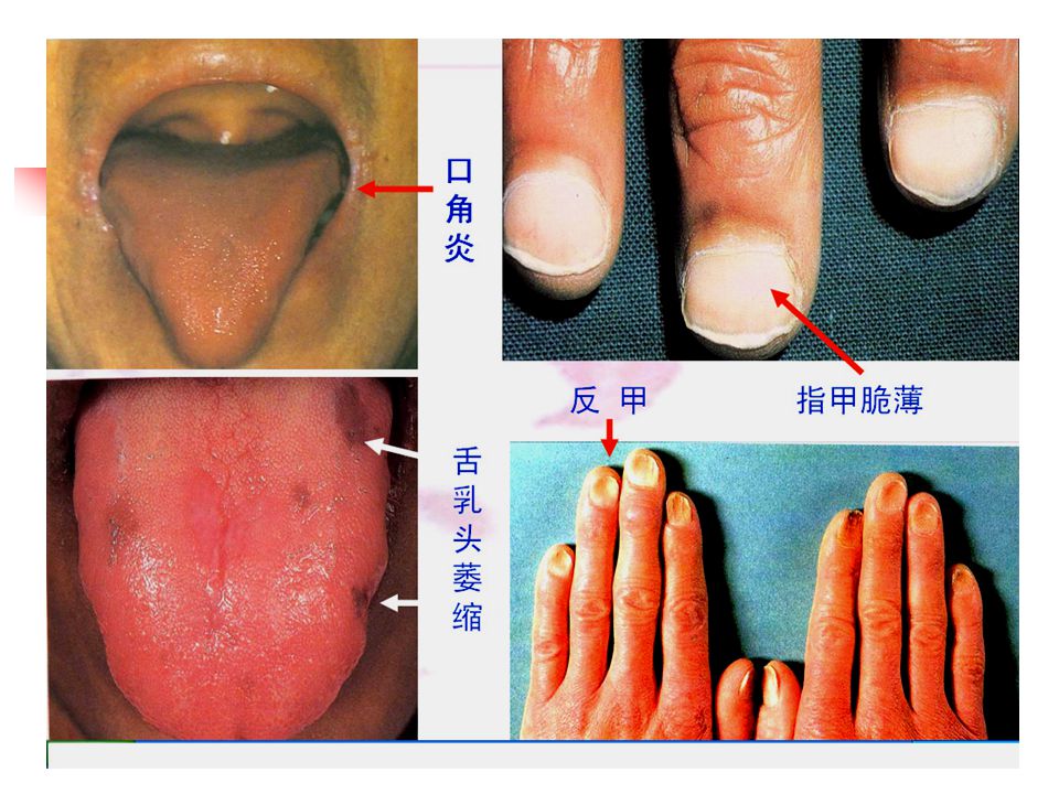
Physical examination Pallor Smooth red tongue, stomatitis Angular cheilitis Koilonychia(rare) Retinal hemorrhages/exudates(severe anemia) Accelerated retinopathy in diabetics Splenomegaly(occasionally)
 Accelerated retinopathy in diabetics. Splenomegaly(occasionally)")
21
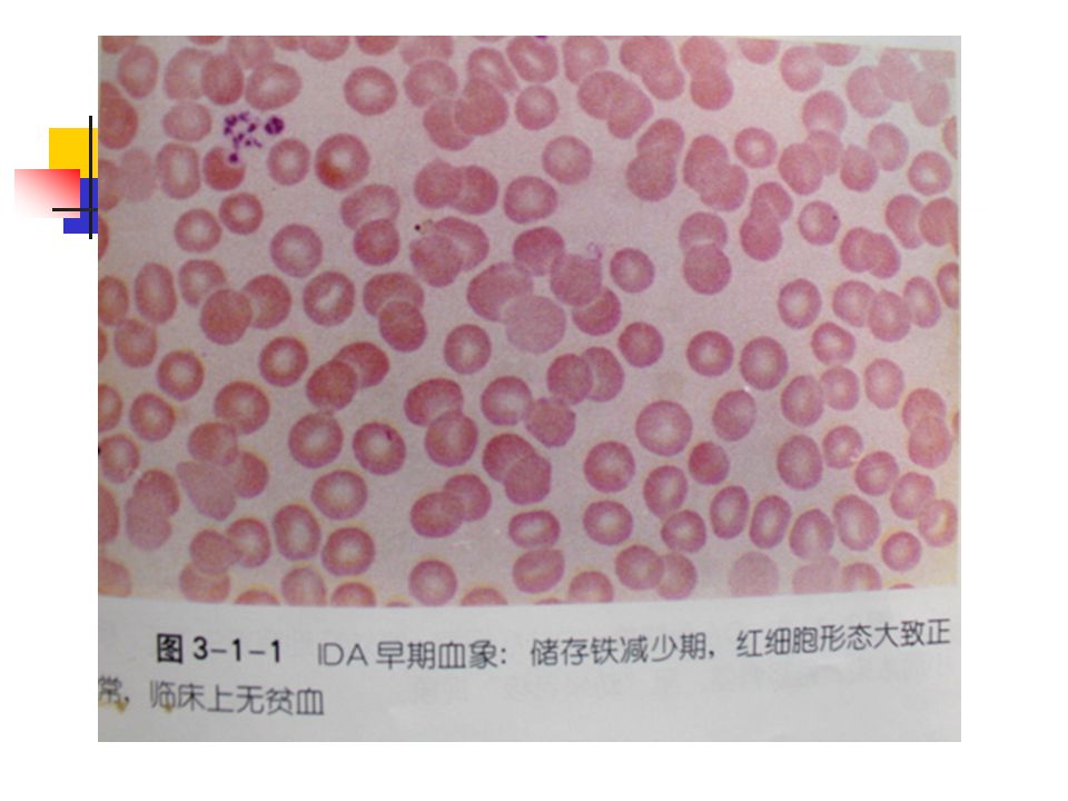
Laboratory Findings 1.Blood : Hypochromic microcytic anemia RBC↓,Hct ↓, Hb ↓ :male<120g/l, female<110g/l, pregnant women<100g/l, MCV<80gl, MCH<26g, MCHC<31% RDW ↑(>14%, earliest change ) morphology: anisocytosis , mild ovalocytosis, target cells, ring cells, elongated hypochromic elliptocytes (pencil cells), nucleated RBC, basophilic stippling RBC Ret normal or reduced Leukocyte normal or decreased Platelets increased or decreased
 morphology: anisocytosis , mild ovalocytosis, target cells, ring cells, elongated hypochromic elliptocytes (pencil cells), nucleated RBC, basophilic stippling RBC. Ret normal or reduced. Leukocyte normal or decreased. Platelets increased or decreased.")
23
IDA早期血象:红细胞生成缺铁期,部分红细胞生理性中心浅染区轻度扩大,临床上无贫血

25
marrow usually hypercellularity with M/E ratio variable.
morphology: erythroid hyperplasia and mainly late normoblasts, which may be small, with narrow rim of ragged cytoplasm and poor Hb formation (polychromatic) and densed nucleus(old nucleated young cytoplasm). Basophiloic stippling and Howell-Jolly body may present in normoblasts.The mature RBCs as in blood. 2. Marrow: hypercellularity anemia
 and densed nucleus(old nucleated young cytoplasm). Basophiloic stippling and Howell-Jolly body may present in normoblasts.The mature RBCs as in blood. 2. Marrow: hypercellularity anemia.")
26
Nucleated cells marked hypercellularity

27
IDA骨髓象:中幼红细胞、晚幼红细胞增生为主,胞体较小,胞浆边缘不规则呈锯齿状、胞浆偏蓝,呈“幼浆老核”现象。

28
HA IDA

29
3 The indexes of iron metaolism
①Marrow stainable iron. Reference value: -extracellular iron (+~++) -intracellular iron (19%~44%) It is a direct and reliable index to reflect the level of storage iron. It decrease (<15%),or absent in ID Sideroblasts decrease in IDE
 -intracellular iron (19%~44%) It is a direct and reliable index to reflect the level of storage iron. It decrease (<15%),or absent in ID. Sideroblasts decrease in IDE.")
30
IDA骨髓铁染色:外铁阴性

31
正常:内铁阳性 IDA骨髓铁染色:内铁阴性

32
② SF(serum ferritin)and EF(erythrocyte alkaline ferritin)
SF Reference value: --< 10µg/l in IDA --10 ~ 20µg/l are presumptive, but not diagnostic. --May be elevated with concomitant inflammation diseases . IDA can be suspected in rheumatoid arthritis if SF is less than 60µg/l or less than 30µg/l in chronic inflammation. Adult male:50~200µg/l; It is a sensitive and reliable index to evaluate total body iron stores, but can be interfered by some conditions. EF reference value: less sensitive than SF, <6.5µg/E in IDA

33
Measurements of iron stores
SF(µg/l ) Marrow iron stain(0-4+) <12 1~300mg 12~20 1+ 300~800mg 20~50 2+ 800~1000mg 50~150 3+ 1~2g 150~300 4+ Iron overload >500
 Marrow iron stain(0-4+) <12. 1~300mg. 12~ ~800mg. 20~ ~1000mg. 50~ ~2g. 150~ Iron overload. >500.")
34
③Serum iron concentration
SI is a direct measure of the amount of iron bound to transferrin. Reference value:50-150µg/dl Adult M:11.6~31.3µmol/l; F:9.0~30.4µmol/l(A:20µmol/l) transporting iron, with a lot of affect factors
 transporting iron, with a lot of affect factors.")
35
Reference value: 360~390µg/l
④TIBC: total iron binding capacity TIBC is a measure of the amount of iron that can be bound by transferrin. Reference value: 360~390µg/l TIBC:male:50~77µmol/l; female 54~77µmol/l Usually increased in ID. decreased in liver disease, malignant tumor, HA, chronic renal disease…

36
⑤TS (Transferrin Saturation): SI TS = ×100% , TIBC
In normal condition, 1/3 transferrin binds to iron. Reference value: 20~50%. ( A:30%) 15% or less in patients with IDA ; >50~60% resulting in iron loading.
 15% or less in patients with IDA ; >50~60% resulting in iron loading.")
37
UIBC SI TIBC

38
⑥sTfR(serum soluble transferrin receptor) :
Reference value: 5~9μg/L(ELISA) sTfR level increase in IDE, IDA (when iron store are exhausted) sTfR level also increase in other disease with ineffective or effective erythroid precursor proliferation
 sTfR level increase in IDE, IDA (when iron store are exhausted) sTfR level also increase in other disease with ineffective or effective erythroid precursor proliferation.")
39
--usually increased in ID --Very sensitive for diagnosis of ID and
⑦ FEP (Free Erythrocyte Protophorphyrin) and ZPP( protophorphyrin binds to zinc) --usually increased in ID --Very sensitive for diagnosis of ID and suitable for large-scale screening of children , detecting both ID and lead poisoning.(why?) FEP less sensitive than SF and EF.
 and ZPP( protophorphyrin binds to zinc) --usually increased in ID. --Very sensitive for diagnosis of ID and. suitable for large-scale screening of children , detecting both ID and lead poisoning.(why ) FEP less sensitive than SF and EF.")
40
Sensitivity of indexes of iron:
SF Marrow stainable iron EF sTfR FEP TIBC TS SI

41
Diagnosis of IDA: ①+more than two of ②~⑧
①Hypochromic microcytic anemia(Hb male < 120 g/l , female<110g/l, pregnant women<100g/l, MCV < 80fl, MCH<26g, MCHC < 31% , RDW > 14% , and morphologic changes) ② Identified causes associated with ID and significant clinical symptoms ③ SI <10.7µmol/L,and TIBC>64.44 µmol/L ④TS<0.15 ⑤ Extracellular iron (-), sideroblasts <15% or absent ⑥ FEP>0.9 µmol/L(blood) ,or ZPP>0.96umol/L(blood), or FEP/Hb >4.5µg/gHb ⑦ SF<14µg/L; ⑧ Effective to therapy with iron
 ② Identified causes associated with ID and significant clinical symptoms. ③ SI <10.7µmol/L,and TIBC>64.44 µmol/L. ④TS<0.15. ⑤ Extracellular iron (-), sideroblasts <15% or absent. ⑥ FEP>0.9 µmol/L(blood) ,or ZPP>0.96umol/L(blood), or FEP/Hb >4.5µg/gHb. ⑦ SF<14µg/L; ⑧ Effective to therapy with iron.")
42
There are three stages of iron deficiency:
1. Iron depletion (ID): It is the earliest stage of iron deficiency. In which storage iron is decreased or absent but serum iron concentration and blood Hb levels are normal Extracellular iron is decreased or absent ---SF concentration falls
: It is the earliest stage of iron deficiency. In which storage iron is decreased or absent but serum iron concentration and blood Hb levels are normal. ---Extracellular iron is decreased or absent ---SF concentration falls.")
43
2.Iron deficiency erythropoiesis (IDE): It is a somewhat more advanced stage of iron deficiency. In which deficit of the functional iron compartment is associated with the development of iron deficiency erythropoiesis It is characterized by decreased or absent storage iron, usually low SI and transferrin saturation, without frank anemia.

44
manifestations --Extracellular iron is absent --Intracellular iron is decreased --Serum soluble transferrin receptor(sTfR) is increased. --TIBC is increased --Serum iron level falls --Transferrin saturation falls --An increase in the RDW --generally asymptoms.
 is increased. --TIBC is increased --Serum iron level falls --Transferrin saturation falls --An increase in the RDW --generally asymptoms.")
45
3.Iron deficiency anemia (IDA): It is a most advanced stage of iron deficiency. It is characterized by decreased or absent iron stores, low SI , low transferrin saturation and low Hb or hematocrit value. Besides the above characteristics, there are: --red cell count decrease --Many symptoms

47
Laboratory studies in three stages if iron deficiency
ID IDE IDA hemoglobin Normal slight decrease Marked decrease (microcytic/hypochromic) Iron stores <100mg(0-1+) SI (μg/dl) normal <60 40 TIBC(μg/dl) >390 >410 TS(%) 20-30 <15 <10 SF((μg/l) ) <20 <12 Percent sideroblasts 40-60 FEP(μg/dlRBC) 30 >100 >200
 Iron stores. <100mg(0-1+) SI (μg/dl) normal. < TIBC(μg/dl) >390. >410. TS(%) <15. <10. SF((μg/l) ) <20. <12. Percent sideroblasts FEP(μg/dlRBC) 30. >100. >200.")
48
Diagnosis process of -Anemia? -Microcytic hypochromic anemia? -IDA?(measurements of iron metabolism) -The cause of IDA!

49
Sideroblastic Anemias (SA)
The sideroblastic anemias are a heterogeneous group of disordes that have as common features the presence of large number of ringed sideroblasts in the marrow, ineffective erythropoiesis, increased levels of tissue iron and varying proportions of hypochronic erythrocytes in the blood.

50
Classification: hereditary: x chromosome-linked ,partially recessive inheritance, males are anemic and females are carriers autosomally-linked, or mitochondria entities acquired : primary: neoplasia (MDS-RAS) secondary: drugs, toxin, alcohol, or coincident to neoplastic or inflammatory disease
 secondary: drugs, toxin, alcohol, or coincident to neoplastic or inflammatory disease.")
51
Pathogenesis: not clear
1.underlying biochemical lesions Sideroblasts appear by a defect of pyridoxine metabolism or other intramitchondrial defect in heme synthesis. 2. anemia: ineffective erythropoiesis

52
Fe 3+ Tf 80% TfR 铁的利用与血红蛋白合成示意图 血浆 骨髓:幼红细胞和网织红细胞 Tf-Fe 3+ Fe 3+ Fe 2+
protophorphyrin + Heme Heme+globin Hb Ferritin 80% Tf TfR 铁的利用与血红蛋白合成示意图

53
Phosphate pyrindoxine
pyridoxine UROI COPRO I UROgen I UROgen I ②Phosphate pyridoxine Gly ③ ①ALA synthetase ALA PBG Succinyl CoA UROIII UROgen III 线粒体 PROTOgenIII COPROgenIII COPROIII ④ PROTO9 Fe ⑤Heme synthetase ⑥ Pathway of heme Biosynthesis Heme

54
Lab Findings --Hypochromic and micro- or normocytic anemia --Marked anisocytosis and poikilocytosis --Normal or elevated SI levels --Erythroid hyperplasia of the BM --Increased siderablasts in BM, ringed sideroblasts --Increased iron stores --Normal or slightly reduced red cell survival time --Hemosiderosis and/or hemochromatosis [Others] Refractory to therapy with iron

57
Anemias of Chronic Disorders(ACD)
Secondary anemia Anemias of Chronic Disorders(ACD) Anemias of chronic disorders are present in chronic infections, inflammatory diseases and neoplastic diseases . It is a common anemia, probably second in incidence to iron deficiency anemia.
 Anemias of chronic disorders are present in chronic infections, inflammatory diseases and neoplastic diseases . It is a common anemia, probably second in incidence to iron deficiency anemia.")
58
Pathogenesis of ACD --Sequestration of iron in the macrophages SI ,TS decrease --reduce the secretion of EPO and impair its action in marrow Tf, sTfR ,TIBC decrease --Shortened RBC life span (slightly hemolysis)
")
59
80% Tf Tf-Fe 3+ Fe 3+ Fe 2+ Fe 3+ Heme+globin Hb ACD
protophorphyrin + Fe 2+ Heme Tf-Fe 3+ 80% Fe 3+ Fe 2+ Ferritin Fe 3+ Tf Heme+globin Hb ACD Reutilization of iron impaired Hypochromatic anemia

60
Hb Macrophages Neutrophils SF, extracellular iron increase globin heme
Hemosiderin ferritin apo-Ferritin Hb globin heme Fe 3+ Fe 2+ Macrophages Neutrophils Inflammatory cells growth factors-IL 1 Precursor cells Cytokines, such as IL 1 , TNF ,gamma interferon.

61
Clinical Features --Signs common to all anemias. --Signs specific to the underlying disease. --Showing no improvement with iron therapy and only improving with correction of the primary disorders.

62
Lab Findings Bone marrow --No distinctive abnormality --Iron stains shows increased stainable iron in the macrophages despite a decrease in sideroblasts. Other Test --SI decrease, Tf decrease --SF is normal or increased --TIBC is normal to decreased --TS is usually decreased

63
Questions: 1.Describe the characteristics of three stages of iron deficiency. 2. Describe the causes of iron deficiency. 3. How to diagnose ID? IDE? IDA? 4. What is IDA? Sideroblast? Ring sideroblast? 5. What is ferritin? Tf ? TIBC? SI? 6.How to differentiate hypochromatic microcytic anemias in Lab?

64
Lab test Thalassemia ACD IDA RBC low low low
Differential Diagnosis in Lab Lab test Thalassemia ACD IDA RBC low low low MCV low in 20-30% rarely to 60-70 Tf N decrease increase SF N/Increased N/Increased low SI N to high low low HbA2,F,H usually present absent absent TIBC N/ low usually low usually high TS N to high > 15%, low low or N Extracelluiar iron N increase decrease Intracellular iron N decrease decrease

65
Case A female, complained of fatigue and headache for a few months since she had functional uterine bleeding and short of breath when climbing stairs The doctor found her face look pale. He ordered an CBC detection. The result is the follow. Ret is 2.5%

66
Result of CBC

67
外周血涂片染色

68
骨髓细胞学检查。

69
Questions: 1.Does the female has anemia? 2. Do you think which type anemia she has? Give your reasons. 3. Which tests do you need to confirm your advice. Do you image the results?

Similar presentations


>")

/ HYPOCHROMIC &/or (NORMO)/ MICROCYTIC ANEMIAS 1. Disorders of iron utilization a. iron deficiency b. anemia of.>")


 Course code :MLHE-201 Supervisor :Prof.Dr Magda Sultan. Date : 5/ 12 / 2013 Outcome : The student will know : The definition.>")
