Download presentation
Presentation is loading. Please wait.

1
Specimen Collection and Culture Work-up
Dr Mohammad Rahbar

2
Objectives Review collection and transport procedures for wound, stool, respiratory, and genitourinary specimens submitted for microbiological culture. Summarize appropriate algorithms for culture workup of wound, stool, respiratory, and genitourinary specimens submitted for microbiology culture. Discuss the importance of the clinician-laboratory interface

3
Routine Stool Culture Q-Probe Study of 601 institutions
Majority of laboratories (99.3%) included Salmonella and Shigella in the routine stool workup 96% routinely included Campylobacter 30-60% of laboratories surveyed also included other organisms such as Aeromonas, Plesiomonas, Yersinia, Escherichia coli O157, & Vibrio Valenstein, P, M. Pfaller, and M. Yungbluth The use and abuse of routine stool microbiology. Arch Pathol Lab Med. 120:
 included Salmonella and Shigella in the routine stool workup. 96% routinely included Campylobacter % of laboratories surveyed also included other organisms such as Aeromonas, Plesiomonas, Yersinia, Escherichia coli O157, & Vibrio. Valenstein, P, M. Pfaller, and M. Yungbluth The use and abuse of routine stool microbiology. Arch Pathol Lab Med. 120:")
4
Role of Rapid Nonculture Screening Tests
Fecal Leukocyte Test Fecal Lactoferrin Assay Gram stain

5
Fecal Leukocyte Test Hines, J and I. Nachamkin Effective use of the clinical microbiology laboratory for diagnosing diarrheal diseases. Clin Infect Dis. 23:1292–1301.

6
Fecal Lactoferrin Assay
To detect Salmonella, Shigella and Campylobacter spp. The sensitivity ranged from 83 to 93%, and specificity ranged from 61% to 100% 1 85% sensitive and 79% specific when compared to culture 2 As a marker for inflammatory bowel disease (IBD) and irritable bowel syndrome (IBS) 3 Significantly higher lactoferrin levels in patients with active and inactive IBD (85.9% sensitivity and 100% specific) Elevated fecal lactoferrin levels – 100% specific in ruling out IBS Choi et. al To culture or not to culture: fecal lactoferrin screening for inflammatory diarrhea. J. Clin. Microbiol. 34: Silletti, R. P. et. al Role of stool screening tests in diagnosis of inflammatory bacterial enteritis in selection of specimens likely to yeild invasive enteric pathogens. J. Clin. Microbiol. 34: Kane, S. V., et. al Fecal lactoferrin is a sensitive and specific marker in identifying intestinal inflammation. Am. J. Gastroenterol. 98:
 and irritable bowel syndrome (IBS) 3. Significantly higher lactoferrin levels in patients with active and inactive IBD (85.9% sensitivity and 100% specific) Elevated fecal lactoferrin levels – 100% specific in ruling out IBS. Choi et. al To culture or not to culture: fecal lactoferrin screening for inflammatory diarrhea. J. Clin. Microbiol. 34: Silletti, R. P. et. al Role of stool screening tests in diagnosis of inflammatory bacterial enteritis in selection of specimens likely to yeild invasive enteric pathogens. J. Clin. Microbiol. 34: Kane, S. V., et. al Fecal lactoferrin is a sensitive and specific marker in identifying intestinal inflammation. Am. J. Gastroenterol. 98:")
7
Gram Stain The only role of Gram stain is in the diagnosis of Campylobacter sp. The sensitivity ranges from 66 to 94% with high specificity No value for detecting other enteric pathogens. Hines, J and I. Nachamkin Effective use of the clinical microbiology laboratory for diagnosing diarrheal diseases. Clin Infect Dis. 23:1292–1301.

8
Number of Specimens Required to Identify a Pathogen
Number of Specimens Collected per patient Cumulative Percent of Infected Patients Detected Bacteria/Fungi (n = 3349) Parasites (n = 1159) 1 96.9 91.9 2 99.0 97.6 3 99.3 99.8 4 99.4 99.9 Valenstein, P, M. Pfaller, and M. Yungbluth The use and abuse of routine stool microbiology. Arch Pathol Lab Med. 120:
 Parasites. (n = 1159) Valenstein, P, M. Pfaller, and M. Yungbluth The use and abuse of routine stool microbiology. Arch Pathol Lab Med. 120:")
9
Bacteriology Results on Inpatients by Day of Hospital Stay
Hospital Day 1 2 3 4 >4 Total number of specimens collected 7924 6886 3369 2022 10009 Total number of 1st positives, with or without previous specimens 374 155 35 19 49 Percentage of total specimens that were patient's 1st positive specimen 4.7 2.3 1.0 0.9 0.5 Number of 1st positive specimens from patients for whom previous negative specimens were collected 6 Valenstein, P, M. Pfaller, and M. Yungbluth The use and abuse of routine stool microbiology. Arch Pathol Lab Med. 120:

10
CAP CHECKLIST Question MIC.22440 PHASE: I Question MIC.22336 PHASE: I
Does the laboratory have guidelines (developed with clinicians) for the number and/or timing of collection of stool specimens submitted for routine bacterial testing? Question MIC PHASE: I Does the final report for routine bacterial stool cultures list the organisms for which the specimen was cultured (e.g., Salmonella, Shigella, Vibrio, etc.)?
 for the number and/or timing of collection of stool specimens submitted for routine bacterial testing Question MIC PHASE: I. Does the final report for routine bacterial stool cultures list the organisms for which the specimen was cultured (e.g., Salmonella, Shigella, Vibrio, etc.)")
11
Acceptable Specimens Fecal sample Fresh Preserved
received within 1 to 2 h of passage Preserved Buffered Glycerol Saline recommended for Salmonella & Shigella but not for Campylobacter or Vibrio sp., unless enriched with CaCl2 Modified Carey-Blair good overall transport media inappropriate for C. difficile

12
Acceptable Specimens Rectal swabs
Duodenal, colostomy or ileostomy contents stool transport vials Rectal biopsy samples sterile container with a small amount of sterile water

13
Unacceptable Specimens
Unpreserved stool samples > 2h old Dry rectal swabs or biopsy specimens Multiple specimens received on the same day Specimens received from inpatients after the third hospital day, without prior consultation .

14
Workup Guidelines for Salmonella, Shigella, Aeromonas & Plesiomonas species
Media Non-selective BAP Aeromonas sp., Plesiomonas shigelloides, Yeasts, Staphylococcus aureus, Pseudomonas aeruginosa Differential enteric agar MacConkey agar, Eosin Methylene Blue (EMB) differentiates lactose-fermenting from non-lactose fermenting colonies Moderately selective Hektoen Agar, Xylose Lysine Deoxycholate Agar (XLD), or Salmonella Shigella Agar allows growth of Salmonella & Shigella sp. while suppressing the growth of most members of family Enterobacteriaceae Enrichment broth GN Broth, Selenite F increases chances of detecting low numbers of pathogens
 differentiates lactose-fermenting from non-lactose fermenting colonies. Moderately selective. Hektoen Agar, Xylose Lysine Deoxycholate Agar (XLD), or Salmonella Shigella Agar. allows growth of Salmonella & Shigella sp. while suppressing the growth of most members of family Enterobacteriaceae. Enrichment broth. GN Broth, Selenite F. increases chances of detecting low numbers of pathogens.")
15
Workup Guidelines for Salmonella, Shigella, Aeromonas & Plesiomonas species
Enteric Screening Procedure Conventional TSI or KIA, LIA, and Urea Commercial kits latex agglutination Full ID of suggestive screening results Serological identification of Salmonella and Shigella sp.

16
Campylobacter sp. Most frequently isolated Selective media
Campy-Thio (enrichment broth) Campy-BAP Skirrow medium Campylobacter-cefoperazone-vancomycin-amphotericin (CVA) Identification growth at 42°C, oxidase and catalase positive, Hippurate positive Nalidixic Acid susceptible, Cephalothin resistant Latex agglutination test
 Campy-BAP. Skirrow medium. Campylobacter-cefoperazone-vancomycin-amphotericin (CVA) Identification. growth at 42°C, oxidase and catalase positive, Hippurate positive. Nalidixic Acid susceptible, Cephalothin resistant. Latex agglutination test.")
17
Campylobacter sp. Rapid Methods
ProSpecT® Campylobacter Microplate Assay detects Campylobacter specific antigens in stool (fresh or in transport medium) utilizes polyclonal anti- Campylobacter specific antigens capture antibody can be read visually or spectrophometrically Evaluated in three studies Sensitivities of 80, 89 and 96% Specificities of 99% Flexiable, easy to use reduces cost, reduce turnaround time Cross-reactivity with C. upsaliensis, C. hyointestinalis, or C. helveticus unknown Endtz, H. P., et. al Evaluation of a New Commercial Immunoassay for Rapid Detection of Campylobacter jejuni in Stool Samples. Eur. J. Clin. Microbiol. Infect. Dis. 19: Hindiyeh, M. et. al Rapid Detection of Campylobacter jejuni in Stool Specimens by an Enzyme Immunoassay and Surveillance for Campylobacter upsaliensis in the Greater Salt Lake City Area. J. Clin. Microbiol. 38: Tolcin, R. et. al Evaluation of the Alexon-Trend ProSpecT Campylobacter Microplate Assay. J. Clin. Microbiol. 38:
 utilizes polyclonal anti- Campylobacter specific antigens capture antibody. can be read visually or spectrophometrically. Evaluated in three studies. Sensitivities of 80, 89 and 96% Specificities of 99% Flexiable, easy to use. reduces cost, reduce turnaround time. Cross-reactivity with C. upsaliensis, C. hyointestinalis, or C. helveticus unknown. Endtz, H. P., et. al Evaluation of a New Commercial Immunoassay for Rapid Detection of Campylobacter jejuni in Stool Samples. Eur. J. Clin. Microbiol. Infect. Dis. 19: Hindiyeh, M. et. al Rapid Detection of Campylobacter jejuni in Stool Specimens by an Enzyme Immunoassay and Surveillance for Campylobacter upsaliensis in the Greater Salt Lake City Area. J. Clin. Microbiol. 38: Tolcin, R. et. al Evaluation of the Alexon-Trend ProSpecT Campylobacter Microplate Assay. J. Clin. Microbiol. 38:")
18
Genus Vibrio Laboratory Diagnosis
Media TCBS (thiosulfate citrate bile salts sucrose) agar sucrose-fermenting (Yellow colonies) V. cholerae, V. alginolyticus, & V. fluvialis non-sucrose-fermenting (green colonies) V. parahaemolyticus, V. vulnificus (Lactose Fermentor) Susceptible to 150 g of vibriostatic agent (O/129)
 agar. sucrose-fermenting (Yellow colonies) V. cholerae, V. alginolyticus, & V. fluvialis. non-sucrose-fermenting (green colonies) V. parahaemolyticus, V. vulnificus (Lactose Fermentor) Susceptible to 150 g of vibriostatic agent (O/129)")
19
Escherichia coli Three paradigms by which diarrhea is produced:
enterotoxin production Enterotoxigenic E. coli (ETEC) Enteroaggregative E. coli (EAEC) invasion Enteroinvasive E. coli (EIEC) intimate adherence with membrane signaling Enteropathogenic E. coli (EPEC) Enterohemorrhagic E. coli (EHEC/ STEC)
 Enteroaggregative E. coli (EAEC) invasion. Enteroinvasive E. coli (EIEC) intimate adherence with membrane signaling. Enteropathogenic E. coli (EPEC) Enterohemorrhagic E. coli (EHEC/ STEC)")
20
Enterohemorrhagic E. coli
Aka Shiga toxin-producing E. coli (STEC) There are at least 100 serotypes of STEC Only one serotype, namely E. coli O157:H7 can be detected in clinical laboratories. Selective media: sorbitol-MacConkey agar confirm by latex agglutination Varied geographic distribution - evaluate prevalence for the need of routine workup Availability of EIA for detection of STEC
 There are at least 100 serotypes of STEC. Only one serotype, namely E. coli O157:H7 can be detected in clinical laboratories. Selective media: sorbitol-MacConkey agar. confirm by latex agglutination. Varied geographic distribution - evaluate prevalence for the need of routine workup. Availability of EIA for detection of STEC.")
21
Yersinia enterocolitica
Detection based on conventional methods Selective media - CIN agar dark red “bull’s eye” with a transparent border

22
Clostridium difficile Testing
Acceptable specimen Unformed stool specimen unless ileus due to C. difficile is suspected. Rejection criteria Specimens that are not liquid or soft Specimens from infants under 1 year old should be discouraged Specimen more than 24 hours old. Rectal swab specimens “Test for cure” or testing from asymptomatic individuals Gerding, D.N. et. al Clostridium difficile-associated diarrhea and colitis.Infect. Control. Hosp. Epidemol. 16: Johnson, S. and D. N. Gerding Clostridium difficile associated diarrhea: a review. Clin. Infect. Dis. 26:

23
Clostridium difficile Testing
Culture – most sensitive Selective medium - Cycloserine-cefoxitin-fructose agar Characteristic horse-dung smell typical yellow-green fluorescence under UV light Limitations does not distinguish between toxigenic and non-toxigenigic strains delayed turn-around time Use of latex agglutination test that detects glutamte dehydrogenase is discouraged

24
Clostridium difficile Testing
Cell Culture Cytotoxicity Assay – most specific detects Toxin B Limitations Requires 24 to 48 hours Tedious Non-commercial versions are not standardized EIAs for toxin A or toxins A and B Rapid Less sensitive than cell cyotoxicity assay Tests that detect only toxin A may miss isolates that are toxin A-B+

25
Wounds

26
Wound Cultures: Controversies
Is sampling a wound for culture relevant? When and how should wounds be sampled? How should samples be transported? What analysis should be requested? Gram stain only? Culture only? Susceptibility testing? Quantitative cultures?

27
Wounds:Classification
Acute Caused by external damage to intact skin Types Surgical Bites Burns Minor cuts Abrasions Severe traumatic Chronic Precipitated by predisposing conditions that lead to compromise of dermal/epidermal tissue Types Impaired venous drainage Impaired arterial supply Metabolic diseases eg. diabetes Bowler PG, et. al Wound microbiology and associated approaches to wound management. Clin Microbiol Rev 14: 244.

28
Wound Infections:Etiology
Surgical wounds Aerobes: S. aureus, coagulase negative staphylococci, Enterococcus spp. E. coli, P. aeruginosa, Enterobacter spp. Anaerobes: Bacteroides spp., Peptostreptococcus, Clostridium spp. Acute soft tissue infections Staph aureus only organism in 30% 30-50% mixed aerobes/anaerobes 20-30% other eg. Group A streptococci, Clostridium spp. Bite wounds Special pathogens: Pasteurella multocida, Capnocytophaga canimorsus, Bartonella henselae, Eikenella corrodens Other mixed aerobes and anaerobes

29
Wound Infections:Etiology
Burn wounds Primarily aerobic organisms: P. aeruginosa, Staphylococcus aureus, E. coli, Klebsiella spp. Enterococcus spp. and Candida spp. Diabetic foot ulcers Aerobes: Staph aureus, Streptococcus spp. P. aeruginosa, Enterococcus spp., enterics Anaerobes: Peptostreptococcus, Bacteroides spp., Prevotella spp. Decubitus ulcers Mixed aerobic and anaerobic bacteria

30
Wound Cultures For open wounds
Clean the wound margins with surgical soap or 70% ethyl or isopropyl alcohol Aspirate from the depth of the wound using a sterile syringe and needle Aspirated fluid should be sent to the laboratory in an appropriate transport system Alternatively, a curette may be used to obtain tissue from base of the wound Swabs are strongly discouraged

31
Wound Cultures For closed wounds
Prepare site as described for obtaining blood culture Aspirate as much purulent material as possible Transport in aerobic/anaerobic transport system

32
Wound Cultures: Gram stain
Pros Useful in estimating organism load from tissue biopsies Presence of microorganisms on smear from swabs correlates with > 106 organisms (burns) Facilitates identification of etiologic agent of wound infection following clean surgery Cons Poor correlation seen between Gram stain and culture results from biopsy of diabetic foot infections In mixed infections, little value although presence of leukocytes indicates infection
 Facilitates identification of etiologic agent of wound infection following clean surgery. Cons. Poor correlation seen between Gram stain and culture results from biopsy of diabetic foot infections. In mixed infections, little value although presence of leukocytes indicates infection.")
33
Wound Specimens: Algorithms
Three approaches PMN predominance Q-Score Q system

34
Wound Specimens: Algorithms
Modified from Sharp SE. Clin Micro Newsletter 21:14, 1999

35
Wound Cultures Culture for aerobic and anaerobic bacteria if appropriately collected Gram stain results suggest adequate collection or presence of inflammation Tissues or aspirates vs. swabs Primary plating media: 5% SBA, Choc agar, MacConkey agar; anaerobic plates and thio if appropriately collected Identify anaerobes to Genus level only Perform susceptibility testing of predominant organisms only

36
Wound Cultures: Extent of Workup
Possible approaches Use Gram stain result Work up organisms seen on stain only List others Work up any potential pathogens to maximum of three, list others present by morphology Work up any quantity S. aureus, P. aeruginosa, beta hemolytic streptococci, enterics and gram-negative anaerobes

37
Wound Cultures: Examples
Gram stain results: (Acceptable) Many neutrophils, no epithelial cells Many gram positive cocci in clusters Many gram negative bacilli Few morphotypes resembling skin flora Work up (identify and perform susceptibility testing): Gram positive cocci in clusters and gram neg bacilli Culture report: Many S. aureus, many Klebsiella pneumoniae, light aerobic bacteria resembling skin flora
 Many neutrophils, no epithelial cells. Many gram positive cocci in clusters. Many gram negative bacilli. Few morphotypes resembling skin flora. Work up (identify and perform susceptibility testing): Gram positive cocci in clusters and gram neg bacilli. Culture report: Many S. aureus, many Klebsiella pneumoniae, light aerobic bacteria resembling skin flora.")
38
Wound Cultures: Examples
Gram stain: many neutrophils, few epithelial cells, Gram positive cocci in clusters, Gram positive cocci in chains, Culture grows: many S. aureus, many Group A streptococci, few enteric bacilli Work up: S. aureus, Group A streptococcus: limited ID and no susceptibility on enteric bacilli; susceptibility testing on Group A strep not required

39
Wound Cultures: Examples
Gram stain: Many neutrophils, few epithelial cells, multiple morphotypes Culture grows: more than 3 potential pathogens Consider source Tissue or aspirate ? Contamination likely ? Type of patient May need to consult with clinician or Infectious Diseases service

40
Q-Score Squamous Epithelial Cells Neutrophils
Q-Score = # of potential pathogens (PP) to work up Squamous Epithelial Cells No Cells 1-9/lpf 10-24/lpf >25/lpf Score -1 -2 -3 Neutrophils 3 +1 +2 1 +3 2
 to work up. Squamous Epithelial Cells. No Cells. 1-9/lpf /lpf. >25/lpf. Score Neutrophils")
41
Workup of Wound Cultures
Q-Score System Good quality specimen (Q3) Up to 3 organisms can be considered as potential pathogens and worked up (ID/AST) Lower quality specimen (Q2, Q1) More SEC Fewer organisms are worked up
 Up to 3 organisms can be considered as potential pathogens and worked up (ID/AST) Lower quality specimen (Q2, Q1) More SEC. Fewer organisms are worked up.")
42
Workup of Wound Cultures
Q-Score System If the Q-score is greater than or equals the PP in culture Workup all potential pathogens If Q-Score is less than the PP in culture Look at the Gram stain Workup all PP that are seen on GS Morphologically ID others If all PP present on GS then only Morph ID all

43
Workup of Wound Cultures
Q/2-3-4 System Culture workup is based on the # of PP present 2PP – ID/AST 3PP Look at the Gram stain Workup two PP if they are seen on GS If all 3 present on GS then Morph ID 4PP Morph ID only

44
Wound Cultures: Example
Gram stain: many neutrophils, few epithelial cells, Gram positive cocci in clusters, Gram positive cocci in chains, Culture grows: many S. aureus, many Group A streptococci, few enteric bacilli Q score = 2 [PMN (+3), few epi (-1)] Q/2-3-4 = 3 PP look at gram stain Work up: S. aureus, Group A streptococcus, Morph ID and no susceptibility on enteric bacilli
, few epi (-1)] Q/2-3-4 = 3 PP. look at gram stain. Work up: S. aureus, Group A streptococcus, Morph ID and no susceptibility on enteric bacilli.")
45
Respiratory Specimens

46
Respiratory Specimens
Upper respiratory tract specimens Throat detection of streptococcal pharyngitis

47
Respiratory Specimens
Upper respiratory tract specimens Nose detection of MRSA carriers Nasopharyngeal swabs diagnosis of Bordetella pertussis Nasopharyngeal swabs and washings diagnosis of viral disease

48
Lower Respiratory Tract Infections

49
“ The culture of lower respiratory specimens may result in more unnecessary microbiologic effort than any other type of specimen.” Raymond C Bartlett

50
Lower Respiratory Tract Infections Epidemiology
Pneumonia is the sixth leading cause of death in US Increasing numbers of patients at risk Aging population Increase in patients with immunocompromising conditions Overtreatment has lead to resistance Multidrug resistant Streptococcus pneumoniae Resistance among hospital acquired pathogens such as Acinetobacter, Pseudomonas aeruginosa and others

51
Cumitech 7B:2003 Lower Respiratory Tract Infections
6 contributing authors Major sections Clinical aspects of diseases of LRT Specimen collection Specimen processing Interpretation of bacterial cultures Most common pathogens Methods for implementing change Guidelines for frequency of testing Public health issues Reimbursement codes

52
Categories of Lower Respiratory Tract Infections
Acute bronchitis Community acquired pneumonia Hospital acquired pneumonia Pneumonia in the immunocompromised host

53
Community Acquired Pneumonia Etiologic Agents
Pathogen Frequency (%) Streptococcus pneumoniae 66 Haemophilus influenzae 1-12 M catarrhalis 10 Legionella species 2-15 Mycoplasma pneumoniae 2-14 Klebsiella species 3-14 Enteric gram negative bacilli 6-9 Staphylococcus aureus Chlamydia species 5-15 Influenza viruses 5-12 Other viruses <1-12 Unknown 23-49 Carroll KC J Clin Microbiol 40: Sharp SE, et.al. Cumitech 2003
 Streptococcus pneumoniae. 66. Haemophilus influenzae M catarrhalis. 10. Legionella species Mycoplasma pneumoniae Klebsiella species Enteric gram negative bacilli Staphylococcus aureus. Chlamydia species Influenza viruses Other viruses. <1-12. Unknown Carroll KC J Clin Microbiol 40: Sharp SE, et.al. Cumitech")
54
Community Acquired Pneumonia Diagnosis
Available Test Methodologies Sputum Gram stain and culture Blood cultures Serologic studies Antigen detection tests Nucleic acid amplification tests

55
Sputum Gram Stain and Culture
Proponents Demonstration of predominant morphotype on Gram stain guides therapy Accuracy is good when strict criteria are used Cheap, so why not? Antagonists Poor specimen collection Intralaboratory variability (Gram stain interpretation) Low sensitivity and specificity
 Low sensitivity and specificity.")
56
Sputum Collection Proper patient instruction
Food should not have been ingested for 1-2 h prior to expectoration The mouth should be rinsed with saline or water Patient should breathe and cough deeply Patient should expectorate into a sterile container Transport container immediately to lab Perform Gram stain and plant specimen as soon as possible

57
Sputum Gram Stain Screen for acceptability
Examine specimen under low power (x 10 objective) Examine 10 representative fields Specimens that show few squamous epithelial cells (< 10/lpf) and many PMNs (> 25/lpf) are acceptable Notify physician of unacceptable samples
 Examine 10 representative fields. Specimens that show few squamous epithelial cells (< 10/lpf) and many PMNs (> 25/lpf) are acceptable. Notify physician of unacceptable samples.")
58
Sputum Gram Stain Unacceptable

59
Sputum Gram Stain Good Quality

60
Sputum Gram Stain Good Quality

61
Sputum Gram Stain Good quality specimens
Quantify number and types of inflammatory cells Note presence of bronchial epithelial cells Concentrate on areas with WBCs when looking for organisms Determine if there is a predominant organism (> 10 per oil immersion field) Semiquantitate and report organism with descriptive If no predominant organism is present, report “mixed gram positive and gram negative flora”
 Semiquantitate and report organism with descriptive. If no predominant organism is present, report mixed gram positive and gram negative flora")
63
Utility of the Gram Stain in Diagnosis of Pneumonia
Roson, B, et. al Clin Infect Dis 31: Prospective study Non immunocompromised patients hospitalized with CAP 1,000 bed hospital in Spain ER physicians instructed on sputum collection for Gram stain and culture Sputum collected under supervision of nurse or resident Samples were processed immediately Screened for epithelial cells Screened for predominant morphotype (> 75% of the organisms seen) Sputum planted to blood agar, chocolate agar and MacConkey agar Strictly defined clinical and diagnostic parameters
 Sputum planted to blood agar, chocolate agar and MacConkey agar. Strictly defined clinical and diagnostic parameters.")
64
Utility of the Gram Stain in Diagnosis of Pneumonia
Roson, B, et. al Clin Infect Dis 31:869-74 Results 190/533 (35.6%) patients had no sputum sample submitted (these patients were included in the calculations) 133/533 (25%) patients had a poor quality specimen 210/533 (39.4%) patients had a good quality specimen Overall sensitivity and specificity for pneumococcal pneumonia: 57% and 97% Overall sensitivity and specificity for H. influenzae pneumonia: 82 % and 99% Gram stain gave presumptive diagnosis in 80% of patients who had a good specimen submitted > 95% of patients in whom a predominant morphotype was seen on Gram stain received monotherapy
 patients had no sputum sample submitted (these patients were included in the calculations) 133/533 (25%) patients had a poor quality specimen. 210/533 (39.4%) patients had a good quality specimen. Overall sensitivity and specificity for pneumococcal pneumonia: 57% and 97% Overall sensitivity and specificity for H. influenzae pneumonia: 82 % and 99% Gram stain gave presumptive diagnosis in 80% of patients who had a good specimen submitted. > 95% of patients in whom a predominant morphotype was seen on Gram stain received monotherapy.")
65
Gram Stain Reports Be as descriptive as possible
Moderate neutrophils Moderate Gram positive diplococci suggestive of Streptococcus pneumoniae Few bacteria suggestive of oral flora Keep report short—avoid line listing of all morphotypes present

66
Sputum and Endotracheal Suction Culture Evaluation
Identify and perform susceptibility testing on 2-3 potential pathogens seen as predominant on Gram stain Alpha strep—rule out S. pneumoniae Yeast—rule out Cryptococcus neoformans only S. aureus, Gram negative bacilli < normal flora, quantify and limit ID; no susceptibility Add comment that organism not predominant on stain ID mould, Mycobacteria or Nocardia spp. Modified from Sharp SE, et. Al Cumitech 7B. ASM Press.

67
IDSA Practice Guidelines Diagnostic Tests for CAP
Outpatients Empiric therapy with a macrolide, doxycycline, or a fluoroquinolone Hospitalized patients with CAP Gram stain and culture of sputum 2 pretreatment blood cultures Studies for Mtb, Legionella in select patients Rationale To improve patient care Advance knowledge of epidemiologically important organisms Prevent antibiotic abuse Reduce antibiotic expense Bartlett JG Clin Infect Dis 31:

68
ATS Guidelines Diagnostic Tests for CAP
Empiric therapy for outpatients Macrolide or tetracycline Hospitalized patients with CAP 2 sets of pre-treatment blood cultures Pleural fluid Gram stain/culture when appropriate Studies for Legionella, Mtb, fungi in select patients Sputum Gram stain/culture only if resistant or unusual pathogen is suspected Avoid extensive testing ATS Am J Respir Crit Care Med 163:

69
Hospital Acquired Pneumonia
Most frequent nosocomial infection (30-33% of cases) among combined medical surgical intensive care units 83% are ventilator associated Etiologic agents Frequency (%) Gram positive cocci S. aureus S. pneumoniae Aerobic gram-neg bacilli 60 Pseudomonas aeruginosa Enterobacter sp. Klebsiella pneumoniae Acinetobacter Legionella Anaerobes Fungi Modified from: Carroll KC J Clin Microbiol 40:
 among combined medical surgical intensive care units. 83% are ventilator associated. Etiologic agents Frequency (%) Gram positive cocci. S. aureus 17. S. pneumoniae Aerobic gram-neg bacilli 60. Pseudomonas aeruginosa. Enterobacter sp. Klebsiella pneumoniae. Acinetobacter. Legionella. Anaerobes Fungi Modified from: Carroll KC J Clin Microbiol 40:")
70
Hospital Acquired Pneumonia Diagnosis
American College of Chest Physicians: Clinical findings are not sufficient for definitive diagnosis Qualitative culture or endotracheal sputum has poor predictive value Bronchoscopy is recommended by many pulmonologists Bronchial brushings Bronchial washes Protected specimen brushing Bronchoalveolar lavage specimens (BAL) Transbronchial biopsy
 Transbronchial biopsy.")
71
Respiratory Specimens
Protected Brush Specimen To procure uncontaminated lower airway secretions Brush within 2 catheters

72
Respiratory Specimens
Bronchoalveolar Lavage (BAL) Samples large area of the lung Performed using a bronchoscope 100 to 250 ml of saline injected Injected saline along with secretions is collected by aspiration Transthoracic Aspiration Involves percutaneous introduction of a needle directly into the infiltrate
 Samples large area of the lung. Performed using a bronchoscope. 100 to 250 ml of saline injected. Injected saline along with secretions is collected by aspiration. Transthoracic Aspiration. Involves percutaneous introduction of a needle directly into the infiltrate.")
73
Bronchoalveolar Lavage (BAL) Specimen Acceptability
Microscopic examination of Gram-stained smear Acceptable <1% of cells present are squamous epithelial cells Unacceptable >1% of cells present are squamous epithelial cells Thorpe JE et. al Bronchoalveolar lavage for diagnosing acute bacterial pneumonia. J. Infect. Dis. 155:

74
Processing Bronchoscopy Specimens
Bronchoscopy brush protected Aerobic bacterial culture and Gram stain Anaerobic bacterial culture Limited volume Bronchoscopy brush, unprotected No anaerobic culture Bronchial washings Useful only for pneumonia caused by strict pathogens Reasonable requests: Mtb, Fungi, Legionella, Pneumocystis Bronchoalveolar lavage No anaerobe culture Amenable to extensive testing for all opportunistic pathogens

75
Interpretation of Quantitative PSB/BAL
Dilution Method Quantify each morphotype present and express as CFU/ml Calibrated Loop Method Quantify each morphotype present and express as log10 colony count ranges Thresholds for significance PSB > 103 CFU/ml BAL > 104 CFU/ml Baselski and Wunderink Clin Micro Rev 7:547

76
Bronchoscopy Samples Quantitative Methods
PSB or BAL Baselski and Wunderink Clin Micro Rev 7: vortex s Final dilutions Plate 0.1 ml Chocolate, blood :10 Dilute 0.1 ml to 9.9 ml saline Plate 0.1 ml Chocolate :1000 blood Plate 0.1 ml Chocolate :100,000 blood Dilute 0.1 ml to 9.9 ml saline

77
Bronchoscopy Samples Quantitative Methods
Calibrated loop method Baselski and Wunderink Clin Micro Rev 7:547 PSB vortex s BAL Plate 0.1 ml Plate 0.01 ml Plate ml Chocolate Chocolate Chocolate Final Dilutions 1: : :1000

78
Immunocompromised Patients Suggested BAL Protocol
Aerobic Gram stain quantitative bacterial culture Fungal stain and culture Mycobacterial stain and culture Viral culture/Respiratory DFA Pneumocystis DFA Legionella culture

79
Genital Specimens

80
GENITAL TRACT SPECIMENS
Patients in high risk situations: Patients known to have gonorrhea Male patients with NGU, PGU, epididymitis, and Reiter's Syndrome Females with mucopurulent cervicitis, urethral syndrome, endometriosis, and salpingitis Neonates born to infected mothers Infertility investigations

81
GENITAL TRACT SPECIMENS
Sexually active asymptomatic females who: Are age 25 years or younger Are pregnant Have evidence of purulent or mucopurulent cervical discharge Exhibit endocervical bleeding, induced by swabbing on examination Have had a new sex partner in the preceding 2 months Use no contraceptives or a non-barrier method for contraception

82
GENITAL TRACT SPECIMENS
For Females Cervical specimens should be collected after removing excess mucous from the cervical os and surrounding mucosa Use a second swab to collect specimen by rotating the swab for 10 to 30 secs. in the endocervical canal Collect vaginal specimens using a speculum without any lubricant

83
GENITAL TRACT SPECIMENS
For males Urethral specimens are collected by inserting a swab 2 to 4 cm. into the urethra and rotating the swab for 2 to 3 seconds

84
GENITAL TRACT SPECIMENS
For HSV lesions Fluid from lesions should be aspirated using a syringe Swab can be used to collect vesicle fluid or cellular material from the base of the lesion before crusting and healing have begun

85
Genital Specimens Specimen source Potential Pathogens
Primary plating media Special considerations Cervix Chlamydia; GC; herpes SBA, Choc, TM; viral transport media for herpes NAT testing recommended for GC, CT Cul-de-sac Anaerobes, GC, CT, enterics SBA, choc, TM, Mac, ana, thio Collect aspirate; Anaerobe transport Endometrium Mixed aerobes /anaerobes Surgical biopsy or sheathed catheter Vagina Group B strep; Mixed aerobes anaerobes BV SBA; LIM or other special broth Culture for Group B strep only; do not culture for BV

86
LABORATORY DETECTION OF BV
Clue Cells: vaginal epithelial cells studded with coccobacilli wet mount pH > 4.5 Whiff test + (10-20% KOH) Scored gram stain Culture = NO G. vaginalis isolated in > 92% women with BV and 70% asymptomatic woman Probe: AFFIRM (Becton Dickinson) agrees well with high count G. vaginalis
 Scored gram stain. Culture = NO. G. vaginalis isolated in > 92% women with BV and 70% asymptomatic woman. Probe: AFFIRM (Becton Dickinson) agrees well with high count G. vaginalis.")
87
Trichomonas vaginalis
Common sexually transmitted disease Disease associations and adverse outcomes Vaginitis Urethritis—men and women Outcomes Adverse pregnancy events Associated with increased HIV shedding

88
Trichomonas vaginalis Diagnosis
Culture- “gold standard” Diamond’s media InPouch TV; BioMed Diagnostics, San Jose CA) Barenfanger J, et. al J Clin Microbiol 40:1387. Wet mount—insensitive (~ 50%) Rapid tests XenoStrip-Tv (GenzymeDiagnostics, Inc. San Antonio, Tex.) more sensitive than wet prep less sensitive than culture useful as a POC test Pillay A, et. al J Clin Microbiol 42:3853. Kurth A, et. al J Clin Microbiol 42:2940.
 Barenfanger J, et. al J Clin Microbiol 40:1387. Wet mount—insensitive (~ 50%) Rapid tests. XenoStrip-Tv (GenzymeDiagnostics, Inc. San Antonio, Tex.) more sensitive than wet prep. less sensitive than culture. useful as a POC test. Pillay A, et. al J Clin Microbiol 42:3853. Kurth A, et. al J Clin Microbiol 42:2940.")
89
Lab detection (cont.) GLC—no longer used
Detection of sialidases (neuraminidases that remove sialic acid from sialogly-coconjugates) In BV, associated with Prevotella and Bacteroides sp. Colorimetric test BVBlue System (Gryphus Diagnostics—91.7% sensitive; 97.8% specific Myziuk L, et. al BVBlue test for diagnosis of bacterial vaginosis. J Clin Microbiol 41:1925.
 In BV, associated with Prevotella and Bacteroides sp. Colorimetric test BVBlue System (Gryphus Diagnostics—91.7% sensitive; 97.8% specific. Myziuk L, et. al BVBlue test for diagnosis of bacterial vaginosis. J Clin Microbiol 41:1925.")
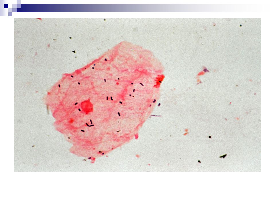
91
Clue Cell of BV

92
BV Scored Gram Stain ( from Nugent RP 1991;29:297)
TYPE Number seen/OIF None <1 1-5 6-30 >30 Lacto 4 3 2 1 Gard/Bact Curved GNR

93
Interpretation of Scored Gram Stain
0-3 = Normal 4-6 = Intermediate may indicate trichomoniasis, GC or CT abnormal gram stain, but not consistent with BV 7-10 = Consistent with Bacterial Vaginosis Significance of results unknown in pre-menarchal girls or post-menopausal women

95
Who Should be Screened for BV?
Women with vaginal symptoms esp. if failed therapy Pregnant women at high risk of preterm birth Pregnant women with genital symptoms rule out trichomoniasis as well Women with gynecologic surgery

96
Thank You Questions ?????

Similar presentations


 Microbiology Manager Wesley Medical Center.>")


 Prof. Dr. Ebtisam.F. El Ghazzawi Medical Research Institute (MRI) Alexandria University.>")


 Intermittent: Associated with abscesses.>")

, and the parenchyma.>")

 Multiplex PCR Assays>")
 wound, and Burn Cultures>")


 Prof. Dr. Ebtisam.F. El Ghazzawi. Medical Research Institute (MRI) Alexandria University.>")
