Download presentation
Presentation is loading. Please wait.

3
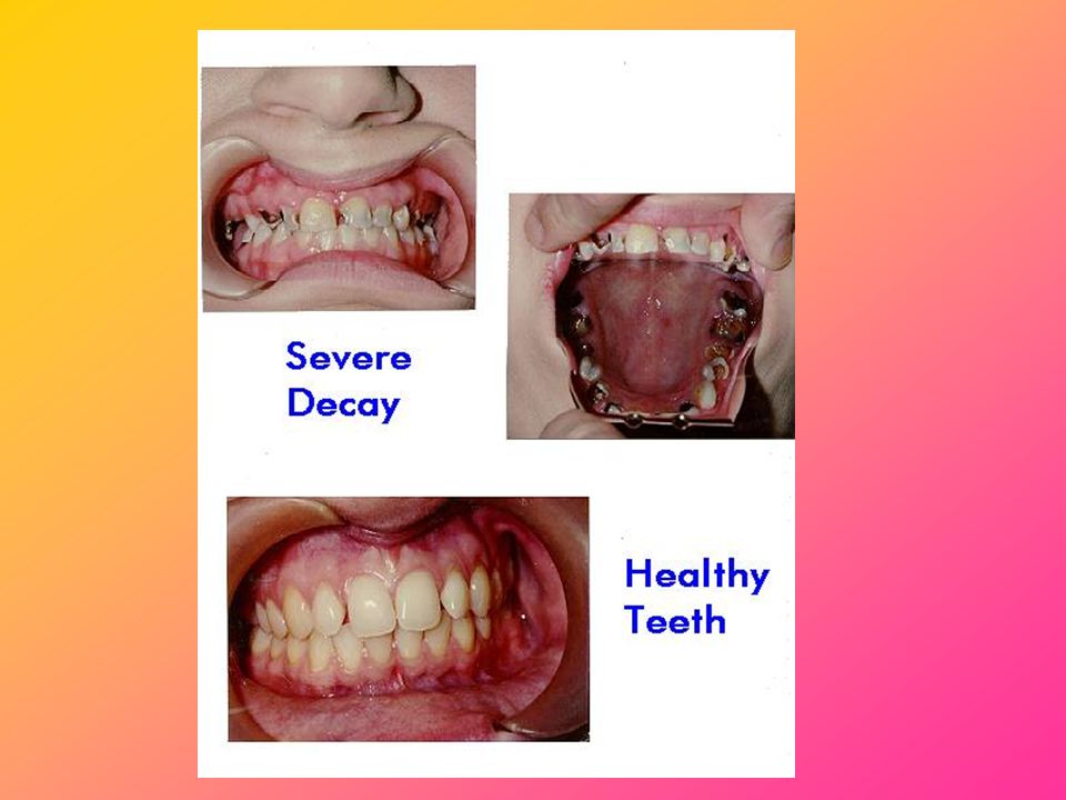
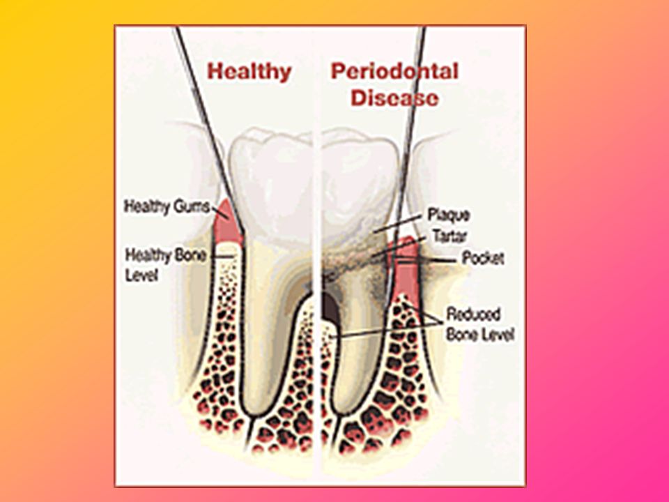
Periodontal/Gum Disease
Periodontal/gum diseases are serious infections that, left untreated, can lead to tooth loss The word periodontal literally means "around the tooth“ Periodontal disease is a chronic bacterial infection that affects the gums and bones supporting the teeth Periodontal disease can affect one tooth or many teeth. It begins when the bacteria in plaque causes the gums to become inflamed

7
Do you have gum disease??? Do you ever have pain in your mouth?
Do your gums ever bleed when you brush your teeth or when you eat hard food? Have you noticed any spaces developing between your teeth? Do your gums ever feel swollen or tender? Have you noticed that your gums are receding (pulling back from your teeth) or your teeth appear longer than before? Do you have persistent bad breath? Have you noticed pus between your teeth and gums? Have you noticed any change in the way your teeth fit together when you bite? Do you ever develop sores in your mouth?
 or your teeth appear longer than before Do you have persistent bad breath Have you noticed pus between your teeth and gums Have you noticed any change in the way your teeth fit together when you bite Do you ever develop sores in your mouth")
8
How to look after your teeth

9
How to Brush Place your toothbrush next to the teeth so that it rests on the gums forming a 45-degree angle against the gums. Move the toothbrush from the gums towards the edge of teeth to move the dental plaque away from the gum line. After brushing, one by one tooth, all the outer teeth surfaces do the same for the inner surfaces. Brush the chewing surfaces of the teeth with horizontal moves.

10
How to Floss

11
Human Torso Model *Please refer to your handout of the human digestive system

12
Human Digestive System
Alimentary Canal + Associated Glands

13
Alimentary Canal and Associated Glands
Mouth Pharynx Oesophagus Stomach Small Intestine Large Intestine Anus Salivary Glands Gastric Glands Pancreas Liver Intestinal Glands

14
Digestion

15
Digestion Digestion is the process of breaking down large, complex substances into smaller, simpler molecules for absorption Carbohydrates -> glucose/fructose/galactose Proteins -> amino acids Fats -> fatty acids and glycerol Vitamins, minerals and water can be absorbed directly without digestion

16
Mechanical Digestion vs. Chemical Digestion
Mechanical process Chewing of teeth Churning of stomach Food is changed physically but not chemically Increase surface area of food substances Chemical process Involves action of digestive enzymes secreted from glands Different types of enzymes break down different food types

17
Protease Breaks down protein molecules
A protein molecule is made of many different amino acids Amino acids

18
Breaks down carbohydrate molecules
Carbohydrase Breaks down carbohydrate molecules A starch molecule is made of many glucose molecules Glucose

19
Lipase Breaks down fat molecules Fatty acids
Glycerol A fat molecule is made up of fatty acids and glycerol molecules

20
Example of Carbohydrase: Amylase
In saliva and pancreatic juice Helps break down starch into simple sugars in mouth and in small intestine

21
Example of Protease: Pepsin
In gastric juice Helps break down proteins into amino acids in stomach

22
Example: Lipase In pancreatic juice
Helps break down oil droplets into fatty acids and glycerol in small intestine

23
Saliva The taste, smell and sight of food can stimulate salivary glands to secrete saliva into the mouth via salivary ducts Saliva – contains water, mucus and salivary amylase. Slightly alkaline Water – moistens and softens food Mucus – lubricate food for swallowing Salivary amylase – starch -> maltose

25
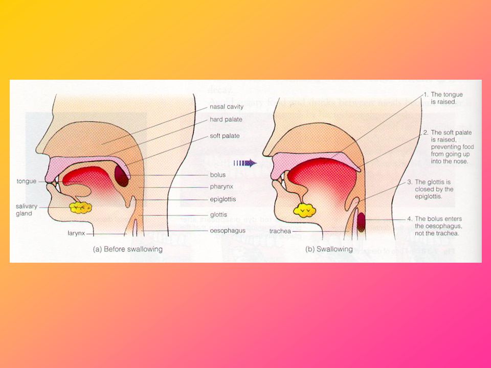
Swallowing Food is chewed and mixed with saliva
Tongue rolls the food into a bolus Food is swallowed down the oesophagus through the pharynx Tongue moves upwards and backwards to prevent food from entering the trachea/nasal cavity The soft palate moves up to block the nasal cavity The larynx moves upwards to so that the glottis (the opening to the larynx) is covered by the epiglottis to prevent food from entering the trachea
 is covered by the epiglottis to prevent food from entering the trachea.")
27
Movement of Food Along the Alimentary Canal
Inner surface of alimentary canal is lined with one to several layers of cells – epithelium Some epithelial cells produce mucus, which acts as a lubricant

28
Peristalsis The small intestine has two muscle layers that work together in peristalsis and segmentation

29
Peristalsis The inner circular muscles contract, tightening the tube and pushing the food forward in the intestine

30
Peristalsis When the circular muscles relax, the outer longitudinal muscles contract, and the intestinal tube is shortened

31
Peristalsis As the circular and longitudinal muscles tighten and relax, the food moves forward

32
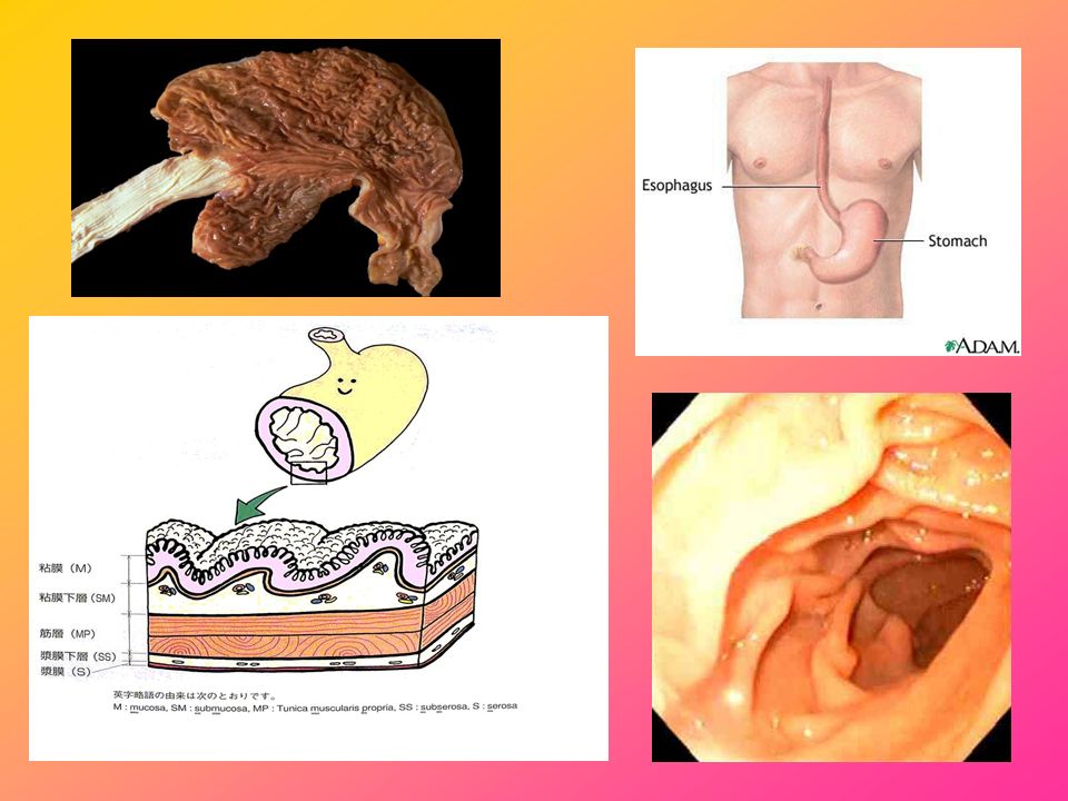
Stomach a muscular, elastic, pear-shaped bag, lying crosswise in the abdominal cavity food enters the stomach from the esophagus. The connection between the stomach and the esophagus is called the cardiac sphincter The other end of the stomach empties into the duodenum, the first section of the small intestine. The pyloric sphincter separates the stomach from the duodenum

35
Sphincter Opened Sphincter Closed

36
Functions of Stomach Storage
Mechanical digestion – turns food into chyme Chemical digestion

38
The lining of the stomach contains deep collections of cells organized into gastric glands
The openings of the gastric glands into the surface of the stomach are called gastric pits The mucous cells in the gastric pits secrete mucus In the deeper part of the gland, the parietal cells secrete hydrochloric acid The chief cells secrete pepsinogen (an inactive form of the protein-digesting enzyme pepsin)
")
39
Rennin In young children, the gastric juice also contains a type of protease called rennin Rennin coagulates milk – allow the proteins to stay in stomach longer for digestion * Rennin can be used to curdle milk to make cheese!

40
Ulcer/Peptic Ulcer A small erosion in the gastrointestinal tract
A weakening of the mucus coating – acid erodes the wall of the GI tract Stomach – gastric ulcer Small intestine – duodenal ulcer Main cause – bacterial infection Can be treated with antibiotics

41
Small Intestine The small intestine is divided into 3 sections:
Duodenum Jejunum Ileum * In the small intestine, both digestion and absorption occur

43
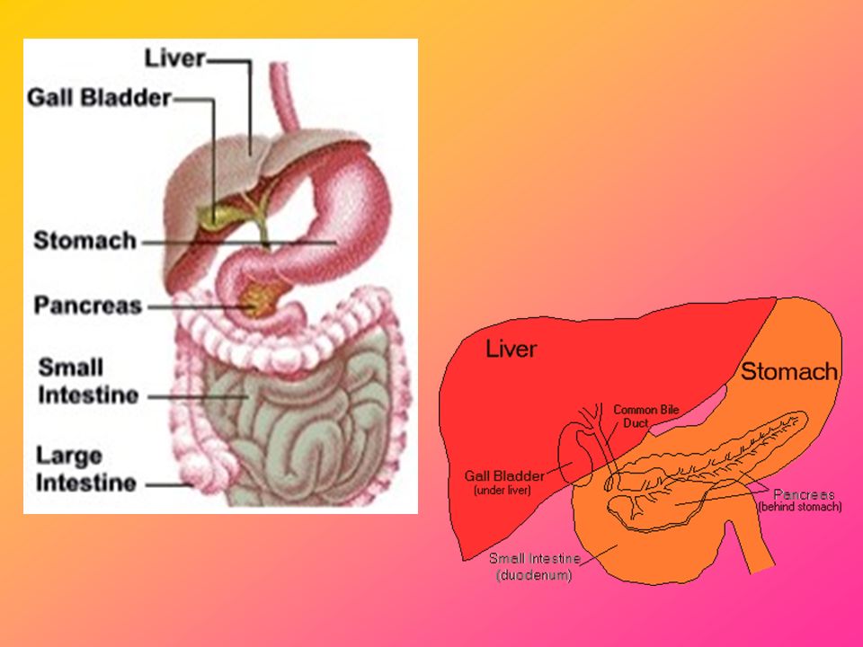
Bile Bile is a dark green fluid containing: 1) bile salts
2) sodium hydrogen carbonate 3) bile pigments Bile does NOT contain digestive enzymes Made by the liver Stored in the gall bladder
 sodium hydrogen carbonate. 3) bile pigments. Bile does NOT contain digestive enzymes. Made by the liver. Stored in the gall bladder.")
45
The gall bladder contracts to release bile into the duodenum via the bile duct
Stimulated by the arrival of chyme in the duodenum

46
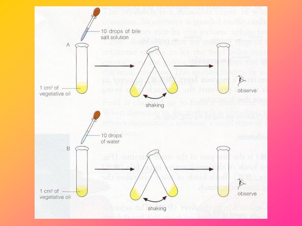
Bile 1) Bile salts – EMULSIFICATION
Bile salts break up (emulsify) lipids into small oil droplets This allows enzymes to have a larger surface area to break down the fat molecules Lipids Small oil droplets Bile salts (emulsification)
 lipids into small oil droplets. This allows enzymes to have a larger surface area to break down the fat molecules. Lipids Small oil droplets. Bile salts (emulsification)")
47
Bile 2) Sodium Hydrogen Carbonate – NEUTRALIZATION
It neutralizes stomach acid to provide the necessary alkaline condition (pH 8) for the pancreatic and intestinal enzymes to work
 for the pancreatic and intestinal enzymes to work.")
48
The pH Scale Acidic Alkaline Neutral More acidic More basic
Acidic Alkaline Neutral More acidic More basic

49
The Need for Different pH Levels
The stomach releases hydrochloric acid to provide an acidic condition (pH1 - 2) for stomach proteases (e.g. pepsin) to work. Acid also kills germs The activity of salivary amylase is stopped in the stomach since it cannot work in acidic conditions. Pancreatic amylase also requires an alkaline condition to work
 for stomach proteases (e.g. pepsin) to work. Acid also kills germs. The activity of salivary amylase is stopped in the stomach since it cannot work in acidic conditions. Pancreatic amylase also requires an alkaline condition to work.")
50
The Need for Different pH Levels
3) The gall bladder releases bile into the small intestine to provide an alkaline condition (pH 8) for the pancreatic and intestinal enzymes to work
 The gall bladder releases bile into the small intestine to provide an alkaline condition (pH 8) for the pancreatic and intestinal enzymes to work.")
51
Bile 3) Bile pigments Waste products formed from the breakdown of old red blood cells in the liver
 Bile pigments Waste products formed from the breakdown of old red blood cells in the liver")
52
Investigation #1: Investigating the effect of bile salts on oil

54
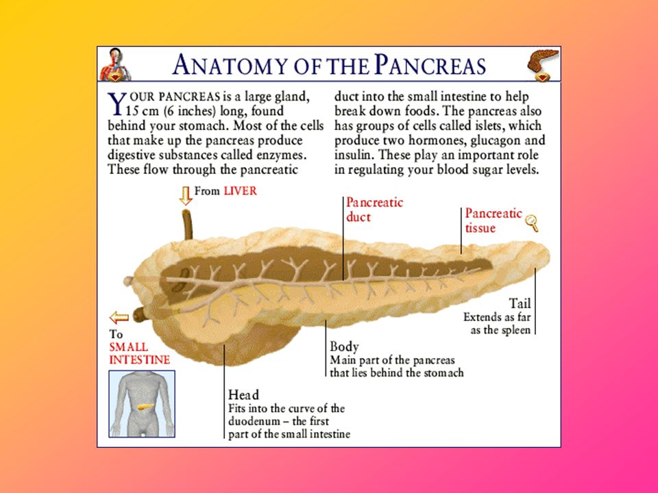
Pancreas A yellowish organ found beneath the stomach and is connected to the small intestine at the duodenum Produces pancreatic juice that flows into the duodenum through the pancreatic duct

57
Pancreas Pancreatic juice contains 3 types of digestive enzymes:
Tryptase Amylase Lipase *Pancreas also produces the hormones glucagon and insulin to regulate the level of blood glucose

58
Intestinal Juice Alkaline solution containing digestive enzymes, hormones, mucus, neutralizing substances, etc. Secreted by intestinal glands in the wall of the duodenum e.g. carbohydrases catalyze the breakdown of double sugars into simple sugars

59
Carbohydrase Maltase: Maltose -> Glucose + Glucose
2) Sucrase: Sucrose -> Glucose + Fructose 3) Lactase: Lactose -> Glucose + Galactose
 Sucrase: Sucrose -> Glucose + Fructose. 3) Lactase: Lactose -> Glucose + Galactose.")
60
Lactose Intolerance Inability to digest significant amount of lactose, the predominant sugar of milk, due to a shortage of the enzyme lactase Common symptoms include nausea, cramps, bloating, gas, and diarrhoea The undigested lactose will serve as food for bacteria found in the large intestine

61
Digestion in Ileum Completion of digestion
Food is churned by peristaltic movement and is mixed with digestive juices Food becomes watery fluid called chyle Food is now present in simplest form

62
Absorption

63
Absorption Absorption is the uptake of simple and small food molecules from the alimentary canal into the blood stream Food molecules can be absorbed into blood by diffusion or active transport Absorption occurs in the stomach, the small intestine and the large intestine

64
Absorption in Stomach Food substances that are absorbed in the
Water Minerals Alcohol Simple sugars Water-soluble vitamins

65
Absorption in Small Intestine
Most of the digested food is absorbed in the small intestine The inner lining of the small intestine is folded to provide a large surface area The inner surface of the small intestine is made up of a large number of finger-like projections called villi (singular: villus) Peristalsis in the small intestine allows the digested food to come into contact with the villi for absorption
 Peristalsis in the small intestine allows the digested food to come into contact with the villi for absorption.")
70
Structure of Villi Epithelium lining
Blood capillaries (transportation of simple sugars, amino acids and minerals) Lacteal (lymph vessel)
 Lacteal (lymph vessel)")
72
Lacteal fatty acids and glycerol recombine in the epithelium of the villus to form fat which then enters the lacteal as fine fat droplets the lymphatic system converges with the circulatory system at a duct located in the neck area

74
They take up the absorbed food and transport them away
4. Each villus contains a dense network of blood capillaries This allows the food to cross the membrane rapidly 3. The wall of villi is thin (one-cell thick) These villi can increase the total surface area for absorption 2. Its inner surface is covered with numerous finger-like projections called villi Food can stay long enough for absorption to occur 1. It is very long (6 m) Adaptation Feature
 These villi can increase the total surface area for absorption. 2. Its inner surface is covered with numerous finger-like projections called villi. Food can stay long enough for absorption to occur. 1. It is very long (6 m) Adaptation. Feature.")
77
Absorption in Large Intestine
Much of the remaining water and minerals is absorbed in the colon The appendix, which has no known functions, is joined to the caecum Appendicitis – food materials trapped in the appendix causing bacterial infection

78
Caecum in Herbivores Do not have digestive enzymes to break down cellulose Rely on bacteria residing in the long caecum to provide the enzyme cellulase Cellulose -> Glucose

79
Investigation #2: A Model Gut

80
What does the content inside the dialysis tubing represent?
What does the dialysis tubing represent? What does the water in the beaker/boiling tube represent?

81
Assimilation

82
Assimilation the process by which absorbed food molecules in the blood are transported to cells for the use of growth, tissue repair and other metabolic activities. The actual destiny of each food molecule depends not only on its type but also on the body requirements at that time (e.g. use immediately or put into storage)
")
85
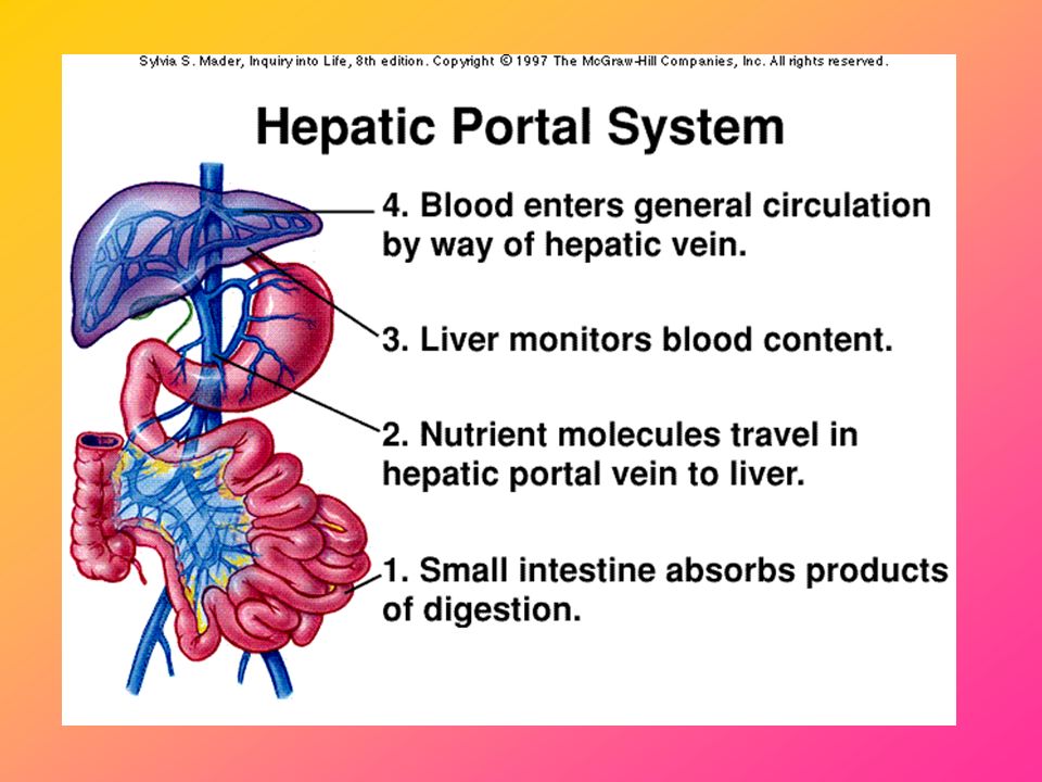
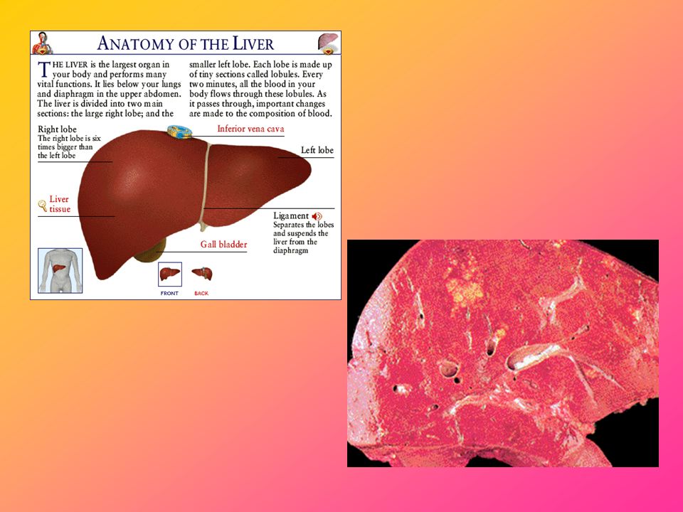
Liver Largest organ of the body Reddish brown in color
Lies on the right side of the abdominal cavity beneath the diaphragm Blood is carried to the liver via two large vessels called the hepatic artery and the hepatic portal vein After processing in the liver, blood leaves the liver through the hepatic vein

88
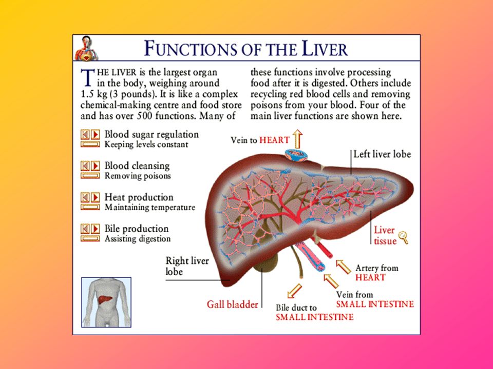
Functions of Liver Carbohydrate metabolism - the liver converts excess glucose into glycogen as a temporary way of storing the glucose. Glycogen can also be converted back to glucose when needed Fat metabolism - the liver converts excess protein and carbohydrate into fat. Excess glycogen is stored as fat for long term storage

89
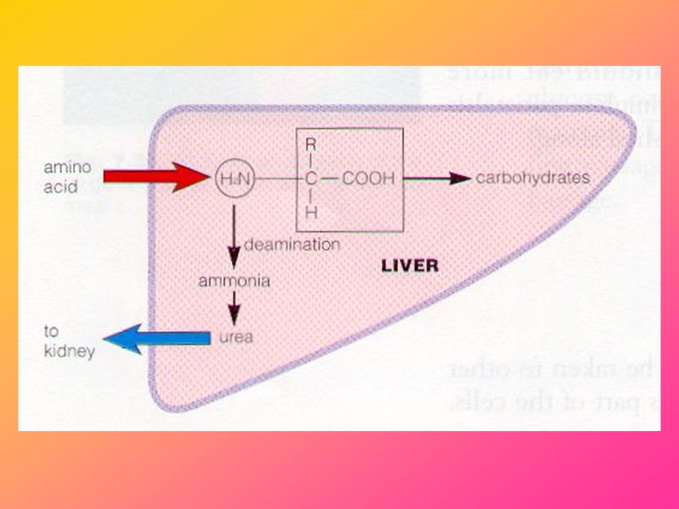
Functions of Liver 3) Protein metabolism – the liver can synthesize new proteins/ amino acids and deaminate excess amino acids
 Protein metabolism – the liver can synthesize new proteins/ amino acids and deaminate excess amino acids.")
90
Deamination Amino group (~NH2) removed Ammonia (NH3) produced (toxic)
Ammonia converted to urea – excreted in urine Carbon skeleton – converted to carbohydrates

92
Functions of Liver 3) Protein metabolism – the liver can synthesize new proteins/amino acids and deaminate excess amino acids 4) Vitamin storage - the liver stores mainly vitamins A, D and B12 5) Iron storage - the liver stores iron which is obtained from the breakdown of red blood cells. The iron salts can be used in the formation of new RBC
 Vitamin storage - the liver stores mainly vitamins A, D and B12. 5) Iron storage - the liver stores iron which is obtained from the breakdown of red blood cells. The iron salts can be used in the formation of new RBC.")
93
Functions of Liver 6) Bile production – emulsification and neutralization 7) Drug/Alcohol metabolism – the liver changes the drug into an excretable and harmless form (detoxification) 8) Disposal of bacteria - The liver filters many bacteria, viruses, and other microorganisms from the blood
 Drug/Alcohol metabolism – the liver changes the drug into an excretable and harmless form (detoxification) 8) Disposal of bacteria - The liver filters many bacteria, viruses, and other microorganisms from the blood.")
94
Egestion

95
Egestion Faeces – semi-solid, greenish brown mass containing undigested and unabsorbed food substances. Also contains bile pigment (hence the color of faeces), dead RBC, cells from intestinal wall, bacteria, etc. Temporarily stored in rectum Anal sphincter – relaxes to allow a person to defaecate
, dead RBC, cells from intestinal wall, bacteria, etc. Temporarily stored in rectum. Anal sphincter – relaxes to allow a person to defaecate.")
96
Constipation and Diarrhoea
Peristalsis too slow Too much water absorbed Some common causes include lack of fibre in diet, not enough liquids, lack of exercise, etc. Lead to dry, hard faeces Difficulty in defaecation May damage wall of rectum and cause bleeding or form piles Peristalsis too fast Too little water absorbed Some common causes include bacterial / viral / parasitic infections, food intolerance, etc. Lead to loose, watery stools More frequent egestion May cause dehydration

97
Haemorrhoids Also referred to as piles
Haemorrhoids are enlarged veins just under the surface tissue of the rectum or the anus Haemorrhoids in the rectum are called internal haemorrhoids; those that occur around the anus are called external haemorrhoids May cause bleeding, pain, itching and a sense of pressure

98
Haemorrhoids Increased pressure in the veins around the anus is thought to be the cause of haemorrhoids: straining to pass a bowel motion because of hard, dry stools (as in constipation) diarrhoea heavy lifting being very overweight sitting or standing for long periods pregnancy
 diarrhoea. heavy lifting. being very overweight. sitting or standing for long periods. pregnancy.")
Similar presentations


>")


 Tract. This is a one way tube. Peristalsis is.>")


