Download presentation
Presentation is loading. Please wait.

1
MR Venography Ivan Pedrosa, M.D. Beth Israel Deaconess Medical Center
Harvard Medical School Boston, MA

2
Why MR Imaging? Conventional venography US Multiple injections
I.V. access in affected edematous extremity Radiation / iodinated contrast US Limited in central veins Limited FOV and anatomic landmarks

3
Why MR Imaging? CT Nephrogenic Systemic Fibrosis (NSF) Radiation
Iodinated contrast Pitfalls due to poor opacification / mixing artifacts Nephrogenic Systemic Fibrosis (NSF) Increased indications for non-contrast MRV
 Increased indications for non-contrast MRV.")
4
MRV Techniques Clinical Applications Dark Blood Imaging
Bright Blood Imaging Gd-enhanced MRV Clinical Applications Chest Abdomen Pelvis

5
MRV techniques Non-contrast MRV Double IR Spin echo TOF
Dark blood Sequences Bright blood Sequences Double IR Spin echo TOF Double IR SSFSE GRE (Cine) Dynamic SSFSE FIESTA (Cine) Phase Contrast Gd-enhanced MRV 3D FS T1-W GRE (VIBE, LAVA, THRIVE)
 Dynamic SSFSE. FIESTA (Cine) Phase Contrast. Gd-enhanced MRV. 3D FS T1-W GRE (VIBE, LAVA, THRIVE)")
6
Spin Echo (“dark blood”)
180º 180º 90º 90º

7
HAlf-Fourier Single shot Turbo Spin Echo (HASTE or SSFSE)
K space 90º 180º One second to collect the whole image Dark blood Protons exit slice Slow flow - ↑↑ SI Thrombus - ↓↑ SI The most important sequence in our MRCP protocol is the HASTE sequence. HASTE stands for half fourier single shot turbo spin echo Depending on the manufacturer this sequence is also named single shot fast spin echo or SSFSE This MR sequence apply a 90 degree excitation pulse followed by a series of 180 degree pulses an it collects all the data for one image. By doing that we acquire the whole data without applying multiple 90 degree excitation pulses and we save time. But we also save time because only half of the K space is acquired. The k space is a virtual space where the MR keeps the data that is collecting during the acquisition time. One characteristic of these space is that is symmetric so if we acquire half of the k space we can assume that the other halh of the k space has similar values. This is how the k space is filled in the HASTE sequence. In fact we acquire a little bit more than half of the k space. This extra lines in the center, at the contralateral side, are obtained for mathematical calculations. So by applying only one 90 degree pulse and collecting only half of the k space we save a lot of time. Typically one image is acquired approximately one second. The second important characteristic about the HASTE sequence is that by changing our echo time or TE we can change the contrast of the image and the signal of the background tissues. SSFSE/HASTE

8
Dynamic HASTE VALSALVA Intravascular signal void Valsalva
intrathoracic P Venous return T2 of blood is long

9
Dynamic HASTE VALSALVA Valsalva T2 of blood is long intrathoracic P
Venous return T2 of blood is long

11
DB HASTE (“dark blood”)
180º 180º TI 180º 180º 90º TI

12
Double IR T1 FSE IR-T1W Cardiac-gated IR-HASTE
1 slice (~16 sec) breath-hold ~20 slices ( sec) breath-hold 2 slices with ASSET
 breath-hold ~20 slices ( sec) breath-hold. 2 slices with ASSET.")
13
Bright blood Sequences
TOF GRE (Cine) FIESTA (Cine) Phase Contrast
 FIESTA (Cine) Phase Contrast.")
14
Time-of-Flight (TOF)
")
15
Time-of-Flight (TOF)
")
16
Time-of-Flight (TOF)
")
17
Time-of-Flight (TOF)
")
18
Time-of-Flight (TOF)
")
19
Time-of-Flight (TOF)
")
20
Time-of-Flight (TOF)
")
21
Time-of-Flight (TOF)
")
22
TOF

23
TOF optimization for slow flow

24
TOF: in-plane saturation
Sagittal Sagittal Axial acquisition Gad-MRV

25
TOF optimization for slow flow
Slice perpendicular to vessel of interest Decrease slice thickness Cardiac gating? ECG Tracing Blood flow (Pulse Oximeter) Systole (arterial)
 Systole (arterial)")
26
True FISP / FIESTA / Balanced FFE
True Fast Imaging with Steady-state Precession Gradients are fully balanced in order to recycle the transverse magnetization in long T2 species Contrast T2 / T1 ratio Blood vessels are bright (T2 of blood is )
")
27
True FISP Pros Cons Fast No breathing artifacts Thrombus
Road map No breathing artifacts Thrombus Filling defect SI Cine True FISP FIESTA Cons Artifacts Pulsatile flow Off-resonace Acute / subacute thrombus

28
True FISP

29
True FISP True FISP Gd-enhanced MRV

30
True FISP L True FISP Gd-enhanced MRV Pedrosa I. AJR 2005

31
Phase Contrast (PC) 2 equal and opposite Venc gradients between the excitation and echo. With stationary protons, phase shifts induced by the first gradient are reversed and canceled by the second gradient. In moving protons, the second gradient does not quite cancel out phase shifts induced by the first gradient These phase shifts are detected and proportional to the amount of motion in the direction of the encoding gradients

32
Phase Contrast (PC) Phase Image Magnitude Image
High velocity flow towards the head (Ascending aorta) Moderate velocity flow towards the head (Pulmonary artery) Venc gradient applied in the slice (superior-inferior) direction In the phase (velocity) image Gray represents stationary background tissues White represents blood flowing caudally (towards feet) Black represents blood flowing cranially (towards head) The intensity of white or black represents the magnitude of velocity in the respective directions Phase Image Moderate velocity flow towards the feet (SVC) High velocity flow towards the feet (Descending aorta) Magnitude Image
 Moderate velocity flow towards the head (Pulmonary artery) Venc gradient applied in the slice (superior-inferior) direction. In the phase (velocity) image. Gray represents stationary background tissues. White represents blood flowing caudally (towards feet) Black represents blood flowing cranially (towards head) The intensity of white or black represents the magnitude of velocity in the respective directions. Phase Image. Moderate velocity flow towards the feet (SVC) High velocity flow towards the feet (Descending aorta) Magnitude Image.")
33
Phase Contrast (PC) If Venc is chosen to be too low, aliasing (“wrap-around artifact”) occurs when velocities exceed that value causing velocities to mimic a “lower” value If Venc is chosen to be too high, sensitivity to slow flow and accuracy of quantitative analysis of velocity/flow are diminished Venc for venous imaging? 40-60 cm/sec Venc set to 140 cm/sec, appropriate for this volunteer Venc set to 70 cm/sec, too low for this volunteer. Aliasing or “wrap-around” results in the high-velocity flow areas of the aorta. Phase Images
 occurs when velocities exceed that value causing velocities to mimic a lower value. If Venc is chosen to be too high, sensitivity to slow flow and accuracy of quantitative analysis of velocity/flow are diminished. Venc for venous imaging cm/sec. Venc set to 140 cm/sec, appropriate for this volunteer. Venc set to 70 cm/sec, too low for this volunteer. Aliasing or wrap-around results in the high-velocity flow areas of the aorta. Phase Images.")
34
Phase Contrast (PC) Venc = 40 cm/sec
 Venc = 40 cm/sec")
35
Phase Contrast (PC) 3D PC
 3D PC")
36
Gadolinium-enhanced MRV
Indirect MRV Direct MRV

37
Indirect Venography I.V. access in any peripheral vein Gadolinium
Antecubital vein (Right UE) Gadolinium Single dose (~20 2 cc/seg Single dose (~ cc/seg 20 cc 0.8 cc/seg 3D GRE T1 Subtractions Venogram-like MIP reconstructions Double dose Gd Single injection/dual rate
 Gadolinium. Single dose (~20 2 cc/seg. Single dose (~ cc/seg. 20 cc 0.8 cc/seg. 3D GRE T1. Subtractions. Venogram-like MIP reconstructions. Double dose Gd. Single injection/dual rate.")
38
Timing arterial phase

39
Indirect Venography VENOUS PHASE ARTERIAL PHASE - SUBTRACTION =

40
Indirect Venography SUBSTRACTION MIP

42
Direct Venography I.V. access in affected extremity or bilateral
Gadolinium 5 cc Gd in 100 cc saline (1:20) Tourniquet in lower extremities 3D GRE T1 Li W et al. J Magn Reson Imaging 1998; 8(3): 630-3
 Tourniquet in lower extremities. 3D GRE T1. Li W et al. J Magn Reson Imaging 1998; 8(3):")
43
Direct Venography

44
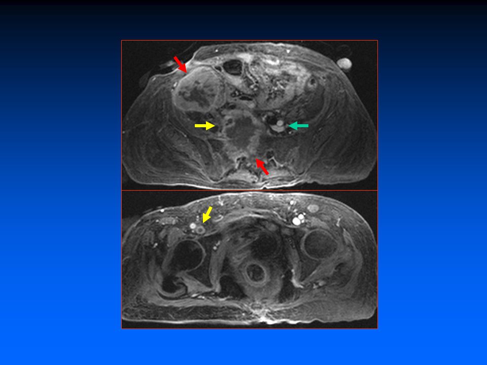
Thrombus Characterization
Bland thrombus No enhancement Variable SI Tumor thrombus Enhancement on Gd-MRV Subtractions! Absence of enhancement does NOT exclude tumor thrombus SI on T2-weighted images

46
Tumor thrombus: Intravenous leiomyomatosis U

47
Staging Acute thrombus Chronic thrombus
Enlargement of vein by intraluminal thrombus SI on T2-weighted images Vessel wall Thrombus Perivascular soft tissue edema SI on T1-weighted images (subacute) Chronic thrombus Vein attenuated or not visible Venous collaterals ↓ SI on all sequences
 Chronic thrombus. Vein attenuated or not visible. Venous collaterals. ↓ SI on all sequences.")
48
Acute thrombosis of the portal vein

49
T2W T1W post-contrast Trombosis aguda/subaguda Primaria
Síndrome de Paget-Von Schroetter (“Effort thrombosis”) Trombosis aguda/subaguda Expansión de la vena con defecto de repleción Edema en la pared de la vena (Señal en T2) Realce de la pared con Gd < 14 d Froehlich JB et al. J Vasc Surg 97;26:809 T1W post-contrast
 Trombosis aguda/subaguda. Expansión de la vena con defecto de repleción. Edema en la pared de la vena (Señal en T2) Realce de la pared con Gd. < 14 d. Froehlich JB et al. J Vasc Surg 97;26:809. T1W post-contrast.")
50
Paget von Schrotter syndrome or “effort” thrombosis

51
Chronic Thrombosis FIGURE 15

52
Venous thrombosis Is the thrombosis acute or chronic?
Do I need to anticoagulate this patient?

53
Acute/subacute thrombosis

54
brachiocephalic vein: chronic occlusion

55
Central catheter malfunction
Fibrin sheath

56
Clinical Indications

57
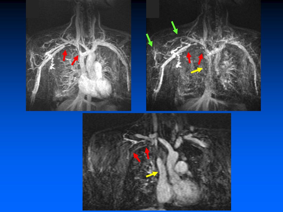
SVC syndrome Estrechamiento / obstrucción VCS
Disnea, edema facial ± EESS, dolor torácico. Tumoral (74–95 %) Pulmón (85%) Otras (3-20%) Catéteres centrales Marcapasos (- frec) Fibrosis mediastínica Aneurisma aórtico
 Pulmón (85%) Otras (3-20%) Catéteres centrales. Marcapasos (- frec) Fibrosis mediastínica. Aneurisma aórtico.")
58
Venous Access Central catheters MRV chest Hemodyalisis Chemotherapy
Parenteral nutrition Thrombosis in first 3 months (10%) MRV chest 15 pts with occlusion or stenosis central veins Venous access possible in 14 pts Shinde TS et al. Radiology 1999;213:
 MRV chest. 15 pts with occlusion or stenosis central veins. Venous access possible in 14 pts. Shinde TS et al. Radiology 1999;213:")
61
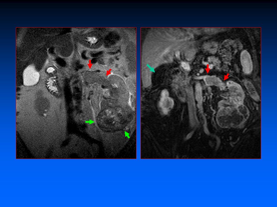
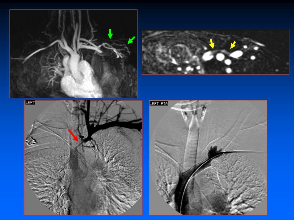
IVC in Renal Cell Carcinoma
51 yo male with PE Papillary carcinoma

62
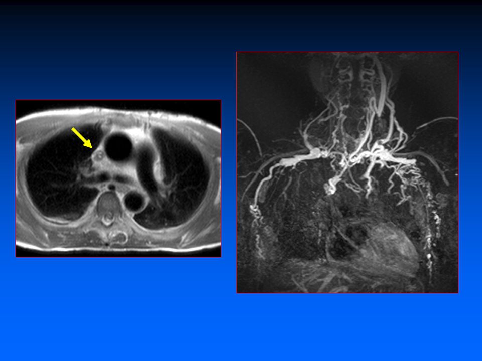
Pulmonary Embolism

63
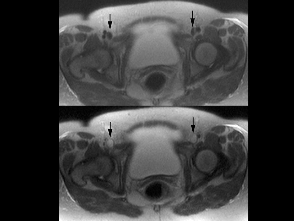
Isolated Iliac Vein DVT

66
Conclusion Central veins of the chest, abdomen and pelvis
Limited evaluation with US Whole-body venous roadmap Vascular access Pregnancy

Similar presentations


 for Pulmonary Embolism Meyer CA, Schiebler ML, Reeder SB, Francois CJ, Nagle SK.>")


>")


 and the portal vein ( 75% of vascularization).>")


