Download presentation
Presentation is loading. Please wait.

1
Hip ultrasound: Why, When, and How?
<?xml version="1.0"?><Settings><answerBulletFormat>Numeric</answerBulletFormat><answerNowAutoInsert>No</answerNowAutoInsert><answerNowStyle>Explosion</answerNowStyle><answerNowText>Answer Now</answerNowText><chartColors>Use PowerPoint Color Scheme</chartColors><chartType>Horizontal</chartType><correctAnswerIndicator>Checkmark</correctAnswerIndicator><countdownAutoInsert>No</countdownAutoInsert><countdownSeconds>10</countdownSeconds><countdownSound>TicToc.wav</countdownSound><countdownStyle>Box</countdownStyle><gridAutoInsert>No</gridAutoInsert><gridFillStyle>Answered</gridFillStyle><gridFillColor>0,0,0</gridFillColor><gridOpacity>100%</gridOpacity><gridTextStyle>Keypad #</gridTextStyle><inputSource>Response Devices</inputSource><multipleResponseDivisor># of Responses</multipleResponseDivisor><participantsLeaderBoard>5</participantsLeaderBoard><percentageDecimalPlaces>0</percentageDecimalPlaces><responseCounterAutoInsert>No</responseCounterAutoInsert><responseCounterStyle>Oval</responseCounterStyle><responseCounterDisplayValue># of Votes Received</responseCounterDisplayValue><insertObjectUsingColor>Blue</insertObjectUsingColor><showResults>Yes</showResults><teamColors>User Defined</teamColors><teamIdentificationType>None</teamIdentificationType><teamScoringType>Voting pads only</teamScoringType><teamScoringDecimalPlaces>1</teamScoringDecimalPlaces><teamIdentificationItem></teamIdentificationItem><teamsLeaderBoard>5</teamsLeaderBoard><teamName1></teamName1><teamName2></teamName2><teamName3></teamName3><teamName4></teamName4><teamName5></teamName5><teamName6></teamName6><teamName7></teamName7><teamName8></teamName8><teamName9></teamName9><teamName10></teamName10><showControlBar>Slides with Get Feedback Objects</showControlBar><defaultCorrectPointValue>100</defaultCorrectPointValue><defaultIncorrectPointValue>0</defaultIncorrectPointValue><chartColor1>187,224,227</chartColor1><chartColor2>51,51,153</chartColor2><chartColor3>0,153,153</chartColor3><chartColor4>153,204,0</chartColor4><chartColor5>128,128,128</chartColor5><chartColor6>0,0,0</chartColor6><chartColor7>0,102,204</chartColor7><chartColor8>204,204,255</chartColor8><chartColor9>255,0,0</chartColor9><chartColor10>255,255,0</chartColor10><teamColor1>187,224,227</teamColor1><teamColor2>51,51,153</teamColor2><teamColor3>0,153,153</teamColor3><teamColor4>153,204,0</teamColor4><teamColor5>128,128,128</teamColor5><teamColor6>0,0,0</teamColor6><teamColor7>0,102,204</teamColor7><teamColor8>204,204,255</teamColor8><teamColor9>255,0,0</teamColor9><teamColor10>255,255,0</teamColor10><displayAnswerImagesDuringVote>Yes</displayAnswerImagesDuringVote><displayAnswerImagesWithResponses>Yes</displayAnswerImagesWithResponses><displayAnswerTextDuringVote>Yes</displayAnswerTextDuringVote><displayAnswerTextWithResponses>Yes</displayAnswerTextWithResponses><questionSlideID></questionSlideID><controlBarState>Expanded</controlBarState><isGridColorKnownColor>True</isGridColorKnownColor><gridColorName>Yellow</gridColorName><AutoRec></AutoRec><AutoRecTimeIntrvl></AutoRecTimeIntrvl><chartVotesView>Percentage</chartVotesView><chartLabelsColor>0,0,0</chartLabelsColor><isChartLabelColorKnownColor>True</isChartLabelColorKnownColor><chartLabelColorName>Black</chartLabelColorName><chartXAxisLabelType>Full Text</chartXAxisLabelType></Settings> <?xml version="1.0"?><AllQuestions /> <?xml version="1.0"?><AllAnswers /> Hip ultrasound: Why, When, and How? Dorothy Bulas M.D. Children’s National Medical Center Washington D.C.

2
Disclosure I have no relevant financial relationships with the manufacturers of any commercial products and/or provider of commercial services discussed in this CME activity I do not intend to discuss an unapproved use of a commercial product/device in my presentation

3
Objectives Review the risk factors for developmental dysplasia of the hip (DDH) Understand the appropriate work up and follow up of DDH
 Understand the appropriate work up and follow up of DDH.")
4
Changes in practice Use appropriateness criteria to assess for developmental dysplasia. Selective screening by ultrasound after 2 weeks of age

5
Introduction Developmental dysplasia of the hip is the preferred term to describe the condition in which the femoral head has an abnormal relationship to the acetabulum. DDH is a spectrum of abnormalities frank dislocation (luxation) partial dislocation (subluxation) unstable - femoral head comes in & out of socket inadequate formation of the acetabulum.
 partial dislocation (subluxation) unstable - femoral head comes in & out of socket. inadequate formation of the acetabulum.")
6
DDH Many of these findings may not be present at birth
SO - the term developmental more accurately reflects the biologic features than the term congenital.

7
Early Diagnosis The earlier a dislocated hip is detected, the simpler and more effective is the treatment.

8
Late Diagnosis Late dx in children may lead to increased surgical intervention and complications. Late dx in adults can result in debilitating end-stage degenerative hip joint disease.

9
Why Screen? Screening decreases the incidence of late diagnosis of DDH. Despite screening programs, DDH continues to be diagnosed later in infancy /childhood, delaying appropriate therapy Substantial number of malpractice claims

10
Incidence F:M 6:1 (?hormonal) 1.5 : 1,000 Caucasian Americans
less frequent African Americans. F:M 6:1 (?hormonal) The reported incidence influenced by FH, race, diagnostic criteria, experience /training of examiner, age.
 The reported incidence influenced by FH, race, diagnostic criteria, experience /training of examiner, age.")
11
Incidence Family History Left hip 3 :1
6% risk - healthy parents & affected child 12% risk - affected parent 36% risk- affected parent & 1 affected child. Left hip 3 :1 (?related to left occiput anterior positioning of most nonbreech newborns. the left hip resides posteriorly against mother's spine, limiting abduction.)
")
12
Embryology Femoral head / acetabulum develop from the same block of primitive mesenchymal cells. A cleft develops at 7- 8 wks' gestation. By 11 wks' gestation, development complete. Acetabulum continues to develop. Fibrocartilaginous labrum surrounds the bony acetabulum deepens the socket.

13
Embryology Development of femoral head /acetabulum related, normal adult hip joints depend on growth of these structures.

14
Embryology Hip dysplasia may occur in utero, perinatally
during infancy childhood

15
Embryology Dislocations divided into 2 types: teratologic/ typical. Teratologic dislocations occur in utero and often associated with neuromuscular disorders - arthrogryposis/myelodysplasia, or syndromes. Typical dislocation occurs in otherwise healthy infant - prenatally or postnatally.

16
Embryology Newborn period- laxity of hip capsule
femoral head may spontaneously dislocate and relocate. If hip spontaneously relocates /stabilizes, hip development is normal. If subluxation/ dislocation persists structural anatomic changes develop.

17
Embryology Need deep concentric position of femoral head in acetabulum. If not, labrum flattens, acetabulum doesn’t grow/remodel and becomes shallow. If dislocates, inferior capsule pulled up over empty socket. Adductors contract, limiting hip abduction. Hip capsule constricts; hip cannot be reduced manually operative reduction necessary.

18
Embryology At risk 4 periods:
1) 12th gest week- fetal lower limb rotates medially. Teratologic. 2) 18th gest week – hip muscles dev. Myelodysplasia/arthrogryposis lead to Teratologic dislocations 3) Final 4 weeks of gestation Oligohydramnios/breech. Breech 3% of births, DDH up to 23%. Frank breech hip flexion /knee extension at highest risk. 4) Postnatal period -swaddling, combined with ligamentous laxity Typical
 12th gest week- fetal lower limb rotates medially. Teratologic. 2) 18th gest week – hip muscles dev. Myelodysplasia/arthrogryposis lead to Teratologic dislocations. 3) Final 4 weeks of gestation Oligohydramnios/breech. Breech 3% of births, DDH up to 23%. Frank breech hip flexion /knee extension at highest risk. 4) Postnatal period -swaddling, combined with ligamentous laxity Typical.")
19
Risk Factors Family history Breech Oligohydramnios Foot deformities
Torticollis

20
Clinical evaluation Evolves - clinical exam changes.
Should be performed at each well-baby visit until 12 months. Newborn relaxed, examined on firm surface.

21
Physical Exam No signs are pathognomonic for a dislocated hip.
Asymmetrical gluteal folds (best observed prone) Apparent limb length discrepancy Restricted motion
 Apparent limb length discrepancy. Restricted motion.")
22
Ortolani Sign- elicits sensation of dislocated hip reducing
supine, index / middle fingers placed at greater trochanter , thumb along inner thigh. The hip is flexed to 90° Gently abducted while lifting the leg anteriorly. "clunk" felt as dislocated head reduces into acetabulum.

23
Barlow Sign- detects unstable hip dislocating from acetabulum
Supine hips flexed to 90°. Leg adducted while posterior pressure on knee. Palpable clunk as head exits acetabulum. Forceful /repeated exam can break the seal b/w labrum /femoral head.

24
Physical Exam after 3 months
By weeks, capsule laxity decreases, muscle tightness increases Barlow /Ortolani maneuvers no longer positive. After 3 mos, limitation of abduction most reliable sign. Discrepancy of leg lengths.

25
Physical Exam False negative exam - Acetabular dysplasia may have no subluxation/ dislocation. False Positive exam - <1 mos NORMALLY increased capsular laxity - subluxation due to maternal estrogens Equivocal examination asymmetric thigh or buttock creases Apparent or true short leg, Limited abduction.

26
Can Radiographs help?

27
Radiographs Radiographs readily available, low cost.
In neonate- femoral heads cartilage, limited Displacement and instability undetectable 4 - 6 months, radiographs more reliable, when ossification center develops.

28
Developmental Dysplasia of the Hip Radiologic Findings
Acetabular index slope of acetabular roof > 30 0 Line of Hilgenreiner triradiate cartilage Perkins line (vertical) Femoral epiphysis in inner lower quadrant Shenton’s curve
 Femoral epiphysis in inner lower quadrant. Shenton’s curve.")
31
Negative radiograph does not R/O dislocation

32
Arthrogaphy

33
Sonographic Evaluation
No sedation, no radiation Rapid Noninvasive Inexpensive Cartilage visualized can assess the stability of the hip and the morphologic features of the acetabulum.

34
Methods Graf method – single coronal plane
Dynamic or real-time method- Harcke- assesses the hip for stability of femoral head in socket, as well as static anatomy. With both techniques, considerable interobserver variability, especially during the first 3 weeks of life.

35
Sonographic Evaluation
Assess Acetabular depth Position of limbus Stability of hip

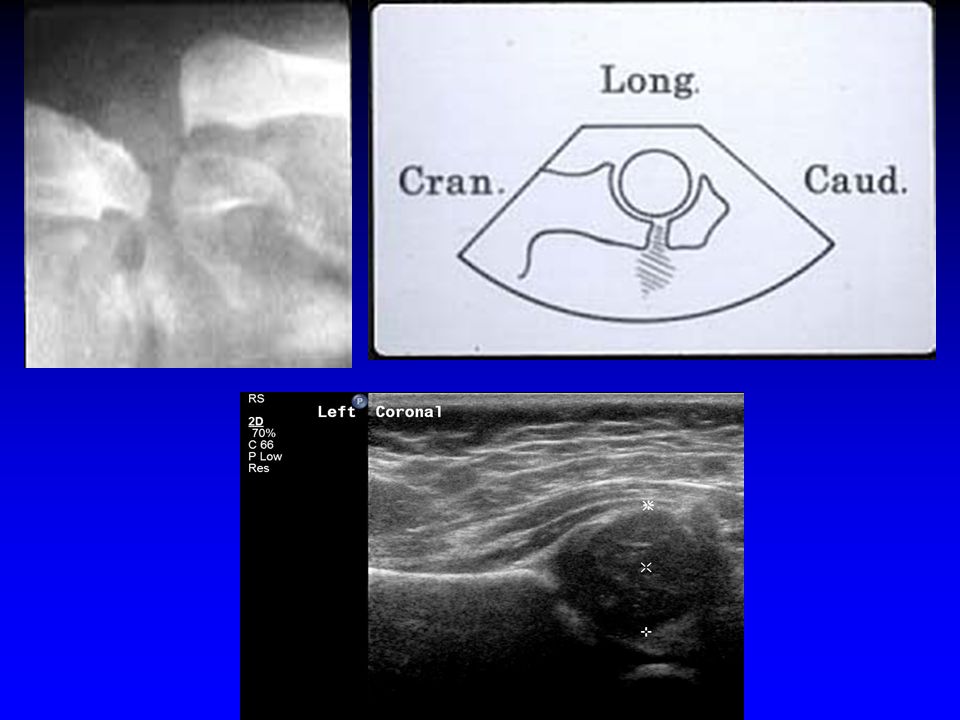
37
Ac Acetabular cartilage
C Capsule G Gluteus muscles GT Greater trochanter H Cartilaginous femoral head IL Ilium Is Ischium L Labrum LT/P Ligamentum teres/ pulvinar complex M Femoral metaphysis Tr Triradiate cartilage

39
Graph Technique Single coronal image emphasizes acetabular development

40
Graf Technique Type 1: normal α angle > 60o

41
Graf Technique -Type II : α 44-60o, β 55-88o
IIa < 3 months immature acetabulum (40-59%) No referral required IIb,c,d require referral for treatment
 No referral required. IIb,c,d require referral for treatment.")
42
Graf Technique Type III : α <44o, β>77o Low displacement
Type IV : completely dislocated Immediate therapy

43
Coronal Harke method

44
Acetabular Coverage >50%

45
Acetabular Coverage >50%

48
40% Coverage

49
33% Coverage

50
20% Coverage

51
Dislocated

53
Dynamic Sonography-Technique
Supine or lateral Coronal view at rest neutral or flexed stress view optional Transverse flexion view with stress

55
A

56
Ac Acetabular cartilage
G Gluteus muscles GT Greater trochanter H Cartilaginous femoral head Is Ischium L Labrum LT/P Ligamentum teres/pulvinar complex M Femoral metaphysis Pu Pubis Tr Triradiate cartilage

57
A A

58
A

62
Stress - Stable

63
Stress - unstable

65
Dislocated

66
Dislocated

68
Calcified femoral epiphysis

69
3 month old

70
Peterlein et al BMC Pediatr. 2010 24;10:98
Peterlein et al BMC Pediatr ;10:98. Reproducibility of different screening classifications in US of the newborn hip. Concordance of 2 classifications of hip morphology and subjective parameters by 3 investigators w/different levels of experience. METHODS: 207 newborns: α-angle and β-angle,"femoral head coverage" (FHC) shape of bony roof and position of cartilaginous roof. RESULTS: shape of bony roof (0.97) and position of cartilaginous roof (1.0) demonstrated high intra-observer reproducibility. Best results were achieved for α-angle, followed by β-angle then FHC. CONCLUSIONS: Higher measurement differences in objective scorings. Variations by every investigator irrespective of level of experience
 shape of bony roof and position of cartilaginous roof. RESULTS: shape of bony roof (0.97) and position of cartilaginous roof (1.0) demonstrated high intra-observer reproducibility. Best results were achieved for α-angle, followed by β-angle then FHC. CONCLUSIONS: Higher measurement differences in objective scorings. Variations by every investigator irrespective of level of experience.")
71
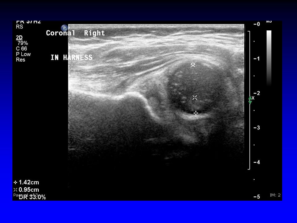
Follow up Can perform exam in Pavlik Harness
Perform out of harness only if requested and/or hip appears stable Once femoral head ossifies difficult to assess position.

73
Treatment Dislocated – treat Stable – don’t treat
Unstable (lax not displaced) ? Early treatment or observation?? 80% normalize
 Early treatment or observation 80% normalize.")
75
1mos old 3 wk FU

76
3 wk old 1 month FU in Harness

77
DDH - 35%

78
One month later

79
FU 6 weeks

80
Follow Up

81
Should we Screen? There is no consensus on imaging screening for DDH.
Screening balanced between the benefits of early detection of DDH and the increased treatment and cost factors.

82
Who? Universal Newborn Screening pro- treat early
con-over treat minor abnormalities that resolve Considerable resources Late cases missed Higher rate of therapy? Higher rate of avscular necrosis?

83
Universal Screening Randomized trials evaluating primary US screening did not find significant decrease in late diagnosis of DDH. This practice is yet to be validated by clinical trial.

84
Who? Selective screening
AAP US recommended as adjunct to clinical evaluation. technique of choice to clarify physical finding, assess high-risk infant, and monitor DDH as is observed or treated. Can guide treatment and may prevent overtreatment

85
Who? In the United States, hip US is selectively performed Club foot
Torticollis Females in breech position Optional males in breech position Optional females with positive FH Inconclusive PE

86
Studies – Selective Screening
British 10 yr prospective of 34,723 2,578 clinical instability or risk factor 77 unstable - 31% risk factor Irish 52,893 infants US – 5,484 with FH, breech, click. 18 dislocatable,153 (2.73%) dysplastic /1000 required Rx 33 center United Kingdom Hip Trial found reduces splinting, and no increase in surgical Rx
 dysplastic 3.2/1000 required Rx. 33 center United Kingdom Hip Trial. found reduces splinting, and no increase in surgical Rx.")
87
Preterm infants DDH may be unrecognized.
When the infant has cardiorespiratory problems, the diagnosis and management are focused on providing appropriate ventilatory and cardiovascular support, careful examination may be deferred until a later date. The most complete examination the infant receives may occur at the time of discharge from the hospital, and this single exam may not detect subluxation or dislocation. critical to examine the entire child.

91
When? PRO - US can detect abnormal position, instability, and dysplasia not evident on clinical examination. CON - during the first month minor degrees of instability and acetabular immaturity. nearly all mild early findings not be apparent on PE, resolve spontaneously without treatment. Newborn screening - high frequency of reexamination and hips being unnecessarily treated. screening with higher false-pos results yields increased prevention of late cases.

92
When? pro con Screen those at risk at 4-6 wks (9%)
less expense,simpler process fewer false positives con miss late cases

93
Hip Evaluation

94
What are the AAP recommendations?
All newborns screened by PE by a properly trained health care provider (Evidence strong.) US of all newborns is not recommended. (Evidence fair; consensus is strong.) Although indirect evidence supports US screening of all newborns, not advocated – operator-dependent, availability is questionable, increases treatment, interobserver variability is high, increased costs.
 US of all newborns is not recommended. (Evidence fair; consensus is strong.) Although indirect evidence supports US screening of all newborns, not advocated – operator-dependent, availability is questionable, increases treatment, interobserver variability is high, increased costs.")
95
3. If positive Ortolani or Barlow sign found in the newborn, refer to an orthopaedist.
4. If results of the PE at birth are "equivocally" positive (ie, soft click, mild asymmetry,), FU hip exam by the pediatrician in 2 weeks is recommended. (Evidence is good; consensus is strong.)
, FU hip exam by the pediatrician in 2 weeks is recommended. (Evidence is good; consensus is strong.)")
96
The hips must be examined at every well-baby visit
(2–4 days for newborns discharged in less than 48 hours after delivery, 1 mos, 2 mos, 4 mos, 6 mos, 9 mos, 12 mos). If DDH is suspected confirmation made by a focused PE, by consultation with another pediatrician, orthopaedist, by US if the infant is < 5 months of age, or by radiography if the infant > 4 months of age.
. If DDH is suspected confirmation made by a focused PE, by consultation with another pediatrician, orthopaedist, by US if the infant is < 5 months of age, or by radiography if the infant > 4 months of age.")
97
Conclusions US has become the standard of care in the evaluation of the neonate with possible developmental dysplasia of the hip. Availability widespread, however, accurate results require training and experience.

98
Changes in practice Use appropriateness criteria to assess for developmental dysplasia. Selective screening by ultrasound after 2 weeks of age

99
AAP Clinical Practice Guideline: Early Detection of DDH
Committee on Quality Improvement, Subcommittee on Developmental Dysplasia of the Hip

Similar presentations


>")


 MB BS BSc MSc (SEM) MRCS (Eng) Diploma in MM (UIAA)>")


 Dysplasia of the Hip. Natural History and Prevention Levels. Nicolas Padilla Professor of Pediatrics School of Nursing and Obstetrics.>")


