Download presentation
Presentation is loading. Please wait.

1
PROF OF OB &GYN. AIN SHAMS UNIVERSITY,GYNEONCOLOGY UNIT.
Cancer vulva MOUNIR M F ELHAO, PROF OF OB &GYN. AIN SHAMS UNIVERSITY,GYNEONCOLOGY UNIT.

2
HISTORICAL. The surgical treatment, back in the early 1900s Basset from France who adopted a Hallstedian concept to the treatment of vulvar cancer very similar what Dr. Hallstead had adopted for breast cancer, felt that wide surgical excision was the best.

3
INCIDENCE. Relatively rare, accounting for about 3 to 5% of all gynecologic malignancies.

4
INCIDENCE. Fourth most common malignancy of the female genital tract

5
INCIDENCE. As the 6th , 7th decade of life and does increase with increasing age.

6
INCIDENCE. very high association with the high risk HPV serotypes, specifically type 16 and 18.

7
PREDISPOSING FACTORS. any chronic inflammatory condition, herpes has been implicated, obesity, diabetes, hypertension, prior squamous cell carcinoma of the cervix, vagina or the anal rectal area as well, and then vulvar dystrophy probably increases a woman’s risk.

8
ASSOCIATIONS. associated vulvar dystrophies, they may even have vulvar intraepithelial neoplasia, they often times are incontinent there is often a delay of 2 to16 months between the onset of symptoms and the initial presentation to the physician

9
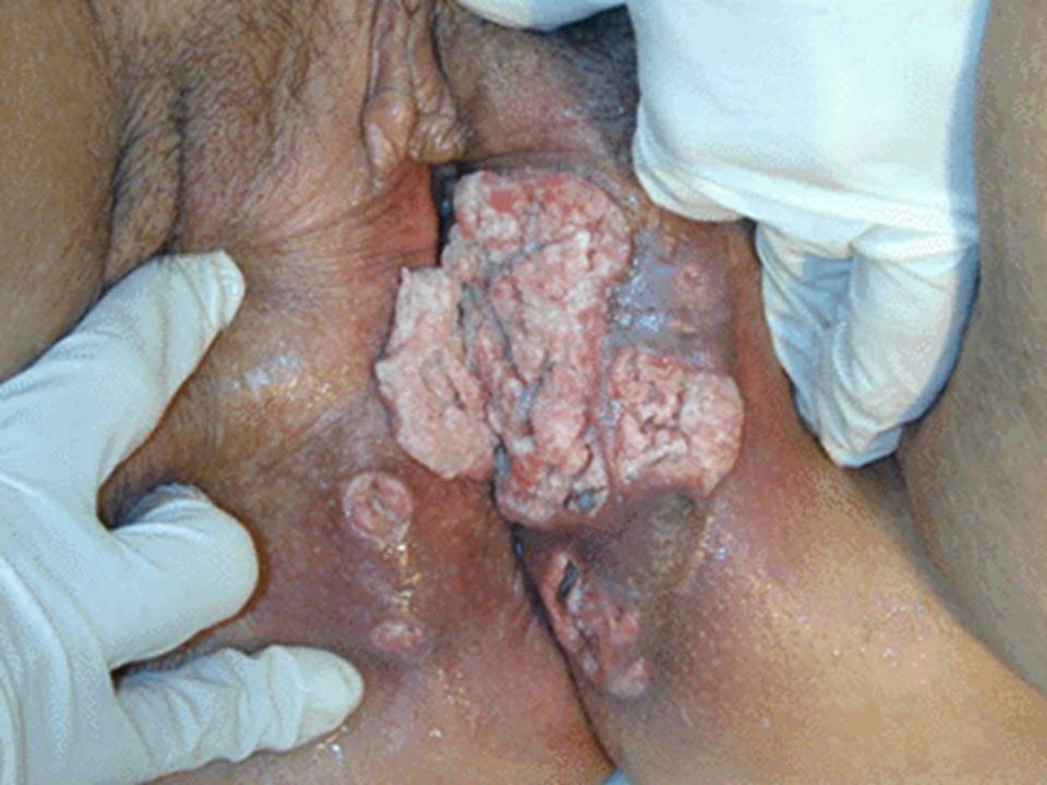
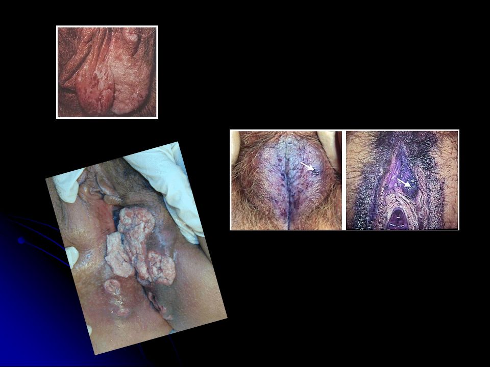
SYMPTOMS. Symptoms include chronic pruritus, a lump or mass, pain, bleeding ulceration, dysuria and leg edema

10
FACT:SURVIVAL Survival rates in most series relates to nodal involvement.

11
Intraepithelial lesions.
are treated a number of different ways, it depends on the site, the age of the patient and the size of the lesion. It is proper to excise with a 3 to 4 mm margin and then primarily close the area.

12
In order to make a diagnosis
you need to get tissue, and wedge biopsy, excisional biopsies, colposcopy, it’s very important to remember that you have to examine the remainder of the genital tract looking for vaginal lesions and also for cervical dysplasia or early invasive cancer because often times these can be metastatic from another site,

13
Squamous cell carcinoma
is the most common followed by melanomas, about just under 6% of the time, Bartholin gland cancers are third, basal cell carcinoma, you see some sarcomas, you can very rarely see invasive Paget’s disease.

14
SITES. The most frequent sites are on the labia majus, followed by the labium minorum, and then some patient’s will have combined lesions about 15%.

15
FACT. the demarcation for micro invasion is actually 1 mm or less.

16
Anatomy The superficial inguinal lymph nodes lie along the saphenous vein, deep to Camper's fascia and superficial to the cribriform fascia which overlies the femoral vessels. They are found in the triangle bounded by the inguinal ligament superiorly, the border of the sartorius muscle laterally, and the adductor longus muscle medially. There are appoximately 10 superficial lymph nodes. The deep inguinal lymph nodes are located medial to the femoral vein and under the cribriform fascia. There are approximately 3 to 5 deep nodes. The superior-most node is located under the inguinal ligament and is called Cloquet's node

18
Drainage The superficial inguinal lymph nodes receive drainage from the vulva and anus. The superficial nodes drain to the deep inguinal lymph nodes, which then drain superiorly to the external iliac lymph nodes, then to the pelvic lymph nodes and to the paraaortic lymph nodes.

32
The TNM staging system is used.
T Pre-malignant change. T-1A A cancer less than 2.0cm in diameter and less than 1.0mm in depth of invasion. T-1B A cancer less than 2.0cm in diameter but greater than 1.0mm in invasion. T-2 Greater than 2.0cm in diameter. T-3 Involves vagina, urethra or anus. T-4 Involves bladder, rectum or pelvic bone. N-0 No lymph nodes involved . N-1 Lymph node metastases to one groin. N-2 Lymph node metastases to both groins. M-0 No distant metastases. M-1 Any distant metastases.

33
The standard treatment ( Hallstedian concept )
, was block radical vulvectomy with bilateral inguinal femoral lymphadenectomies and we did selective pelvic lymphadenectomies through separate extra peritoneal incisions and this basically is what has been called the butterfly incision or the Texas longhorn incision.

34
Why conservative surgery?
The rationale for conservative surgery is that most of the metastases occur by embolization and the early advocates of the more conservative procedures in their series found no metastatic lesions in the skin bridge between the vulva and the groin,

35
FACT: Current place of pelvic lymphadenectomy?
No patients with negative groin nodes Had positive pelvic nodes. Positive bilateral groin nodes five year survival <20 %

36
We omitted routine pelvic lymphadenectomy,
patient’s who have positive nodes, , end up getting radiation therapy to the whole pelvis anywhay.

37
Role of adjuvent radiotherapy?
Review of recurrence studies of in Homesley,s study suggests that adjuvent RT is more effective largely because groin recurrences are reduced.

38
Should we do separate groin incisions ?
Understanding that the mode ofmetastatic spread is embolic rather than by contiguous grouth allowed for three-incision technique.. Less morbidity. No impact on survival. (54% BREAKDOWN RATE WITH BUTTERFLY TECH.)
")
39
Is there a place for unilateral inguinofemoral lymphadenectomy?
May be indicated in well lateralised early tumors. No lymph-capillary space involvement. Negative groin nodes by frozen section.

40
What is the place of superficial inguinal lymphadenectomy?
Above the cribriform fascia , mainly those associated with great saphenous and superficial epigastric veins. ONLY with low risk for LN metastasis. Tumors confined to labia majora. Negative superficial nodes on frozen section.

41
Can we omit groin node dissection in superficial diseases?
Stage 1a have <1% for groin node metastasis.

42
we do give postoperative radiation for groin nodal metastases

43
Is there a place for preoperative radiotherapy?
we give preop radiation therapy for advanced disease.

44
Conclusions. Now to run through management, again
for stage I, it’s pretty much radical local excision, and you want to try to maintain at least a 1 cm margin and if it’s truly a small lesion with less than a mm invasion, it is felt that most of those patient’s do not need to have lymph nodes removed.

45
CONCLUSIONS. For large stage II lesions, again, depending on where it’s located, we do a radical vulvectomy and bilateral inguinal femoral lymphadenectomy, if there are more than two lymph nodes positive, the patient’s will get postoperative whole pelvic radiation.

46
For stage III tumors, it depends on what’s involved, you can do a radical excision which often times becomes extended and you have to take the distal vagina and even sometimes the distal urethra and if you are going to treat it surgically it needs to be combined with the bilateral inguinal femoral lymphadenectomy and again, if there is lymph node involvement POST OPERATIVE RADIOTHERAPY.

47
Conclusions. For advanced disease, again you have to individualize, add up with surgical clearance for disease sometimes involves the anus, rectum, proximal urethra and requires an exenterative procedure with radical vulvectomy and bilateral groin nodes and that particular circumstance is very important that patient’s are evaluated either with MRI, CAT scans and possibly even a PET scan for metastatic disease prior to undertaking such a large procedure. The operative mortality is about 5 to 10%.

48
Conclusions. Survival is also determined whether or not the nodes are positive or negative, and by which nodes are involved. If patient’s have negative groin nodes, the five year survival is 90% and that’s for stage I and stage II. If they have positive groin nodes, survival drops about 57%.

49
If they have positive pelvic lymph nodes, it drops to 20%
If they have positive pelvic lymph nodes, it drops to 20%. Unilateral positive groin nodes is about 70% five year survival, bilateral positive groin nodes, however, drops down to 25% five year survival, and then the increasing number of positive nodes.

50
CONCLUSIONS. and also the tumor diameter affects nodal involvement, lymphatic vascular space involvement and then overall survival.

51
Conclusions. The Cochrane Database of Systematic Reviews 2006 Issue 1 Copyright © 2006 The Cochrane Collaboration. Published by John Wiley & Sons, Ltd. Surgical interventions for early squamous cell carcinoma of the vulva Ansink A, van der Velden J, Collingwood M Plain language summary Less extensive surgery for vulvar cancer appears safe and limits mutilation Vulvar cancer is rare, affecting mainly older women. Until the 1980s, affected women underwent extensive, mutilating surgery. Groin nodes on both sides as well as all vulvar tissue were removed. Recently surgeons have carried out a smaller operation, leaving as much vulvar tissue as possible behind. No randomized controlled trials have been conducted on the safety of this reduced surgery, but from the available evidence it appears to be safe to perform this smaller operation in most patients.

52
CONCLUSIONS. The Cochrane Library Cochrane review abstract and plain language summary This is an abstract and plain language summary of a regularly updated, systematic review prepared and maintained by The Cochrane Collaboration. The full text of the review is available in The Cochrane Library (ISSN X). The Cochrane Database of Systematic Reviews 2006 Issue 1 Copyright © 2006 The Cochrane Collaboration. Published by John Wiley & Sons, Ltd. Primary groin irradiation vs primary groin surgery for early vulvar cancer van der Velden J, Ansink A Plain language summary Insufficient evidence that radiotherapy works as well as surgery for vulvar cancer. Cancer of the vulva is mainly a disease of elderly women. Surgery involves removal of the tumour and surrounding lymph nodes, occasionally followed by radiotherapy. Although survival rates are high if the tumour is found early enough, removal of the lymph nodes causes swelling, particularly in the legs. Wound healing and sexual problems are also common. While radiotherapy is effective in the short term, there is not enough evidence from trials to show that it is as effective as surgery in preventing tumour regrowth in the groins.
. The Cochrane Database of Systematic Reviews 2006 Issue 1 Copyright © 2006 The Cochrane Collaboration. Published by John Wiley & Sons, Ltd. Primary groin irradiation vs primary groin surgery for early vulvar cancer. van der Velden J, Ansink A. Plain language summary. Insufficient evidence that radiotherapy works as well as surgery for vulvar cancer. Cancer of the vulva is mainly a disease of elderly women. Surgery involves removal of the tumour and surrounding lymph nodes, occasionally followed by radiotherapy. Although survival rates are high if the tumour is found early enough, removal of the lymph nodes causes swelling, particularly in the legs. Wound healing and sexual problems are also common. While radiotherapy is effective in the short term, there is not enough evidence from trials to show that it is as effective as surgery in preventing tumour regrowth in the groins.")
53
What is the place of modified radical vulvectomy?
main morbidity of radical vulvectomy is sexual dysfunction and compromised function of the anus and urethra. The main fear of about the modified operation is the multicentricity of the tumor.(20-30%). So reservethe operation to well localised tumors,with 2 cm free margin.
. So reservethe operation to well localised tumors,with 2 cm free margin.")
54
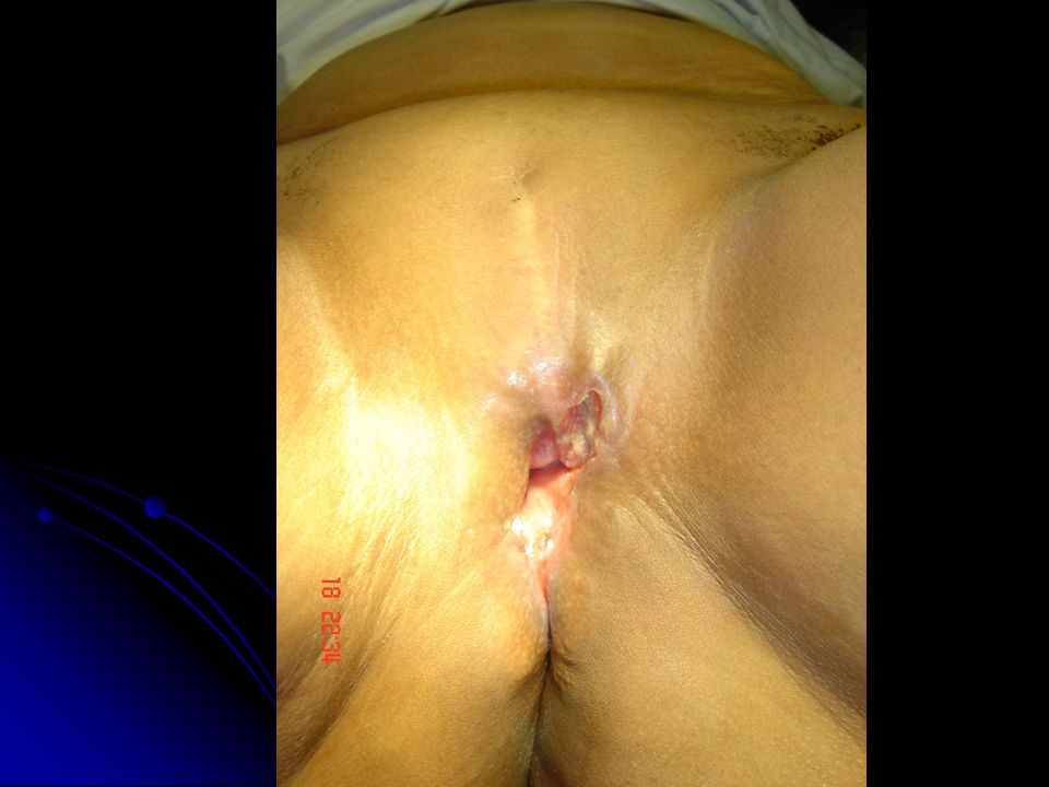
How should we treat vulvar carcinoma with perianal involvement?
The main problem in these cases is to do adequate resection with maintaining sphincteric function.sometimes we may need to do more radical resection and colostomy or preoperative radiotherapy.

55
what is the place of ultraradical surgery?
Only in patients with clearly resectable lesions and negative or one or two microscopicaly positive nodes.

56
what is the place of neoadjuvent chemotherapy?
Resuts are not encouraging for time being.

57
CONCLUSION. 1.Standard radical vulvectomy and bilateral lymphadenectomy(Hallstedian concept.)has compromised the life of many women with cancer vulva.
has compromised the life of many women with cancer vulva.")
58
2.In many well selected patients wide excision with 2 cm margin with or without node selection may suffice.

59
3.modified radical vulvectomy with bilateral groin node dissection will give equaly good results in the majority of cases.

60
4.Pelvic lymphadenectomy should be abondoned except in a minority of selected cases.

61
5.Radiotherapy should be given to the groins and pelvis postoperatively only if
more than one groin nodesis positive for metestatic disease.

62
6.ultraradical surgery selective .

63
7. In situ stage is almost 100% curable
7.In situ stage is almost 100% curable.and FIGO stage 1 disease is 90% curable and 5 year survival rate.

Similar presentations


 ASSOCIATE PROFESSOR OF GYNECOLOGICAL ONCOLOGY King Abdulaziz University Hospital, Jeddah, Saudi Arabia.>")


