Download presentation
Presentation is loading. Please wait.

1
Anatomy of the Digestive System u Functions of the Digestive System u Organs of the GI Tract u Layers of the GI Tract u Gross and Microscopic Anatomy of Specific Regions

2
Functions of the Digestive System u Ingest and masticate food u Move food through the gastrointestinal (GI) tract u Mix and digest food u Absorption of nutrients u Elimination of undigested food
 tract u Mix and digest food u Absorption of nutrients u Elimination of undigested food")
4
Organs of the GI tract oral cavity pharynx esophagus stomach small intestine large intestine salivary glands liver gall bladderpancreas rectum

5
Layers of the GI Tract u The GI tract is composed of four major layers u Mucosa: - mucous epithelium - lamina propria - muscularis mucosa u Submucosa u Muscularis Externa: - inner circular - outer longitudinal u Covering: serosa or adventitia

7
Layers of the GI Tract mucous epithelium lamina propria muscularis mucosa submucosa inner circular outer longitudinal serosa

8
The Oral Cavity u Boundaries are: - lips (anteriorly) - cheeks (laterally) - palate (superiorly) - fauces (posteriorly) u Vestibule: space between the teeth and lips/cheeks u Mucosa: lining of moist, stratified squamous epithelium
 - cheeks (laterally) - palate (superiorly) - fauces (posteriorly) u Vestibule: space between the teeth and lips/cheeks u Mucosa: lining of moist, stratified squamous epithelium")
10
The Oral Cavity u The oral cavity is important in: - mastication (chewing): teeth, tongue, cheeks - secretion of saliva for digestion, coating food - parotid gland - submandibular gland - sublingual gland - speech: tongue, teeth, lips
: teeth, tongue, cheeks - secretion of saliva for digestion, coating food - parotid gland - submandibular gland - sublingual gland - speech: tongue, teeth, lips")
12
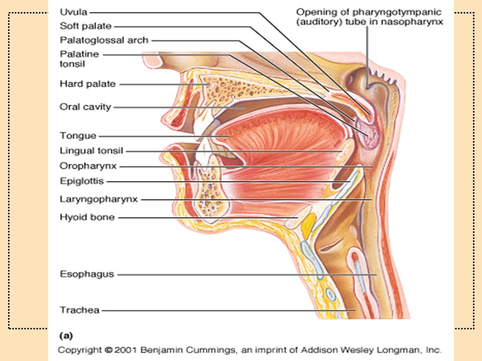
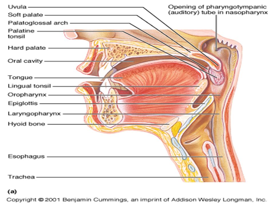
The Pharynx u The pharynx is the passageway from the nose and mouth to the esophagus and respiratory tract (larynx) u Three parts: - nasopharynx: from nares to uvula, lined with respiratory epithelium - oropharynx: from uvula to epiglottis, lined with stratified squamous epithelium - laryngopharynx: from epiglottis to openings of the larynx and esophagus, lined with stratified squamous epithelium
 u Three parts: - nasopharynx: from nares to uvula, lined with respiratory epithelium - oropharynx: from uvula to epiglottis, lined with stratified squamous epithelium - laryngopharynx: from epiglottis to openings of the larynx and esophagus, lined with stratified squamous epithelium")
15
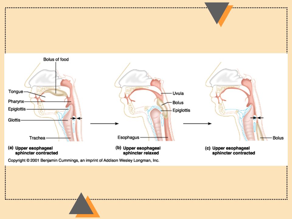
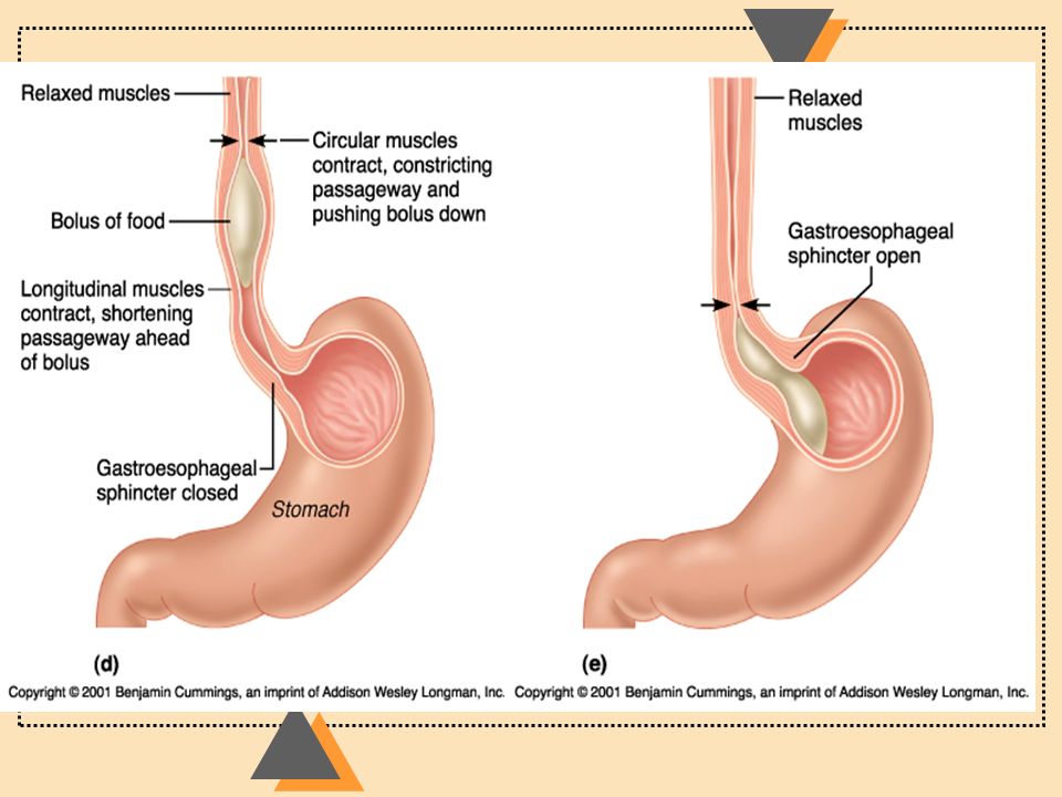
The Esophagus u The esophagus connects the pharynx to stomach u Contains two sphincters: upper and lower esophageal u Mucosal lining: stratified squamous u Muscularis layer: superior portion is skeletal (voluntary), inferior portion is smooth muscle u Outer covering: adventitia
, inferior portion is smooth muscle u Outer covering: adventitia")
17
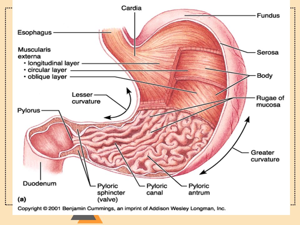
The Stomach u Function: mixing of food, secretion, little absorption u Gross Anatomy of the Stomach esophagus cardiac sphincter fundus body serosa outer longitudinal middle circular inner oblique submucosa mucosa pyloric region cardiac region pyloric sphincter

18
The Stomach Histological Features u Mucosal layer: simple columnar epithelium - gastric pits, gastric glands - five cell types: - surface mucous cells - mucous neck cells - parietal cells (produce HCl) - chief cells (produce pepsinogen) - endocrine cells (gastrin)
 - chief cells (produce pepsinogen) - endocrine cells (gastrin)")
20
The Stomach u Submucosa and mucosa form folds called rugae u Muscularis layer: - outer longituninal - middle circular - inner oblique u Outer covering: serosa

21
The Stomach gastic pit gastric gland mucosa submucosa muscularis externa serosa surface mucous cells mucous neck cells parietal cells chief cells endocrine cells muscularis mucosa oblique muscle layer circular muscle layer longitudinal muscle connective tissue visceral peritoneum

23
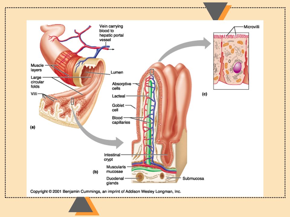
Small Intestine Function: major site of absorption, digestion Gross Anatomy: u Connects stomach with large intestine u Three parts: duodenum, jejunum, ileum u Bile duct and pancreatic duct empty into duodenum

24
Small Intestine Histological Features u Specializations to increase surface area for absorption: - plicae circulares - villi - microvilli (brush border)
")
26
Small Intestine Histological Features: Mucosal epithelium: simple columnar, w/ specialized cells - absorptive cells with microvilli - goblet cells, produce mucus - granular cells (Paneth cells; protect against bacteria) - endocrine cells (gastrin, CCK, secretin) The mucosa invaginates to form intestinal glands (crypts of Lieberkuhn; between villi); cells originate in base of villi
 - endocrine cells (gastrin, CCK, secretin) The mucosa invaginates to form intestinal glands (crypts of Lieberkuhn; between villi); cells originate in base of villi")
27
Histological Differences between Duodenum, Jejunum, and Ileum u The submucosa of duodenum and ileum have specializations which allow them to be recognized u Duodenum: Brunner’s glands (mucus glands) u Ileum: Peyer’s patches (lymphoid tissue)
 u Ileum: Peyer’s patches (lymphoid tissue)")
28
The Large Intestine u Functions: absorption of water and salts, secretion of mucus u Gross Anatomy: ascending colon cecum appendix transverse colon haustra descending colon taenia coli sigmoid colon rectum

29
The Large Intestine Histology: u mucosal lining: simple columnar epithelium u large numbers of goblet cells u outer longitudinal layer: taenia coli

30
Rectum & Anal canal Rectum u Joins the sigmoid colon with the anal canal u simple columnar epithelium u thick muscularis layer Anal Canal u epithelium changes from simple columnar (superiorly) to stratified squamous (inferiorly) u internal sphincter: thick smooth muscle u external sphincter: skeletal muscle
 to stratified squamous (inferiorly) u internal sphincter: thick smooth muscle u external sphincter: skeletal muscle")
32
Next Lecture….. Mechanical & Chemical Digestion

Similar presentations


![Anatomy Practical [PHL 212]](/14/4428258/big_thumb.jpg "Anatomy Practical [PHL 212]>")


