Download presentation
Presentation is loading. Please wait.

1
Spectroscopy 3: Magnetic Resonance CHAPTER 15

2
Conventional nuclear magnetic resonance Energies of nuclei in magnetic fields Typical NMR spectrometer The chemical shift (effect of nearby nuclei) Fine structure (nuclear spin-spin coupling) Pulsed techniques in FT-NMR
 Fine structure (nuclear spin-spin coupling) Pulsed techniques in FT-NMR")
4
Fig 15.1 Interactions between m s states of an electron and an external B field precession ν L ≡ the Larmor freq m s = +1/2 m s = −1/2 where γ e ≡ magnetogyric ratio B o ≡ applied magnetic field

6
Fig 15.3 Nuclear spin states of a spin-1/2 nucleus (e.g., 1 H or 13 C) in a magnetic field = h ν radio Typically: A 100 MHz NMR employs a 2.35 T field Resonance is achieved when ν radio = energy separation between levels
 in a magnetic field = h ν radio Typically: A 100 MHz NMR employs a 2.35 T field Resonance is achieved when ν radio = energy separation between levels")
7
Fig 15.4 Layout of a typical NMR spectrometer

8
The Chemical Shift Nuclear magnetic moments interact with the local field In most cases, B loc ≠ B 0 due to electronic orbital ang momentum The Larmor frequency ν L (frequency of precession) differs for nuclei in different environments Resonance frequencies expressed as the chemical shift TMS
 differs for nuclei in different environments Resonance frequencies expressed as the chemical shift TMS")
9
Fig 15.5(a) Range of typical chemical shifts for 1 H TMS Deshielded nuclei (low field) Shielded nuclei (high field)
 Range of typical chemical shifts for 1 H TMS Deshielded nuclei (low field) Shielded nuclei (high field)")
10
Fig 15.5(b) Range of typical chemical shifts for 13 C TMS Deshielded nuclei (low field) Shielded nuclei (high field )
 Range of typical chemical shifts for 13 C TMS Deshielded nuclei (low field) Shielded nuclei (high field )")
11
Fig 15.6 The 1 H-NMR spectrum of ethanol Integrated signal singlet quartet triplet 1 3 2

12
Fig 15.7 Variation of the chemical shift with electronegativity Trend due to magnetic anisotropy Trend due to electronegativity

13
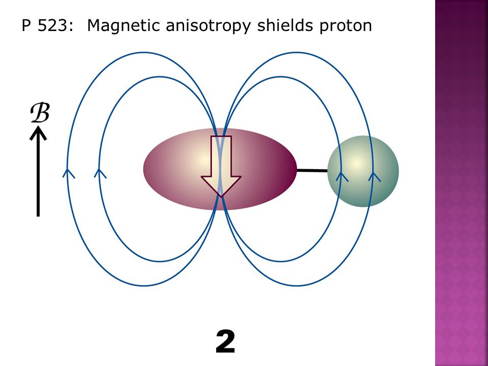
P 523: Magnetic anisotropy shields proton

15
H B loc

16
Fig 15.9 Ring current deshields ring protons and shields substituent protons Special case of neighboring group effect in aromatics deshielded shielded

17
Fig 15.6 The 1 H-NMR spectrum of ethanol Integrated signal singlet quartet triplet 1 3 2 Fine structure

18
Fine Structure Each magnetic nucleus may contribute to the local field of adjacent nuclei ∴ Resonance frequencies are modified Strength of interaction given by the coupling constant, J (Hz) J is independent of applied mag field, B o
 J is independent of applied mag field, B o")
19
Margin pg 526 n equivalent nuclei split adjacent spin(s) into n+1 lines with intensity distribution given by Pascal’s triangle :
 into n+1 lines with intensity distribution given by Pascal’s triangle :")
20
Fig 15.15 Origin of the 1:2:1 triplet in the proton resonance of a –CH 3 species e.g., CH 3 CH 2 OH ⇇⇉ ⇆ ⇄ B0B0

21
Fig 15.16 Origin of the 1:3:3:1 quartet in the proton resonance of a -CH 2 - species e.g., CH 3 CH 2 OHB0B0

Similar presentations

>")
>")


 A photon generates both an electric and a magnetic field A current passing through a wire also generates both an electric and a magnetic.>")
 FID represents the time-domain response of the spin system following application.>")


 Spectroscopy>")


 Spectroscopy Structure Determination>")