Download presentation
Presentation is loading. Please wait.

1
LAB 304 Lecture \ 9

2
Learning objectives To recognize urinalysis procedures: Physical Chemical Microscopic List some of urine crystals List some of urine casts.

3
Results interpreting

4
1 2 3

5
1- Physical examination Color Appearance Volume Specific gravity (SG)
")
6
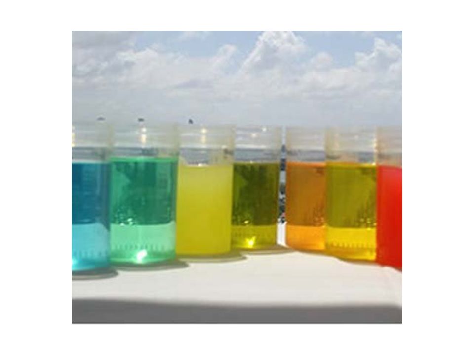
NORMAL COLOR Including color and clarity Normal urine color ranges from pale yellow to deep amber

7
NORMAL COLOR

8
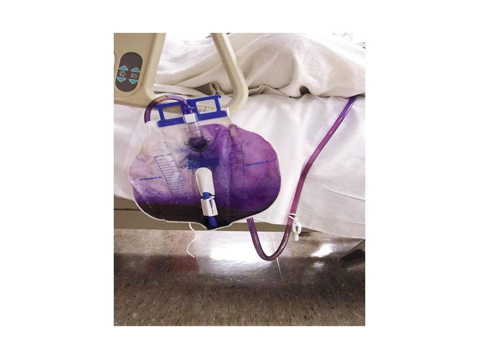
ABNORMAL COLOR Abnormal color : some drugs and urinary tract infection cause color changes reddish urine 1. reddish urine: hematuria, hemoglobinuria yellow-brown or green-brown urine 2. yellow-brown or green-brown urine: bilirubin cause : obstructive jaundice greenish urine 3- greenish urine : infection blue,orange,purple Other abnormal colors : blue,orange,purple

11
Appearance Clarity: normally, clear Abnormal appearance: POSSIBLE CAUSEAPPEARANCE BACTERIAL URINARY INFECTION CLOUDY 1- URINARY SCHISTOSOMIASIS 2- BACTERIAL INFECTION DUE TO RBCRED & CLOUDY 1- ACUTE VIRAL HEPATITIS 2- OBSTRUCTIVE JAUNDICE DUE TO BILIRUBIN YELLOW-BROWN OR GREEN-BROWN 1- HAEMOLYSIS 2- HEPATOCELLULAR JAUNDICE DUE TO UROBILINYELLOW-ORANGE

12
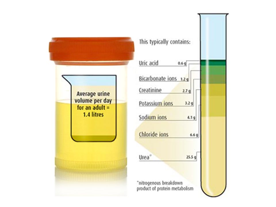
Urine volume The average adult : 1000ml to 2000ml/24h Increased POLYURIA---more than 2500ml of urine in 24 hours 1. physiological states: water intake, some drugs ( diuretics ), intravenous solutions 2. pathologic states: diabetes mellitus, diabetes insipidus
, intravenous solutions 2. pathologic states: diabetes mellitus, diabetes insipidus.")
13
Urine volume Decreased OLIGURIA : less than 400ml of urine in 24 hours ANURIA : less than 100ml of urine in 24 hours 1. pre-renal: hemorrhage, dehydration, congestive heart failure 2. post-renal: obstruction of the urinary tract (may be stones, carcinoma)
.")
14
Specific gravity (SG) Reflect the density of the urine Range of 1.001 to 1.040 Increased: Dehydration, Fever, Vomiting, Diarrhea, Diabetes Mellitus (urine volume ↓ and SG ↑ ) Decreased: diabetes insipidus (urine volume ↑ and SG ↓ )
 Reflect the density of the urine Range of to Increased: Dehydration, Fever, Vomiting, Diarrhea, Diabetes Mellitus (urine volume ↓ and SG ↑ ) Decreased: diabetes insipidus (urine volume ↑ and SG ↓ )")
15
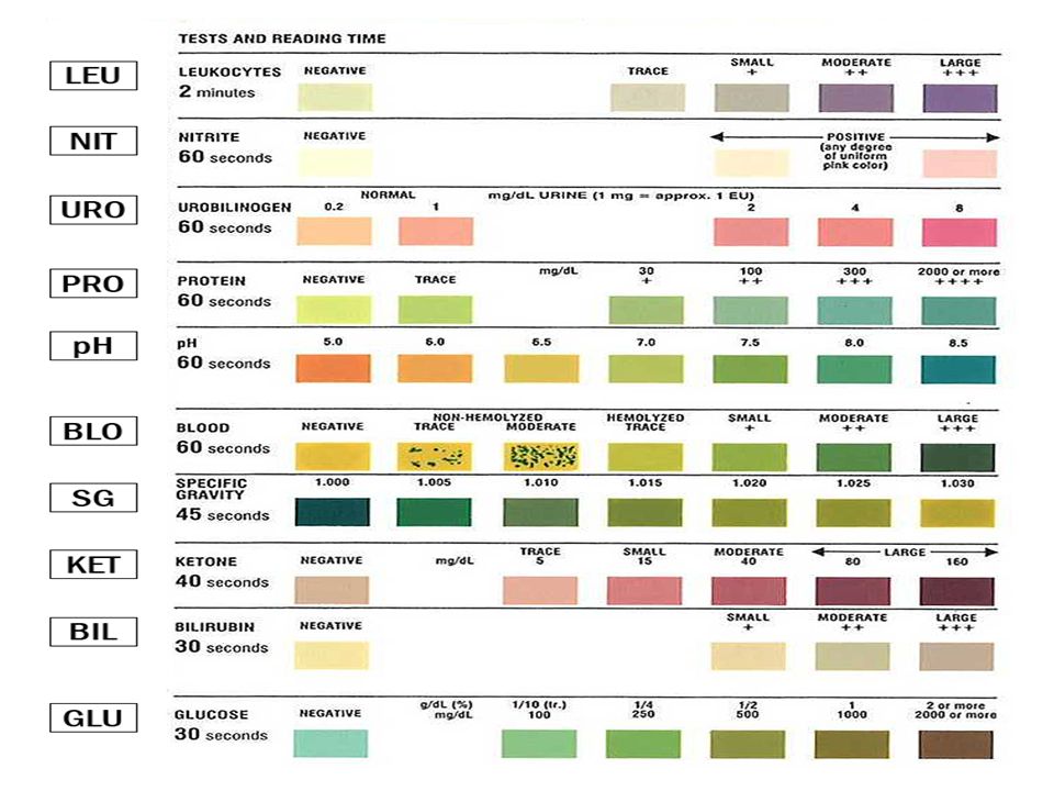
2-Chemical examination Urine PH Protein Glucose Ketones Occult blood Bilirubin Urobilinogen Nitrites

18
Urine PH Normal PH The average is about 6 Range from 5~7 (depends on diet) Higher PH---alkaline urine ( 7.8 – 8.0 ) 1.drugs: sodium bicarbonate 2.vegetarian 3.alkalosis (metabolic or respiratory) 4. urinary tract infection Lower PH---acid urine ( 4.5 – 5.5 ) 1. drugs: ammonium chloride 2. diabetes 3. acidosis (metabolic or respiratory)
 1. drugs: ammonium chloride 2. diabetes 3. acidosis (metabolic or respiratory).")
19
Protein in urine Reference value Qualitative method: negative Quantitative method: < 150mg of protein in 24 hours Urine proteins come from plasma proteins e.g. albumin and Tamm-Horsfall (T-H) glycoprotein
 glycoprotein.")
20
Protein in urine The two most common risk factors for proteinuria are: 1. Diabetes 2. hypertension Proteinuria---- > 150 mg /24 hours or qualitative test is positive Proteinuria quantification (amount of protein ) heavy proteinuria---- > 4 g/24 hours moderate proteinuria----1 - 4 g/24 hours minimal proteinuria---- < 1 g/24 hours
 heavy proteinuria---- > 4 g/24 hours moderate proteinuria g/24 hours minimal proteinuria---- < 1 g/24 hours.")
21
Protein in urine TYPE PATHOPHYSIOLOGIC FEATURES CAUSE Glomerular glomerular capillary permeability to protein Primary or secondary glomerulopathy e.g. IgA nephropathy, lupus nephritis Tubular tubular reabsorption of proteins in glomerular filtrate Tubular or interstitial disease due to: Uric acid nephropathy Heavy metals, NSAIDs Overflow production of low-molecular-weight proteins Monoclonal gammopathy, leukemia Classification of Proteinuria

22
Glucose in urine Reference value Qualitative method: negative Glycosuria--- qualitative test is positive 1. with hyperglycemia ( most common ): diabetes mellitus, Cushing’s syndrome 2. without hyperglycemia (Renal glycosuria ): renal tubular dysfunction, such as pyelonephritis
: diabetes mellitus, Cushing’s syndrome 2. without hyperglycemia (Renal glycosuria ): renal tubular dysfunction, such as pyelonephritis.")
23
Ketones in urine Including three ketone bodies: 1. acetone 2% 2. acetoacetic acid 20% 3. β-hydroxybutyric acid 78% The products of fat catabolism ( breakdown ) Reference value: qualitative method: negative Ketonuria--- qualitative test is positive
 Reference value: qualitative method: negative Ketonuria--- qualitative test is positive.")
24
Ketones in urine Ketonuria 1. diabetic ketonuria : 1. diabetic ketonuria : I. Poorly controlled diabetes II. Diabetic ketoacidosis (DKA) 2. nondiabetic ketonuria: 2. nondiabetic ketonuria: I. Acute or severe illness II. Burns III. Fever IV. Hyperthyroidism V. Pregnancy & lactation VI. Abnormal food or nutrition intake due to: Anorexia, fasting, high protein or low carbohydrate diets, starvation, vomiting over a long period of time
 2. nondiabetic ketonuria: 2. nondiabetic ketonuria: I. Acute or severe illness II. Burns III. Fever IV. Hyperthyroidism V. Pregnancy & lactation VI. Abnormal food or nutrition intake due to: Anorexia, fasting, high protein or low carbohydrate diets, starvation, vomiting over a long period of time.")
25
3-Microscopic examination Sample preparation 1- Obtain fresh urine sample 2- shake the container to mix the sample 3- pipette suitable amount to test tube 4- Centrifuge it at 1500 to 3000 rpm for 5 minutes 5- Decant supernatant part 6- from the sediment Place 1 drop of urine on slide and apply cover slip 7- examine it under microscope ( 10x, 40x )
")
26
Microscopic examination Examination A- Cells B- Bacteria C- Crystals D- Casts

27
Microscopic examination A- Cells : 1- White Blood Cells (pus cell) Normal <2/ HPF in men and <5/ HPF in women Few : up to 10/ HPF Moderate : 11-40 / HPF Many : > 40 / HPF HPF = high power field
 Normal <2/ HPF in men and <5/ HPF in women Few : up to 10/ HPF Moderate : / HPF Many : > 40 / HPF HPF = high power field")
28
Microscopic examination 2- Red Blood Cells : smaller and more refractile than white cells Normal <3/ HPF Dysmorphic RBCs suggest glomerular disease

29
Microscopic examination 3- Epithelial cells : 3 types 1. Transitional epithelial cells are normally present

30
Microscopic examination 2. Squamous epithelial cells suggest contamination

31
Microscopic examination 3. Renal tubule epithelial cells suggest renal disease

32
Microscopic examination B- Bacteria : B- Bacteria : Diagnostic for Urinary Tract Infection Men: Any bacteria Women: 5 or more bacteria per HPF

33
Microscopic examination C- Crystals 1- Calcium oxalate crystals (square envelope shape)
")
34
Microscopic examination 2- Triple phosphate crystals (coffin lid shape) Associated with increased Urine pH (alkaline) Associated with Proteus Urinary Tract Infection
 Associated with increased Urine pH (alkaline) Associated with Proteus Urinary Tract Infection")
35
Microscopic examination 3- Uric Acid crystals (diamond shape)
")
36
Microscopic examination D- Casts 1- Epithelial cell casts of renal tubule

37
Microscopic examination 2- Red Blood Cell casts

38
Microscopic examination 3- White Blood Cell casts

39
Microscopic examination 4- Hyaline or mucoprotein casts

40
Microscopic examination 5- Granular casts

41
Microscopic examination 6- Waxy casts

42
Microscopic examination 7- Fatty casts Oval fat bodies ( OFB )
")
Similar presentations


>")
; Small mwt proteins (such as peptide hormones,Insulin glucagon, growth hormone) can appear.>")


 AMIT KAUSHAL. MACROSCOPIC ANALYSIS Colour, clarity, and cloudiness may suggest conditions such as: dehydration dehydration infection infection.>")


 –Urea (from amino acids) –Creatinine (from muscle creatine.>")

