Download presentation
Presentation is loading. Please wait.

1
Frog Dissection

2
Why do you think we are dissecting frogs??

3
Frog Dissection Frogs and humans have similar organ systems, so dissecting frogs helps us learn about human anatomy. It’s also a great way to review the characteristics of life!

4
Frog Dissection Safety Comes First: Rules for Dissection Rule #1: Listen for the “3-2-1” direction and respond immediately so you don’t miss instructions!

5
Frog Dissection Safety Comes First: Rules for Dissection Rule #2: Absolutely no horseplay!

6
Frog Dissection Safety Comes First: Rules for Dissection Rule #3: You must remain at your lab station at all times.

7
Frog Dissection Safety Comes First: Rules for Dissection Rule #4: Always wear goggles, apron, and gloves.

8
Frog Dissection Safety Comes First: Rules for Dissection Rule #5: Always keep your hands away from your face, eyes, and mouth.

9
Frog Dissection Safety Comes First: Rules for Dissection Rule #6: All specimen parts must be disposed of in the trash can, not the sink.

10
Frog Dissection Safety Comes First: Rules for Dissection Rule #7: Clean up your area completely using soap and sponge.

11
Frog Dissection Safety Comes First: Rules for Dissection Rule #8: Wash your hands thoroughly with soap and water when you finish.

12
Frog Background Information

13
Classification of Frogs - complex cells w/a nucleus - heterotrophic, multicellular, no cell walls -backbone -?

15
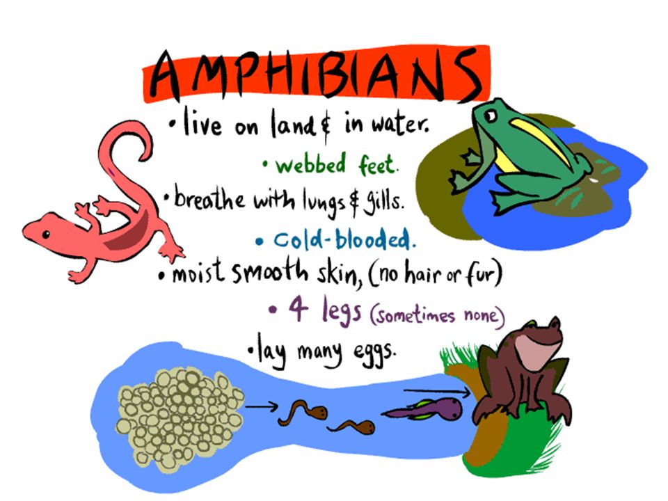
Amphibians Anura “no tail” Urodela “visible tail” Apoda “no legs” frogs and toads newts and salamanders caecilians Phylum Order

16
FROG DOMAIN _____________ KINGDOM _____________ PHYLUM ____________________________ CLASS _______________________________ ORDER _____________________________ ANIMALIA CHORDATA AMPHIBIA “double life” ANURA “without a tail” EUKARYA

17
External Anatomy

18
Nictitating membrane

19
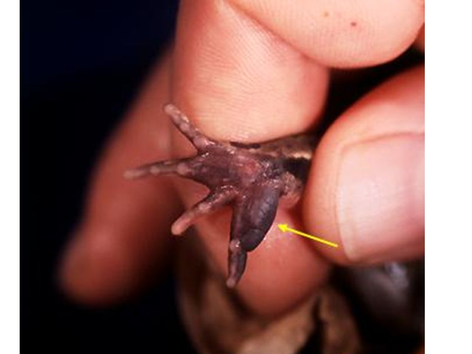
NO CLAWS

20
EXIT OPENINGS OPENING SHARED BY EXCRETORY, REPRODUCTIVE, & DIGESTIVE = ______________ VENT (cloaca)
")
21
What sex is it?

23
AMPLEXUS “firm embrace” Sperm and egg released @ same time and place Increases chances of fertilization

24
External Anatomy: The Oral Cavity

25
The TONGUE is attached at the front (not the back like yours!) When you first open the mouth, you will discover…
 When you first open the mouth, you will discover…")
26
Muscular Back of throat Pulls food into digestive system

27
Connect ears to back of throat

28
image from: http://www.spc.cc.tx.us/biology/jmckinney/Studyimages/frog/frogdissectlist.html GLOTTIS Opening to respiratory GULLET Opening to digestive

29
Internal Anatomy

30
Incision 1: Skin First Place your frog on its back (ventral side up) and pin it to the dissecting tray. Lift the frog’s skin with forceps between the rear legs near the cloaca. Make a small cut through the lifted skin with the scalpel. Take care to cut only the skin. Use the scissors to continue the incision up to the midline all the way through the frog’s skin. Stop cutting when your scissors reach the frog’s skin.

31
Incision 2: Skin Horizontal Use the scissors to make sideways incisions in the skin. The first incisions are made between the front legs. The next incisions are made just above the rear legs Be careful to only cut through the skin, not the muscle.

32
Incision 3: Separate Skin Pick up the flap of skin with the forceps. Use a scalpel to help separate the skin from the muscle layer below. After you've opened the flaps of skin, pin them to the dissection tray.

33
Image from; http://faculty.clintoncc.suny.edu/faculty/Michael.Gregory/files/Bio%20102/Bio%20102%20 Laboratory/frog%20dissection/frog%20dissection_files/frame.htm

34
Incision 4: First Muscle Incision Repeat the incisions, this time through the abdominal muscle. You will find it easier to begin the vertical incision by lifting the muscle layer with the forceps. Do this between the rear legs of the frog near the cloaca. Make a small cut with the scalpel. Using the scissors, continue the incision up the midline to a point just below the front legs. Be careful that you don't cut too deeply. The muscle is thin. It is easy to damage the organs underneath.

35
Incision 5: Chest Bone Cut through the chest bones. When you reach the point just below the front legs, turn the scissors blades sideways, so that you only cut through the bones in the chest. Be careful that you don't cut too deeply. This should prevent damage to the heart or other internal organs. When the scissors reach a point just below the frog's neck, you have cut far enough.

36
Incision 6: Muscle Horizontal Make the horizontal incisions. Just as you did with the skin, make a sideways incision in the muscle with the scalpel. Make the first incision between the front legs. The next incision is just above the rear legs. Again, be careful that you don't cut too deeply.

37
Incision 7: Muscle Separate Separate the muscle flaps from the organs below. Pull back and hold the muscle flaps with the forceps. Use the scalpel to separate the muscle from the organ tissue. Pin the muscle flaps back far enough to allow easy access to the internal organs.

38
Females may have black & white eggs

39
Incision 8: Triangular Flaps Pin the triangular flaps of the skin and muscle to the pan. Pick up the triangular flap of muscle that is just above the legs with the forceps. Use the scalpel, if needed, to help separate the muscle flap from the tissue underneath. Pin the flaps back far enough to allow access to the body cavity.

40
Organs 1: Introduction We are now ready to explore the frog's anatomy. To make our exploration easier, we will look at the organs in four different layers, beginning with the liver and heart layer. As we get deeper into the frog's anatomy, we will reveal new layers. We'll even explore the differences between male and female reproductive anatomy.

41
Organs 2: Liver When we pull back the muscles and skin, the first organs we can see are the liver and heart. The liver is a large, brownish colored organ covering most of the body cavity.

42
Organs 3: Heart You should also be able to see the heart in Layer 1. It is a small triangular shaped organ between the front legs, just above the liver. The frog’s heart has three chambers. The picture (right) shows only the lower chamber of the heart. Can you find all three chambers? How many chambers do you have in your heart?
 shows only the lower chamber of the heart. Can you find all three chambers. How many chambers do you have in your heart .")
43
Organs 4: Layer 2 Reveal layer two. The heart and liver in layer one hide some of the organs below them. Use the forceps and the probe to pick up the liver and reveal layer two. Layer two includes the gall bladder, the stomach, and the small intestine.

44
Organs 5: Gall Bladder Examine the gall bladder. Under the liver, we see a small, greenish sac. This is the gall bladder. You might also see it by separating the right and middle lobes of the liver. The gall bladder can be hard to find.

45
Organs 6: Stomach Examine the stomach. The stomach looks like a sac on the frog's left side (on your right). It is a large firm organ.
. It is a large firm organ..")
46
Organs 7: Small Intestine Examine the small intestine. The small intestine is a long, folded, tube like organ that is posterior the stomach. It is similar in color to the stomach, but smaller in diameter.

47
Organs 8: Layer 3 Reveal layer three. Remove the liver to see the organs in layer three. The liver is easier to remove if you remove the gall bladder and heart at this time. Place the organs in the appropriate circle on your dissection worksheet.

48
Organs 9: Lungs In this layer, we will take a close look at the lungs and pancreas. The lungs are difficult to locate in a preserved frog. They're at the anterior end of the body cavity on either side of the heart. Remove the lungs and place the organ in the appropriate circle on your dissection worksheet.

49
Organs 10: Pancreas You can't see the pancreas without lifting the stomach and intestines with the forceps. The pancreas is a thin, yellowish ribbon. The intestines are held in place by thin, transparent tissue called the mesentery.

50
Female Frogs If you have a female frog, you will need to remove the ovaries before you can see layer four. Place the organs in the appropriate circle on your dissection worksheet.

51
Organs 11: Layer 4 To see layer four, you need to remove the stomach, small intestine, large intestine, and pancreas. Place the organs in the appropriate circle on your dissection worksheet. In layer four, we'll look at the procedures required to see the different organs in both male and female frogs.

52
Organs 12: Spleen Examine the spleen. Locate the spleen in the male frog. It is a small, round reddish organ. It is a little more difficult to find the spleen in a female frog

53
Organs 12: Male Kidneys The kidneys are elongated, brownish colored organs found in the lower part of the frog's abdomen. The kidneys (Y) are situated on each side of the middle of the frog just underneath the testes (X). Female kidneys are located in the same place but can only been seen after removal of the ovaries and oviducts. Remove the organ and place in the appropriate circle on your dissection worksheet.
 are situated on each side of the middle of the frog just underneath the testes (X). Female kidneys are located in the same place but can only been seen after removal of the ovaries and oviducts. Remove the organ and place in the appropriate circle on your dissection worksheet..")
54
Organs 13: Frog Testes Locate the testes in the male frog. The testes are tan colored, bean shaped organs near the anterior end of each kidney. Remove the testes and place the organs in the appropriate circle on your dissection worksheet.

Similar presentations
>")


 – to hear 3. Hind Leg - jump 4. External Nare - breath.>")


 – to hear 3. Hind Leg - jump 4. External Nare - breath 5. Mouth - eat 6. Front Leg – movement 7. Nares.>")


