Download presentation
Presentation is loading. Please wait.

1
Role of cardiac CT in coronary artery diseases
Dr. Ahmed Refaey MBBCh, MS, FRCR

2
Format of the lecture Normal anatomy of coronary arteries
MSCT coronary angio Clinical application of CTA Illustrated cases

3
Coronary arteries anatomy

4
LCA “ left coronary artery “
Normally arises from the left sinus of Valsalva Courses posterior to the right ventricular outflow tract (RVOT), and bifurcates into the left anterior descending (LAD), and the left circumflex (LCX) branches.
, and bifurcates into the left anterior descending (LAD), and the left circumflex (LCX) branches.")
5
Right Coronary Artery (RCA)
Normally arises from the right coronary sinus (CS) and courses in the right AV groove toward the crux of the heart
 and courses in the right AV groove toward the crux of the heart.")
6
Of CAD Diagnosis Clinical Presentation ECG Echocardiography
Stress Test Thallium Study Coronary cathetrization Multislice Coronary CT Scan

7
Methods of imaging of coronary arteries

8
Coronary catheterization
Multislice cardiac CT

9
Coronary catheterization

11
CORONARY CATHETERIZATION
Advantages High resolution Option for intervention Disadvantages X-ray exposure Hospitalization Invasive complications

12
Figure 21.8d Copyright © 2009 Pearson Education, Inc., publishing as Pearson Benjamin Cummings

13
Multislice CT coronary angiography

14
What is Coronary CTA? Coronary CTA is a non-invasive minimal risk procedure to directly visualize the coronary arteries through administration of IV contrast It allows visualization of the coronary arteries similar to a cardiac catheterization with additional information about the WALL of the artery and composition of plaque (calcified or non-calcified)
")
15
Clinical application of CTA
Diagnosis of CAD * intermediate liklihood of disease * after equivocal/discordant stress imaging * coronary anomalies * before vascular surgury * nonischemic vs ischemic cardiomyopathy * acute chest pain * bypass graft patency / location

16
Patient Preparation No Caffeine for 12 hours prior to exam
Everyone gets Beta-Blockers (Verapamil can be substituted)
")
17
Goal Heart Rate < 60 bpm makes us happy

18
Contraindications Atrial Fibrillation Tachycardia
Beta Blockade Contraindication Heart Block Renal Failure (Creat>1.5) Contrast Allergy
 Contrast Allergy.")
19
The Examination

20
Computed Tomography (CT)
X-ray tube and detector rotate around the patient, transversal slices are constructed following each rotation by computer

21
continuous scanning instead separated slices
Spiral multislice CT continuous scanning instead separated slices

22
Entire heart imaged in 5-15 seconds
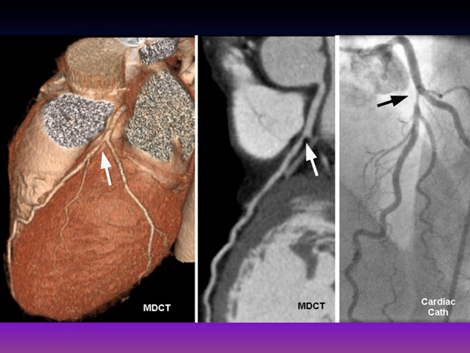
CT images that are used come from mid to end diastole due to relative motion free period

23
CT Angiography

24
Timing

25
CT-Angio Advantage Excellent for Coronary vessel, bypass vessels, LV wall thickness and function, cardiac anatomy and pericardium assessment

26
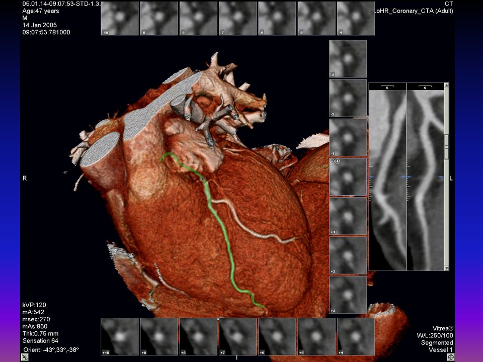
Coronary Vessel Analysis

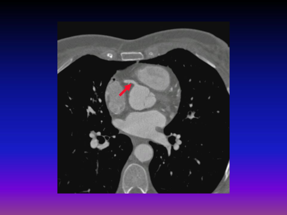
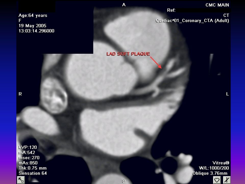
27
Maximum Intensity Projection Soft Plaque in Proximal LAD

28
Curved Planar Image

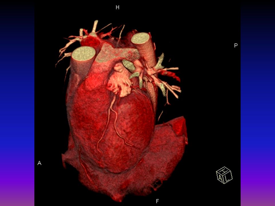
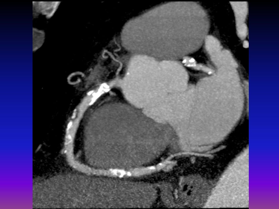
31
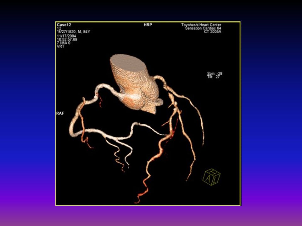
3-D Volume Rendered Image

33
Effective Radiation Doses for Various Tests
Bone Density mSv CXR: mSv Mammogram: mSv CT of the head: mSv CT colonoscopy mSv CT of the abdomen: mSv Stress Gated Myocardial Perfusion Scan SPECT: mSv CT chest: mSv MSCT angiogram: mSv Coronary angiography: 30mSv CT chest/abd/pelvis: mSv Dose allowed for radiological personnel: 20 mSv/year

34
CLINICAL APPLICATION OF CARDIAC CT ANGIO
Examine plaque components Evaluate coronary vessels Evaluate stent patency Assess cardiac function

35
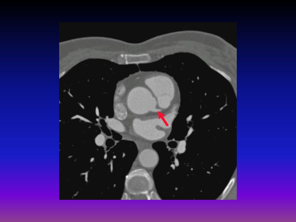
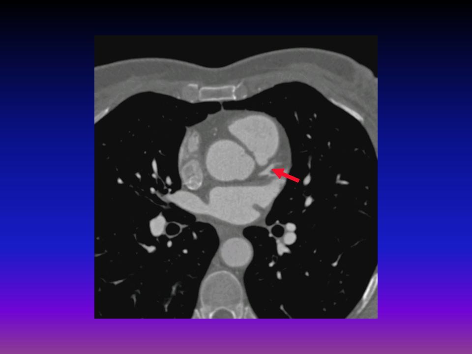
Examine plaque components

36
Plaque Characterization
Calcified vs. Soft Plaque composition rather than the degree of lumen stenosis determines the risk of plaque rupture. Vulnerable or “high-risk” plaques have thin fibrous cap with extracellular lipid core. Not visible by catheterization, but is being explored with CT angio. Plaques initially grow extrinsic and bulge adventitia, then grow into the lumen resulting in stenosis

38
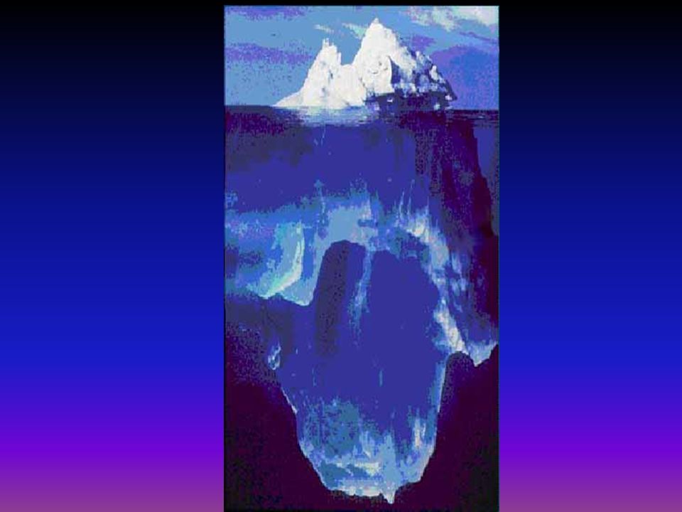
Coronary Artery Plaque:
approximate amounts of lipid rich, fibrotic and calcified plaque Fibrotic & Calcified 20% 66% The “Tip of the Atherosclerotic Iceberg” Fibrotic 80% 33% Lipid Rich

40
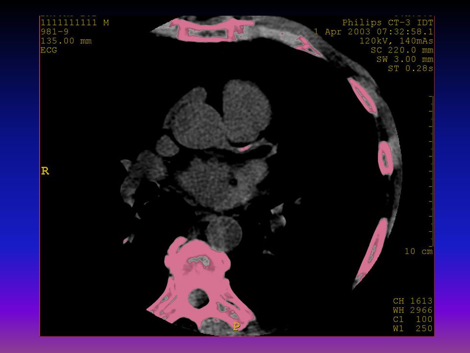
What does coronary calcification mean?
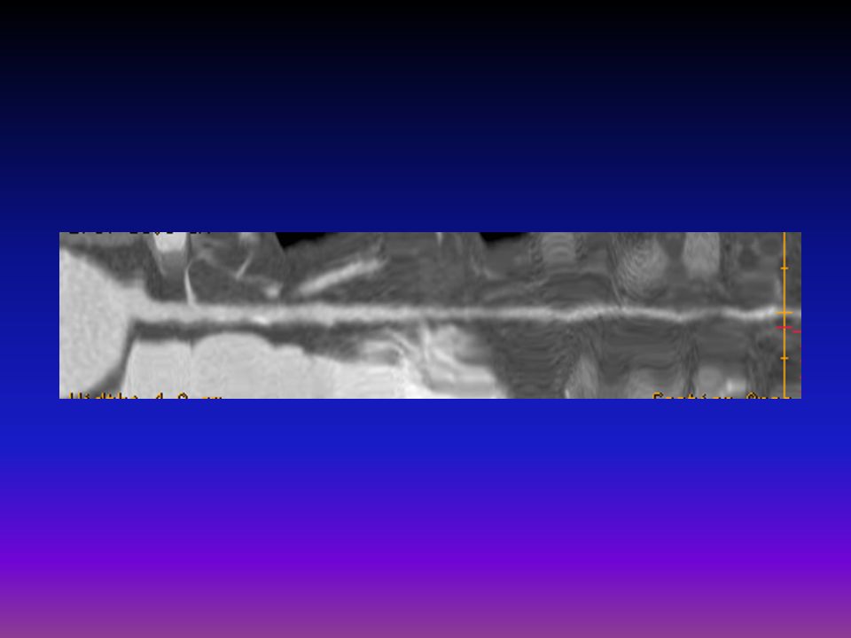
Calcium score correlates extremely well with coronary event risk If multi-vessel CAC, then risk increases Zero calcification suggests a very low probability of obstructive disease Curved MPR reformatted image of Right Coronary 17

41
Calcium Scoring “ Agatston score”

42
The Calcium Scale 1–99 mild 100–400 moderate >400 severe
The calcium scale is a linear scale with 4 calcium score categories: 0 none 1– mild 100– moderate > severe *Calcium score correlates directly with risk of events and likelihood of obstructive CAD*

45
Agatston-90

47
Examples of Coronary Artery Scans
NO CALCIFICATION MODERATE CALCIFICATION SIGNIFICANT CALCIFICATION Images courtesy of HeartScan San Frasco 15

48
Coronary Artery Calcium Scans
Task: Detect Calcium in Coronary Artery 130 kVp 625 mA .1 sec 3 mm

49
Coronary Artery Calcium Scans

50
Coronary Artery Calcium Scans

51
Coronary Artery Calcium Scans

52
Coronary Artery Calcium Scans

53
Coronary Artery Calcium Scans

54
Coronary Artery Calcium Scans

55
Coronary Artery Calcium Scans

56
Coronary Artery Calcium Scans

57
Coronary Artery Calcium Scans

58
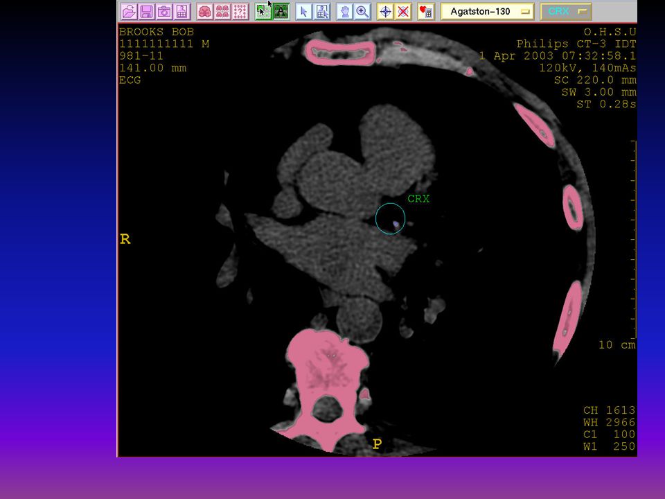
Calcification in LAD 13

59
Calcification in RCA 15

60
EVALUATING CORONARY VESSELS

61
It can look even better than a conventional angiogram

62
Left Main Coronary Artery

63
Left Main, LAD, & Circumflex
Obtuse Marginal

64
Diagonal Branch off LAD

65
Right Coronary Artery Acute Marginal Right Coronary Artery Sinoatrial

66
Right Coronary Artery

67
Evaluate stent patency

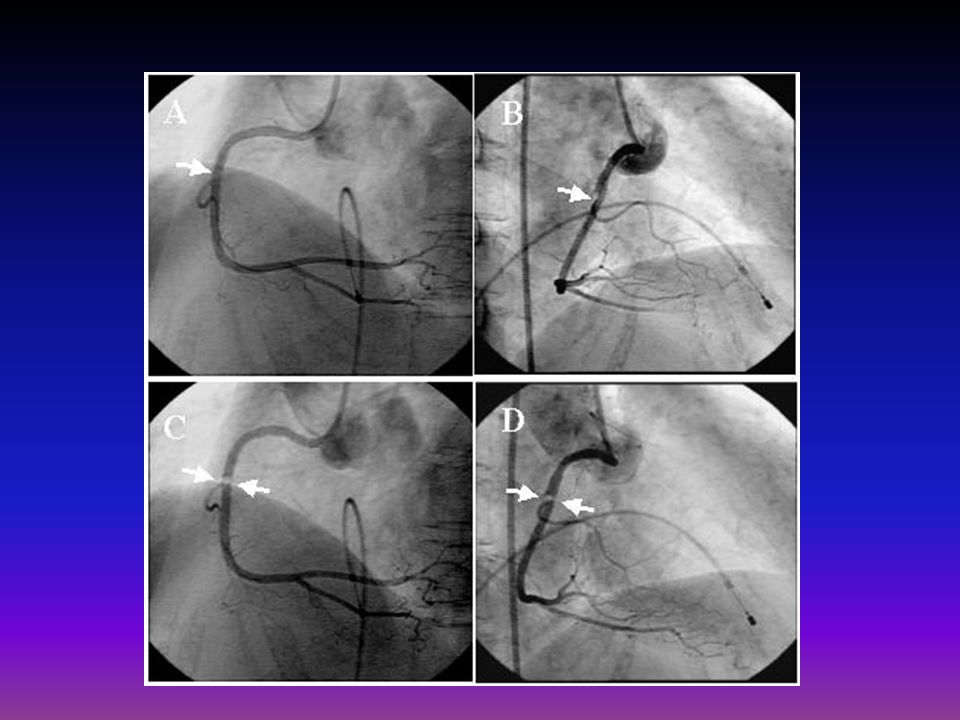
69
LAD Stent from Top to Bottom (1 mm)
")
70
LAD Stent from Front to Back (1 mm)
")
71
Cardiac function Recent studies show good correlation between function parameters derived from MDCT and levocardiography. DETERMINING EJECTION FRACTION

72
FUTURE OF CARDIAC CT One-stop shopping—
( cardiac function, coronary artery evaluation, plaque analysis, calcium quantification.) Non-invasive
 Non-invasive.")
73
Illustrated cases

74
High-resolution Imaging
1 LM LAD 1 2 2 3 4 3 RCA LCx LM 4 DSCT 74

75
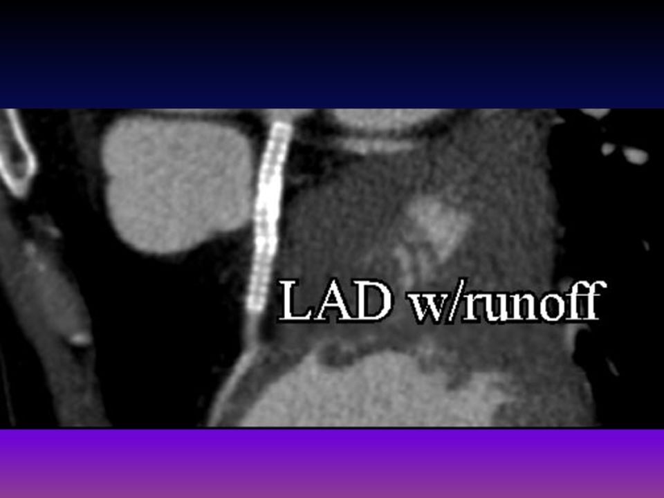
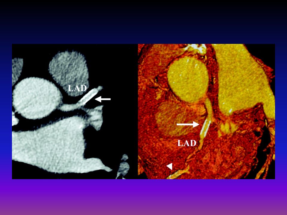
Significant stenosis of the left anterior descending artery

82
Soft Plaque Visualization

85
stent in LAD,LCx & RCA I

86
Aortic Coarctation Visualized
Fröhlich, G et al. Circulation. 2005;112:e81.

87
Pericardial Calcification Multi-Slice CT Scanning Superior to MRI
Hoffmann et al. Circulation 108 (7): 48e Figure IG1
: 48e Figure IG1.")
88
Mild CAD, and…

89
Pulmonary Emboli

90
Teaching Points

91
Cardiac Cath: Lumen only-no wall information. Evaluate stenosis
Cardiac Cath: Lumen only-no wall information. Evaluate stenosis. Cannot characterize plaque. Better delineates small vessels What is needed is a non-invasive, minimal-risk, outpatient procedure to detect early signs of CAD

92
Coronary CTA- Strengths
Noninvasive. Can measure HU of plaques and characterize them as fatty, atheroma, fibrosis, calcium. Can evaluate status of bypass grafts. Can determine stent patency. Evaluates portions of mediastinum and lungs.

93
Coronary CTA- Weaknesses
Cannot accurately measure stenosis with heavy, calcified plaque burden. Occlusions can be missed by brisk collateral flow.

94
What do I do with this information?
Reports will be classified in one of four categories of severity: Normal Mild Plaque with No stenosis Moderate Plaque with mild/Mod stenosis Severe Plaque and stenosis: Cardiac Cath

95
Thank you

Similar presentations


>")


>")
 Dynamic scanning implies 15 or more scans in rapid sequence within one.>")


 Electrons –Atomic particles –Have mass Wouldn’t a beam of particulate.>")


