Download presentation
Presentation is loading. Please wait.

1
The Urinary System

2
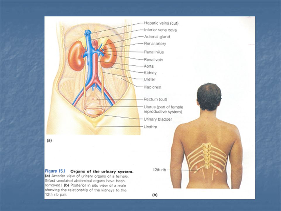
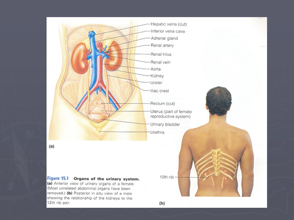
Kidney Small, dark red organs with a kidney-bean shape lie Retroperitonealy in superior lumbar region. against the dorsal body wall. The kidneys extend from the T12 to the L3 vertebra. The right kidney is positioned slightly lower than the left.

4
Adult kidney 12 cm (5 inches) long, 6 cm(2.5 inches) wide, and 3 cm (1 inches) thick. It is convex laterally and has a medial indentation called the hilus. Adrenal gland (part of endocrine system) is on the tope of each kidney.
 is on the tope of each kidney..")
6
Renal capsule (fibrous, transparent) encloses each kidney (glistening appearance). Adipose capsule (fatty mass) surrounds each kidney and holding it against trunk muscles.
 surrounds each kidney and holding it against trunk muscles..")
8
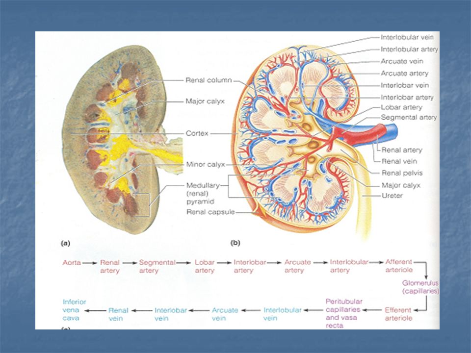
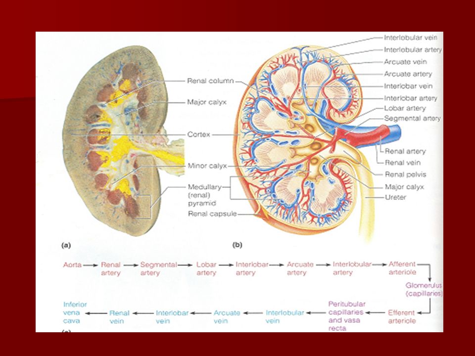
Renal cortex (outer region, light in color). Renal medulla (darker reddish-brown area). medullary pyramids (basically triangular regions with a striped appearance). medullary pyramids (basically triangular regions with a striped appearance). the pyramids are separated by extensions of cortex-like tissue, the renal columns. the pyramids are separated by extensions of cortex-like tissue, the renal columns. Renal pelvis is a flat, basin like cavity medial to the hilus. Calyces are extension of the pelvis, form cup-shaped areas that enclose the tips of the pyramids
. medullary pyramids (basically triangular regions with a striped appearance). the pyramids are separated by extensions of cortex-like tissue, the renal columns. the pyramids are separated by extensions of cortex-like tissue, the renal columns. Renal pelvis is a flat, basin like cavity medial to the hilus. Calyces are extension of the pelvis, form cup-shaped areas that enclose the tips of the pyramids.")
10
Blood Supply Kidneys have a very rich blood supply. Each minute, one-quarter of the total blood supply of the body passes through the kidneys. Renal artery-Segmental-lobar- interlobar - arcuate - interlobular arteries. Venous blood draining from the kidney flows through veins that trace the pathway of the arterial supply but in a reverse direction.

12
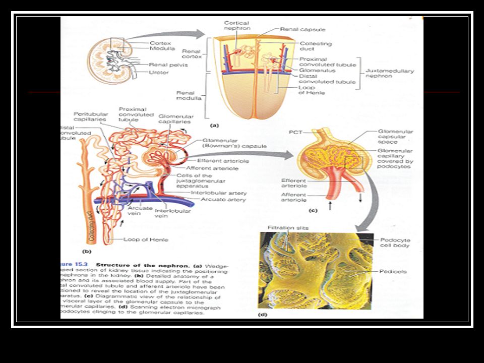
Nephron Are structural and functional units of the kidneys. Glomerulus & Renal tubules. Glomular or Bowman’s capsule, it is a closed end of the renal tubule is enlarged and cup- shaped and surrounds the Glomerulus.

13
The rest of the tubule is about 3 cm (1.25 inches). Proximal convoluted tubule (PCT), loop of Henle, distal convoluted tubule (DCT). Cells of PCT are covered with dense microvilli, which increases their surface area.
, loop of Henle, distal convoluted tubule (DCT). Cells of PCT are covered with dense microvilli, which increases their surface area..")
15
Cortical nephrons. Juxtamedullary nephrons. Collecting duct, it receives urine from many nephrons running through the medullary pyramids givining them their stripped appearance Two capillary sets: Glomerulus - peritubular capillary bed.

16
Glomerulus: Afferent arteriole (feeder vessel)- Efferent arteriole. Specialized for filtration high-pressure

17
Peritubular capillary bed. arises from efferent arterioles Specialized for absorption low-pressure drain in to interlobular veins.

19
Ureters,Urinary Bladder, and Urethra

21
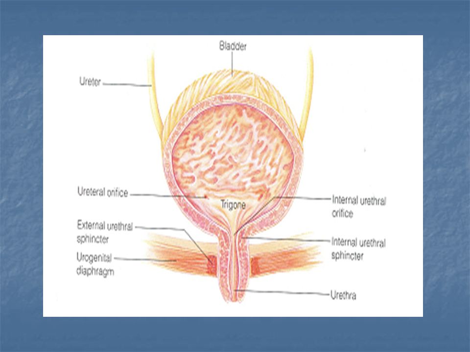
Ureters Slender tubes- 25 to 30 cm (10-12 inches) long, and 6 mm (1/4 inches) in diameter. Each ureter runs behind the peritoneum from the hilus of the kidney to the posterior aspect of the bladder, which enters at a slight angles. The ureters are passgeways that carry urine from kidneys to the bladder. Gravity, and peristalsis movement of the ureter are helping urine drain to the bladder. Small valve like folds of bladder mucosa is preventing urine from flowing back into ureter.

22
Urinary Bladder A smooth, collapsible, muscular sac that stores urine temporarily. It is located retroperitoneally in the pelvis. Two ureter opening and the single opening of the urethra. Trigone is the smooth triangular region of the bladder base outlined by these three openings. The bladder wall contains three layers of smooth muscle (detrusor muscle), and it ’ s mucosa (transitional epithelium).
, and it ’ s mucosa (transitional epithelium)..")
24
It is collapsed, 5 to 7.5 cm (2 to 3 inches) long, and it ’ s wall are thick and thrown into folds. It is collapsed, 5 to 7.5 cm (2 to 3 inches) long, and it ’ s wall are thick and thrown into folds. A moderately full bladder is about 12.5 cm (5 inches) long and holds 500 ml. Distended bladder becomes firm and pear-shaped may be felt above the pubic symphysis.
 long, and it ’ s wall are thick and thrown into folds. A moderately full bladder is about 12.5 cm (5 inches) long and holds 500 ml. Distended bladder becomes firm and pear-shaped may be felt above the pubic symphysis..")
25
Urethra The urethra is a thin-walled tube that carries urine by peristalsis from the bladder to the out side of the body. Internal urethral sphincter (a thickening of the smooth muscle,an involuntary sphincter that keeps the urethra closed. External urethral sphincter is fashioned by skeletal muscle as the urethra passes through pelvic floor.

27
In females, it is 3 to 4 cm (1 1/2 inches) long. Lies anterior to vaginal opening. It ’ s function is to conduct urine In male, it is 20 cm (8 inches) long Prostatic Membranous Spongy, penile It ’ s function is to conduct urine, also it provides the passage-ways through which the sperm is ejected from the body.
 long Prostatic Membranous Spongy, penile It ’ s function is to conduct urine, also it provides the passage-ways through which the sperm is ejected from the body..")
Similar presentations


 2 Ureters (Passage tubes ?) Bladder (Storage) Urethra (excretion)>")


