Download presentation
Presentation is loading. Please wait.

1
Cell Structure and Function

2
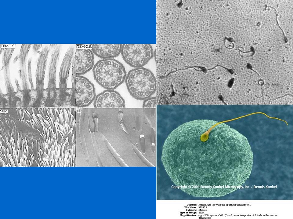
Cell Size 1. 1–100µm 2. Why is there a limit to cell size?
a. Surface-to-volume ratio b. Distance from surface to center

3
diameter of DNA double helix
tallest trees adult human chicken egg frog embryo most eukaryotic cells mitochondrion Figure: 04-01 Title: Relative sizes. Caption: Dimensions commonly encountered in biology range from about 100 meters (the height of the tallest redwoods) through a few micrometers (the diameter of most cells) to a few nanometers (the diameter of many large molecules). most bacteria virus proteins diameter of DNA double helix atoms
 through a few micrometers (the diameter of most cells) to a few nanometers (the diameter of many large molecules). most bacteria. virus. proteins. diameter of DNA. double helix. atoms.")
4
Cell types Prokaryotic—no nucleus, circular DNA, ribosomes
Eukaryotic—larger, nucleus, linear chromosomes, membranous organelles

5
Prokaryotic Cells Have no membrane-bound organelles
Include true bacteria On earth 3.8 million years Found nearly everywhere Spores in each breath; intestines Naturally in soil, air, hot springs

6
nucleoid (DNA) ribosomes food granule cytoplasm cell wall
prokaryotic flagellum Figure: 04-10 Title: A generalized prokaryotic cell. Caption: plasma membrane cytoplasm cell wall

7
Eukaryotic Cells Have numerous internal structures
Various types & forms Plants, animals, fungi, protists Multicellular organisms

8
nuclear pore chromatin (DNA) nucleus nucleolus nuclear envelope
flagellum intermediate filaments cytoplasm plasma membrane rough endoplasmic reticulum ribosome lysosome microtubules Figure: 04-02 Title: A generalized animal cell. Caption: smooth endoplasmic reticulum Golgi complex free ribosome vesicle mitochondrion vesicle

9
(part of cytoskeleton)
microtubules (part of cytoskeleton) mitochondrion chloroplast Golgi complex central vacuole smooth endoplasmic reticulum vesicle cell wall rough endoplasmic reticulum plasma membrane Figure: 04-03 Title: A generalized plant cell. Caption: nucleolus nuclear pore nucleus chromatin nuclear envelope intermediate filaments ribosomes free ribosome
 mitochondrion. chloroplast. Golgi complex. central vacuole. smooth endoplasmic. reticulum. vesicle. cell wall. rough endoplasmic. reticulum. plasma. membrane. Figure: Title: A generalized plant cell. Caption: nucleolus. nuclear pore. nucleus. chromatin. nuclear envelope. intermediate. filaments. ribosomes. free ribosome.")
10
Eukaryotic Cell Structure
Cytoplasm is the clear, gelatinous fluid inside of a cell

11
Eukaryotic cell structure
Nucleus is control center of the cell 1. Membrane bound (nuclear envelope) 2. Contains nucleoli; synthesizes ribosomal RNA 3. DNA in chromosomes (DNA and proteins)
 2. Contains nucleoli; synthesizes ribosomal RNA. 3. DNA in chromosomes (DNA and proteins)")
12
nuclear envelope nucleolus nuclear pores chromatin Figure: 04-04a
Title: The nucleus. Caption: (a) The nucleus is bounded by a nuclear envelope. Inside are chromatin (DNA and associated proteins) and a nucleolus. chromatin
 The nucleus is bounded by a nuclear envelope. Inside are chromatin (DNA and associated proteins) and a nucleolus. chromatin.")
13
nucleus nuclear pores Figure: 04-04b Title: The nucleus. Caption:
(b) An electron micrograph of a yeast cell that was frozen and broken open to reveal its internal structures. The large nucleus, with nuclear pores penetrating its nuclear envelope, is clearly visible.
 An electron micrograph of a yeast cell that was frozen and broken open to reveal its internal structures. The large nucleus, with nuclear pores penetrating its nuclear envelope, is clearly visible.")
14
chromatin chromosome Figure: 04-05 Title: Chromosomes. Caption:
Chromosomes, seen here in a light micrograph of a dividing cell (on the right) in an onion root tip, are the same material (DNA and proteins) as the chromatin seen in nondividing cells adjacent to it, but in a more compact state. chromosome
 in an onion root tip, are the same material (DNA and proteins) as the chromatin seen in nondividing cells adjacent to it, but in a more compact state. chromosome.")
15
Eukaryotic cell structure
Organelles Endoplasmic reticulum consists of folded membranes attached to the nucleus Rough ER is site of protein synthesis and protein secretion

16
rough endoplasmic reticulum smooth endoplasmic reticulum
ribosomes 0.5 micrometers smooth endoplasmic reticulum Figure: 04-07 Title: Endoplasmic reticulum. Caption: There are two types of endoplasmic reticulum: rough ER, coated with ribosomes, and smooth ER, without ribosomes. Although in electron micrographs the ER looks like a series of tubes and sacs, it is actually a maze of folded sheets and interlocking channels. 0.5 micrometers vesicles

17
Eukaryotic Cell Structure
Organelles (cont.) Ribosomes assemble amino acid into polypeptide chains a. Associated with the ER b. Composed of RNA and proteins
 Ribosomes assemble amino acid into polypeptide chains. a. Associated with the ER. b. Composed of RNA and proteins.")
18
rough endoplasmic reticulum
ribosomes Figure: 04-07R-1 Title: Rough endoplasmic reticulum. Caption: rough endoplasmic reticulum 0.5 micrometers

19
smooth endoplasmic reticulum
vesicles Figure: 04-07R-2 Title: Smooth endoplasmic reticulum. Caption: smooth endoplasmic reticulum 0.5 micrometers

20
Eukaryotic Cell Structure
Organelles (cont.) Golgi apparatus are membranous sacs associated with ER a. Processing and transport of proteins, lipids b. Synthesis and transport of polysaccharides
 Golgi apparatus are membranous sacs associated with ER. a. Processing and transport of proteins, lipids. b. Synthesis and transport of polysaccharides.")
21
vesicles from ER vesicles leaving Golgi complex Golgi complex
Figure: 04-08 Title: The Golgi complex. Caption: The Golgi complex is a stack of flat membranous sacs. Vesicles transport material from the ER to the Golgi (and vice versa) and from the Golgi to plasma membrane, lysosomes, and vesicles. Departing vesicles bud off from the Golgi on one face; arriving vesicles join it on the opposite face.
 and from the Golgi to plasma membrane, lysosomes, and vesicles. Departing vesicles bud off from the Golgi on one face; arriving vesicles join it on the opposite face.")
22
Eukaryotic Cell Structure
Vacuoles: membrane-bound compartments that are temporary storage of materials Animal cells do not usually contain vacuoles, if they do they are very small Plant cells usually use them for water storage

23
Eukaryotic cell structure
Organelles (cont.) Lysosomes are Golgi-derived vesicles containing digestive enzymes Digest excess or worn out organelles, food particles, and engulfed viruses or bacteria
 Lysosomes are Golgi-derived vesicles containing digestive enzymes. Digest excess or worn out organelles, food particles, and engulfed viruses or bacteria.")
24
vesicles from ER vesicles leaving Golgi complex Golgi complex
Figure: 04-08 Title: The Golgi complex. Caption: The Golgi complex is a stack of flat membranous sacs. Vesicles transport material from the ER to the Golgi (and vice versa) and from the Golgi to plasma membrane, lysosomes, and vesicles. Departing vesicles bud off from the Golgi on one face; arriving vesicles join it on the opposite face.
 and from the Golgi to plasma membrane, lysosomes, and vesicles. Departing vesicles bud off from the Golgi on one face; arriving vesicles join it on the opposite face.")
25
Eukaryotic Cells: Organelles Energy sources for cell activities
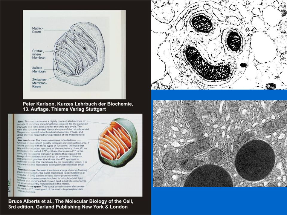
Mitochondria provide energy for cellular functions (respiration) a. Membrane bound, numerous b. Matrix/cristae c. Have their own DNA and ribosomes; self-replicate
 a. Membrane bound, numerous. b. Matrix/cristae. c. Have their own DNA and ribosomes; self-replicate.")
27
5 micrometers Figure: 04-E4-2d Title: An SEM photo. Caption:
(d) An SEM photo at much higher magnification, showing mitochondria, many of which are sliced open. 5 micrometers
 An SEM photo at much higher magnification, showing mitochondria, many of which are sliced open. 5 micrometers.")
28
Eukaryotic Cells: Organelles Energy sources for cell activities
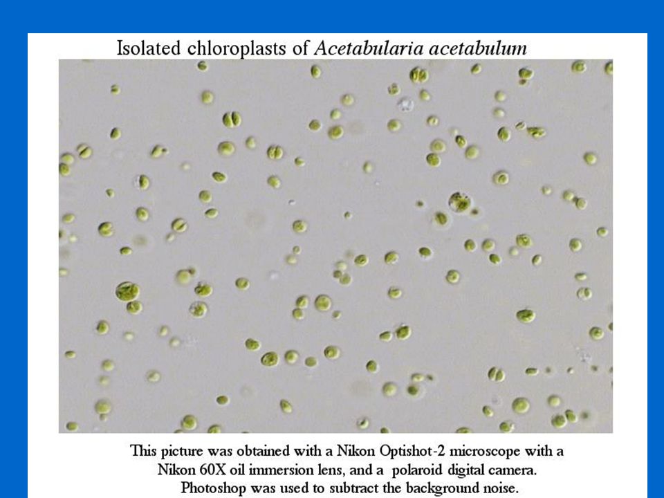
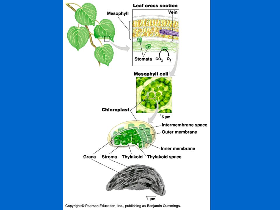
Chloroplasts—function in photosynthesis 1) Green—contain chlorophyll pigment 2) Stroma/grana (thylakoid stacks) 3) Have their own DNA and ribosomes; self-replicate 4) Up to 100 per cell
 Green—contain chlorophyll pigment. 2) Stroma/grana (thylakoid stacks) 3) Have their own DNA and ribosomes; self-replicate. 4) Up to 100 per cell.")
31
Eukaryotic Cells: Organelles
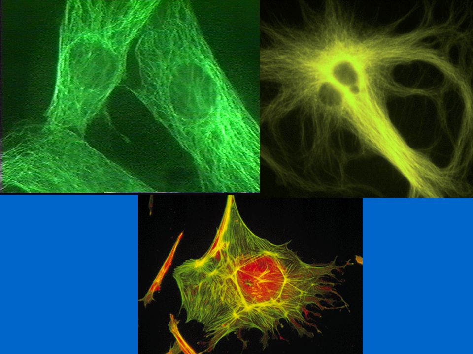
Cytoskeleton Internal infrastructure Surface structures extensions of the plasma membrane aid in movement of simple organisms

32
actin subunits subunit tubulin subunit Figure: 04-2 Title:
Cytoskeleton components. Caption: tubulin subunit

33
Cytoskeleton Microtubules-thin hollow cylinders made of protein
Microfilaments- smaller, solid protein fibers They work together to maintain the shape of the cell

36
Centrioles Organelles found in the cells of animals and most protists
Occur in pairs and are made up of microtubules Play an important role in cell division

37
Cilia and Flagella Cilia are short, numerous projections that look like hairs Flagella are longer projections that move with a whip-like motion Both are used for locomotion or feeding

38
Prokaryotes & Eukaryotes
Similarities & differences Both surrounded by plasma membrane, but very different Prokaryotes – Archaebacteria and Eubacteria Eukaryotes – everything else

39
Plant & Animal Cells Similarities
Both constructed from eukaryotic cells Both contain similar organelles Both surrounded by cell membrane

40
Plant & Animal Cells Differences Animals have Plants have
Cell wall – provides strength & rigidity Have chloroplasts, photosynthetic Animals have Other organelle not found in plants (lysosomes formed from Golgi) Centrioles, important in cell division
 Centrioles, important in cell division.")
41
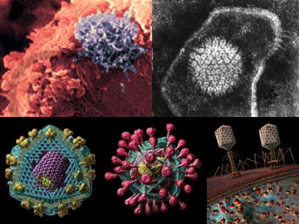
Brief Look at Viruses Viruses are acellular
Not considered to be living Cause serious diseases in most organisms

Similar presentations


 The McGraw-Hill Companies, Inc.>")


