Download presentation
Presentation is loading. Please wait.

1
Essentials of Human Anatomy & Physiology Copyright © 2003 Pearson Education, Inc. publishing as Benjamin Cummings Slides 15.1 – 15.20 Seventh Edition Elaine N. Marieb Chapter 15 The Urinary System Lecture Slides in PowerPoint by Jerry L. Cook

2
Functions of the Urinary System Elimination of waste products Nitrogenous wastes Toxins Drugs

3
Functions of the Urinary System Regulate aspects of homeostasis Water balance Electrolytes Acid-base balance in the blood Blood pressure (by producing the enzyme renin) Red blood cell production (by producing the hormone erythropoietin) Activation of vitamin D
 Red blood cell production (by producing the hormone erythropoietin) Activation of vitamin D")
4
Organs of the Urinary system Kidneys Ureters Urinary bladder Urethra

5
Location of the Kidneys Against the dorsal body wall At the level of T 12 to L 3 The right kidney is slightly lower than the left Attached to ureters, renal blood vessels, and nerves at renal hilus Atop each kidney is an adrenal gland

6
Coverings of the Kidneys Renal capsule Surrounds each kidney Glistening appearance Adipose capsule Surrounds the kidney Provides protection to the kidney Helps keep the kidney in its correct location

7
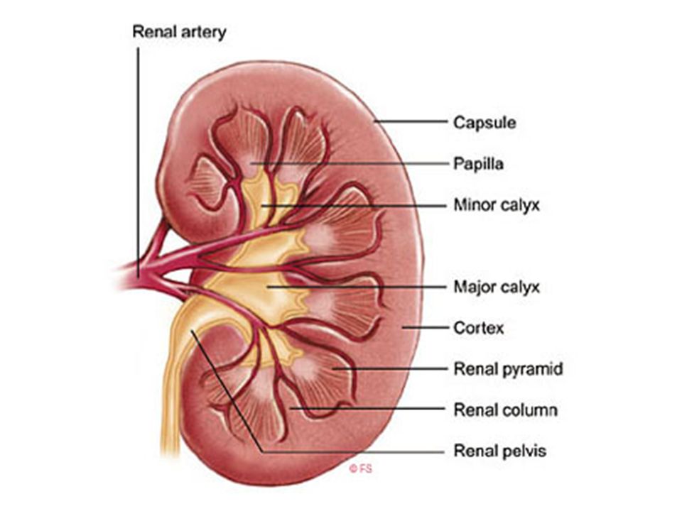
Regions of the Kidney Renal cortex – outer region Renal medulla – inside the cortex Renal pelvis – inner collecting tube

8
Kidney Structures Medullary pyramids – triangular regions of tissue in the medulla Renal columns – extensions of cortex- like material inward separating the medullary pyramids Calyces – cup-shaped structures that funnel urine towards the renal pelvis

10
Blood Flow in the Kidneys

11
Nephrons The structural and functional units of the kidneys Each kidney contains over a million nephrons Responsible for forming urine Main structures of the nephrons Glomerulus Renal tubule

12
Glomerulus A specialized capillary bed Attached to arterioles on both sides (maintains high pressure) Large afferent arteriole Narrow efferent arteriole
 Large afferent arteriole Narrow efferent arteriole")
13
Glomerulus The glomerulus sits within a glomerular (Bowman’s) capsule (the first part of the renal tubule) Capillaries are covered with podocytes from the renal tubule
 capsule (the first part of the renal tubule) Capillaries are covered with podocytes from the renal tubule")
14
Renal Tubule Glomerular (Bowman’s) capsule Proximal convoluted tubule Loop of Henle Distal convoluted tubule
 capsule Proximal convoluted tubule Loop of Henle Distal convoluted tubule")
15
Types of Nephrons Cortical nephrons Located entirely in the cortex Includes most nephrons

16
Types of Nephrons Juxtamedullary nephrons Found at the boundary of the cortex and medulla

17
Peritubular Capillaries Arise from efferent arteriole of the glomerulus Normal, low pressure capillaries Attached to a venule Cling close to the renal tubule Reabsorb (reclaim) some substances from collecting tubes
 some substances from collecting tubes")
18
Urine Formation Processes A. Filtration B. Reabsorption C. Secretion

19
Filtration Nonselective passive process Water and solutes smaller than proteins are forced through capillary walls Blood cells cannot pass out to the capillaries Filtrate is collected in the glomerular (Bowman’s) capsule and leaves via the renal tubule
 capsule and leaves via the renal tubule")
20
Reabsorption The peritubular capillaries reabsorb several materials Some water Glucose Amino acids Ions Some reabsorption is passive, most is active Most reabsorption occurs in the proximal convoluted tubule

21
Materials Not Reabsorbed Nitrogenous waste products Urea Uric acid Creatinine Excess water

22
Secretion – Reabsorption in Reverse Some materials move from the peritubular capillaries into the renal tubules Hydrogen and potassium ions Creatinine Materials left in the renal tubule move toward the ureter

23
Formation of Urine

24
Characteristics of Urine Used for Medical Diagnosis Colored somewhat yellow due to the pigment urochrome (from the destruction of hemoglobin) and solutes Sterile Slightly aromatic Normal pH of around 6 Specific gravity of 1.001 to 1.035
 and solutes Sterile Slightly aromatic Normal pH of around 6 Specific gravity of to 1.035")
25
Ureters Slender tubes attaching the kidney to the bladder Continuous with the renal pelvis Enter the posterior aspect of the bladder Runs behind the peritoneum Peristalsis aids gravity in urine transport

26
Urinary Bladder Smooth, collapsible, muscular sac Temporarily stores urine

27
Urinary Bladder Wall Three layers of smooth muscle (detrusor muscle) Mucosa made of transitional epithelium Walls are thick and folded in an empty bladder Bladder can expand significantly without increasing internal pressure
 Mucosa made of transitional epithelium Walls are thick and folded in an empty bladder Bladder can expand significantly without increasing internal pressure")
28
Urethra Thin-walled tube that carries urine from the bladder to the outside of the body by peristalsis Release of urine is controlled by two sphincters Internal urethral sphincter (involuntary) External urethral sphincter (voluntary)
 External urethral sphincter (voluntary)")
29
Urethra Gender Differences Length Females – 3–4 cm (1 inch) Males – 20 cm (8 inches) Location Females – along wall of the vagina Males – through the prostate and penis Function Females – only carries urine Males – carries urine and is a passageway for sperm cells
 Males – 20 cm (8 inches) Location Females – along wall of the vagina Males – through the prostate and penis Function Females – only carries urine Males – carries urine and is a passageway for sperm cells")
30
Micturition (Voiding) Both sphincter muscles must open to allow voiding The internal urethral sphincter is relaxed after stretching of the bladder Activation is from an impulse sent to the spinal cord and then back via the pelvic splanchnic nerves The external urethral sphincter must be voluntarily relaxed
 Both sphincter muscles must open to allow voiding The internal urethral sphincter is relaxed after stretching of the bladder Activation is from an impulse sent to the spinal cord and then back via the pelvic splanchnic nerves The external urethral sphincter must be voluntarily relaxed")
31
Developmental Aspects of the Urinary System Functional kidneys are developed by the third month Urinary system of a newborn Bladder is small Urine cannot be concentrated

32
Developmental Aspects of the Urinary System Control of the voluntary urethral sphincter does not start until age 18 months Urinary infections are the only common problems before old age

33
Aging and the Urinary System There is a progressive decline in urinary function The bladder shrinks with aging Urinary retention is common in males

Similar presentations