Download presentation
Presentation is loading. Please wait.

1
THE SKULL SIMPLY, AMAZING!

2
Most complex bony structure 22 bones in all Mostly flat bones, but not all!

3
Functions of Cranial Bones Enclose and protect the fragile brain and furnish attachment sites for head and neck muscles

4
Functions of Facial Bones 1. form framework of face 2. contain cavities for special sense organs 3. openings for food/air passage 4. secure the teeth 5. anchor the facial muscles of expression

5
ALL BONES OF THE SKULL ARE FIRMLY LOCKED IN PLACE BY JOINTS CALLED SUTURES Four major sutures

6
THE CRANIUM (8) 1 frontal bone 2 parietal bones 2 temporal bones 1 occipital bone 1 sphenoid bone 1 ethmoid bone
 1 frontal bone 2 parietal bones 2 temporal bones 1 occipital bone 1 sphenoid bone 1 ethmoid bone")
7
THE FRONTAL BONE

8
Parietal Bones: Form most of the superior and lateral aspects of the skull Figure 7.3a

9
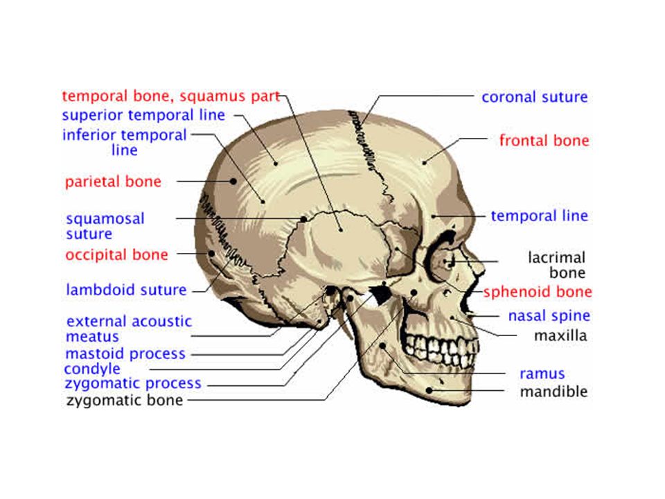
Parietal Bones and Major Associated Sutures Four sutures mark the articulations of the parietal bones –Coronal suture – articulation between parietal bones and frontal bone anteriorly –Sagittal suture – where right and left parietal bones meet superiorly –Lambdoid suture – where parietal bones meet the occipital bone posteriorly –Squamosal or squamous suture – where parietal and temporal bones meet

10
Occipital Bone and Its Major Markings Forms most of skull’s posterior wall and base Major markings include the posterior cranial fossa, foramen magnum, occipital condyles, and the hypoglossal canal Figure 7.2b

11
Temporal Bones Form the inferolateral aspects of the skull and parts of the cranial floor Divided into four major regions – squamous, tympanic, mastoid, and petrous Major markings include the zygomatic, styloid, and mastoid processes, and the mandibular and middle cranial fossae

12
Temporal Bones Major openings include the stylomastoid and jugular foramina, the external and internal auditory meatuses, and the carotid canal

13
Temporal Bones Figure 7.5

14
Sphenoid Bone Spans the width of the middle cranial fossa. Has a butterfly like shape. Articulates with all other cranial bones.

15
Markings and regions to know: Sphenoid sinuses Hypophyseal fossa: a snug enclosure for the pituitary gland. Greater and Lesser Wings Optic Canals: (opening for optic nerves) Superior Orbital fissures: long slit between greater and lesser wings, allows cranial nerves to control eye movement. Pterygoid processes: anchors muscles used in chewing Foramen rotundum and Foramen ovale
 Superior Orbital fissures: long slit between greater and lesser wings, allows cranial nerves to control eye movement. Pterygoid processes: anchors muscles used in chewing Foramen rotundum and Foramen ovale.")
19
Ethmoid Bone Lies between the sphenoid and nasal bones of the face. Most deeply situated. Markings/Regions Cribriform plates: help form roof of nasal cavities and cranial fossa. Olfactory foramina: allow olfactory nerves to pass from nasal cavities to the brain. Crista galli Ethmoid sinuses

24
Maxillary Bones Medially fused bones that make up the upper jaw and the central portion of the facial skeleton Facial keystone bones that articulate with all other facial bones except the mandible Their major markings include palatine, frontal, and zygomatic processes, the alveolar margins, inferior orbital fissure, and the maxillary sinuses

25
Maxillary Bone Figure 7.8b

26
Zygomatic bones Paired bones aka Cheek bones Form prominences of the cheeks and parts of the inferolateral margins of orbits. Articulates with temporal, frontal, and maxillary bones.

29
Nasal Bones Form bridge of nose Articulates with frontal, maxillary, and ethmoid bones.

31
Lacrimal Bones Located in the medial wall of the orbits. Articulates with the frontal, ethmoid, and maxillary bones.

33
Palatine Bones Complete the posterior part of the hard palate. Forms part of the posterolateral walls of the nasal cavity. Small part of the orbits.

35
The Vomer Forms part of the nasal septum dividing the nasal cavity in left and right halves.

36
The Inferior Nasal Conchae Thin curved bones in the nasal cavity. Forms the lateral wall of the nasal cavity.

39
Application Question Mr. and Mrs. Walkabots have recently given birth to a beautiful baby girl, Zeplin. After a few months of great joy they notice that their daughter vomits often, sleeps a lot, is irritable all the time and cannot look them in the eye. Zeplin does not meet developmental milestones over the next six months. HELP!

Similar presentations