Download presentation
Presentation is loading. Please wait.

1
UROGENITAL SYSTEM

2
The kidney starts out as the pronephros, a structure similar to that found in primitive vertebrates, followed by the mesonephros, a more advanced system found in fish and amphibia, and finally forms the metanephros which elaborates into the final human form.

3
The pronephros The pronephros does not function, but it marks at the fourth week of development the start of renal development. This first stage forms in the cervical region of the embryo, at the cranial end of the nephrogenic cord.

5
The mesonephros The mesonephros appears at the end of the fourth week, developing caudal to the degenerating pronephros.. This tissue forms a column in the posterior body wall extending cranially from the cervical region. It contains a primary duct, the mesonephric duct, which opens into the urogenital sinus. Glomeruli are present with short tubules connected to the mesonephric duct.

6
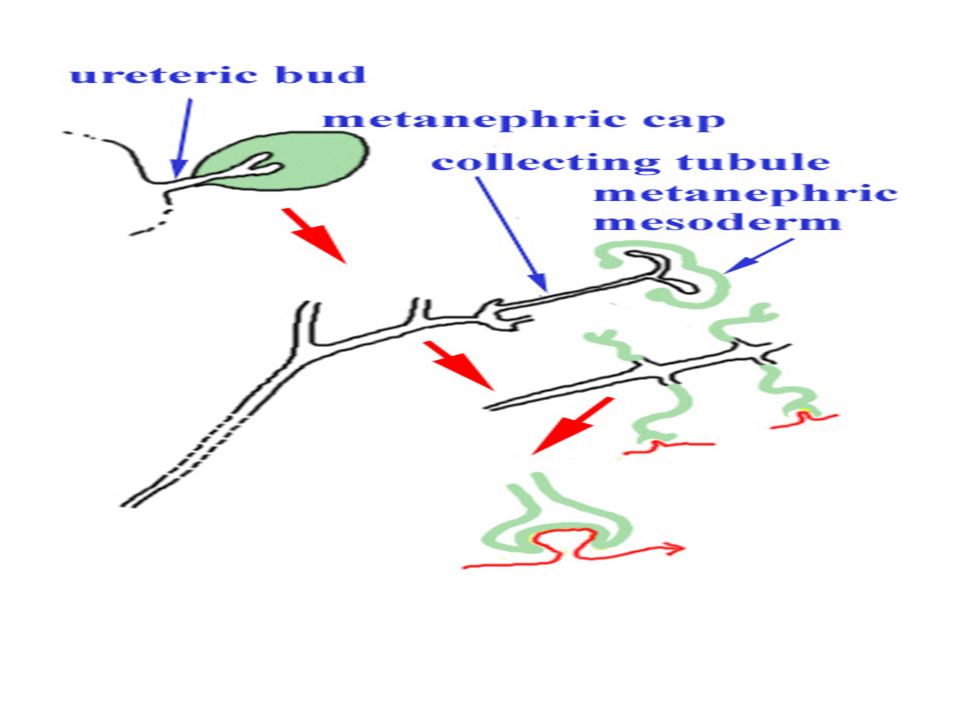
The metanephros The final kidney begins to develop about the fifth week and is fully functional by about the ninth week of development. Development is initiated by the growth of a bud from the base of the mesonephric duct. The metanephric or ureteric bud grows into the metanephric mesoderm. The ureteric bud induces development of the kidney from mesoderm which condenses around it.

8
. As growth proceeds the ureteric bud elongates to keep pace with body growth. In relative terms the kidney ascends out of the pelvis into the abdomen. The ureteric bud bifurcates to form the major calyces, and further subdivides to form the minor calyces and collecting ducts. The metanephric mesoderm differentiates into metanephric tubules which at first are not connected to the collecting ducts.

9
One end of each metanephric tubule becomes invaginated by a glomerulus, while the other connects with a collecting duct. The ureteric bud is essential for induction of differentiation in the metanephric mesoderm. The metanephric cap is essential for bifurcation of the ureteric bud. The collecting ducts are essential for differentiation of the nephrons.

10
Congenital anomalies During the long process of renal development, problems can arise at any stage: Renal agenesis Abnormal rotation and ectopic kidneys Horseshoe kidney Congenital polycystic disease

13
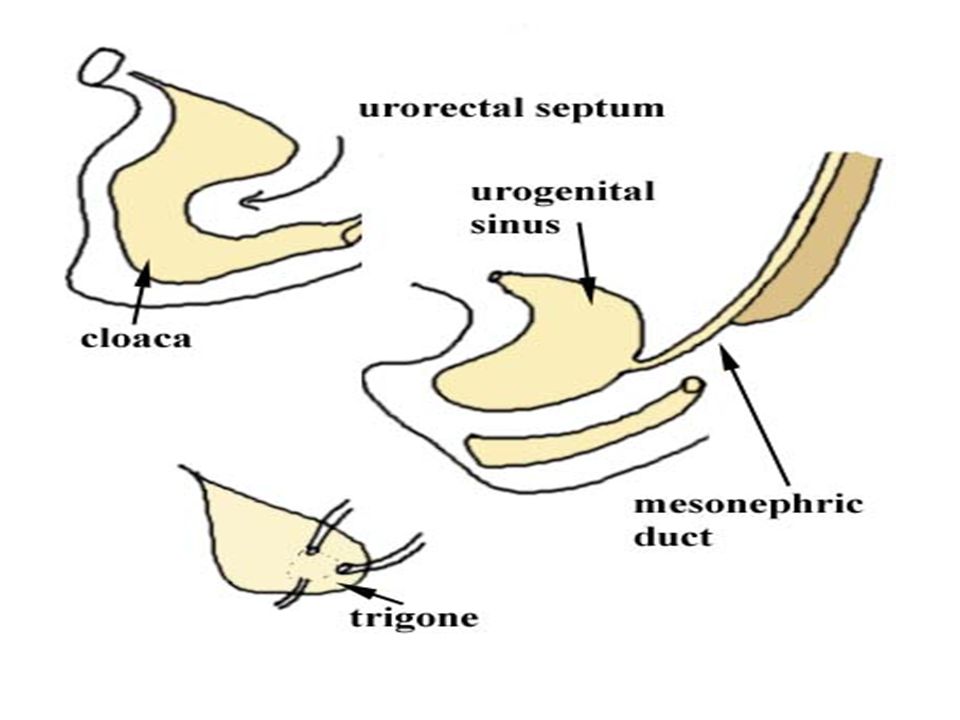
The Bladder The bladder is derived from two sources, the cloaca and mesonephric ducts. The primitive cloaca is divided by the urorectal septum into the urogenital sinus and rectum. The bladder largely develops from the vesicle part of the urogenital sinus. The mesonephric ducts are drawn into the floor of the bladder as it expands, to form the trigone.

16
Reproductive Development Sex determination occurs first in the gonads and is followed by appropriate development of the ducts, glands and associated genitalia.

17
Up until the seventh week of development the gonads are undifferentiated. The undifferentiated gonad has developed from the mesenchyme and mesothelium over the mesonephros. Proliferation of the mesothelial cells forms the gonadal ridge. Primordial germ cells migrate from the wall of the yolk sac into the undifferentiated gonad and become associated with finger- like processes produced by proliferation of the mesothelium, the primary sex cords

18
Further development of the primary sex cords results in differentiation of an outer cortex and inner medulla. The medulla disappears in females and the cortex develops into the ovary. In female embryos the undifferentiated gonad takes longer than in the male to begin differentiation and to become a recognizable ovary. The initial development of primary sex cords results in the formation of insignificant and temporary structures, the rete ovarii.

19
Secondary sex cords subsequently develop from the surface epithelium. The primordial germ cells which have migrated from the yolk sac migrate into the secondary sex cords. The secondary sex cords then break up into clusters of cells surrounding primordial germ cells, the primordial follicles. Division of primordial germ cells and the production of all primordial follicles occurs before birth. Once formed the ova of primordial follicles are arrested at the first prophase of meiotic division.

20
The Fallopian Tubes and Uterus In female embryos the mesonephric ducts regress. The paramesonephric ducts come together in the median plane and fuse into the uterovaginal primordium. The dilated free ends of the tubes open into what will eventually be the peritoneal cavity.

21
The uterovaginal primordium invaginates the dorsal surface of the urogenital sinus to become the epithelium and glands of the uterus. The fallopian tubes form from the unfused portions of the paramesonephric ducts. Failure of the paramesonephric ducts to fuse results in a double uterus. Partial fusing results in the formation of a bicornuate uterus (two horned).
..")
22
Genitalia The development of the external genitalia follows the development of the gonads in that at first they are undifferentiated. In the fourth week a genital tubercle develops ventral to the cloacal membrane, associated with paired labioscrotal swellings and urogenital folds.

23
The genital tubercle elongates to form a phallus with a urethral groove on its ventral surface. In the female the phallus becomes the relatively small clitoris, without fusion of the urogenital folds. The urethra therefore opens directly through the cloacal membrane. The urogenital folds and labioscrotal swellings do not fuse across the midline and remain to form the labia minora and majora.

24
Male Reproductive Development The Testes As in the female, the initial stages of development begin in the fifth week with development of the mesoderm forming the gonadal ridge. Within the ridge the structure is formed into a cortex and medulla. At this stage the gonad is indifferent.

25
. By the sixth week primordial sex germ cells migrate from the yolk sac to seed the sex cords of the gonadal cortex. The testes development factor of the Y chromosome directs the undifferentiated gonad to proceed through a series of changes resulting in the development of testes. An early step in differentiation is the development of a thick capsule, the tunica albuginea separating the sex cords from the outer cortex.

26
. The sex cords extend into the medulla to form the seminiferous tubules, tubuli recti, and rete testis. Mesenchyme develops between the epithelial seminiferous tubules. By about eight weeks the mesenchymal cells differentiate into interstitial cells and start to produce testosterone. Testosterone promotes deverlopment of the male genitalia. Mullerian inhibiting hormone secreted by Sertoli cells of the seminiferous tubules suppresses development of the paramesonephric ducts which in females contribute to formation of the fallopian tubes and uterus.

27
The Prostate The prostatic urethra gives rise to endodermal ducts wich grow into the surrounding mesenchyme. The glands of the prostate differentiate from the ducts and the stroma of the prostate develops from the associated mesenchyme.

28
Genitalia The external genitalia develop under the influence of testosterone produced by the testes. The penis develops by growth of the indifferent phallus. On its ventral aspect a urethral groove develops. In the male testosterone promotes elongation of the phallus into the penis and the urogenital folds fuse and cause the urethra to be incorporated into the corpus spongiosum, with the urethral orifice at the glans penis.

29
The labioscrotal swellings grow towards each other and fuse to form the scrotum. Descent of the testes through the inguinal canals begins during the twenty-eighth week and takes about three days. By thirty-two weeks the testes have entered the scrotum.

Similar presentations