Download presentation
Presentation is loading. Please wait.

1
Neural Communication Biological Psychology Phrenology (Franz Gall)
branch of psychology concerned with the links between biology and behavior some biological psychologists call themselves behavioral neuroscientists, neuropsychologists, behavior geneticists, physiological psychologists, or biopsychologists Phrenology (Franz Gall) Study of the bumps on your head Bumps reveal a person’s abilities and traits
 Study of the bumps on your head. Bumps reveal a person’s abilities and traits.")
2
Phrenology

3
Neural Communication Neuron Soma a nerve cell the basic building
block of the nervous system Soma cell body; serves as neuron’s control center

4
Neural Communication Dendrite Axon Myelin [MY-uh-lin] Sheath
the bushy, branching extensions of a neuron that receive messages and conduct impulses toward the cell body Axon the extension of a neuron, ending in branching terminal fibers, through which messages are sent to other neurons or to muscles or glands Myelin [MY-uh-lin] Sheath a layer of fatty cells segmentally encasing the fibers of many neurons enables vastly greater transmission speed of neutral impulses
![Neural Communication Dendrite Axon Myelin [MY-uh-lin] Sheath](http://slideplayer.com/slide/772957/2/images/4/Neural+Communication+Dendrite+Axon+Myelin+%5BMY-uh-lin%5D+Sheath.jpg "the bushy, branching extensions of a neuron that receive messages and conduct impulses toward the cell body. Axon. the extension of a neuron, ending in branching terminal fibers, through which messages are sent to other neurons or to muscles or glands. Myelin [MY-uh-lin] Sheath. a layer of fatty cells segmentally encasing the fibers of many neurons. enables vastly greater transmission speed of neutral impulses.")
5
Neural Communication

6
Neural Communication Action Potential Threshold
a neural impulse; a brief electrical charge that travels down an axon generated by the movement of positively charged atoms in and out of channels in the axon’s membrane Threshold the level of stimulation required to trigger a neural impulse

7
Neural Communication Cell body end of axon
Direction of neural impulse: toward axon terminals

9
Action Potential Within a Neuron

10
How Neurons Communicate
Neurons communicate by means of an electrical signal called the Action Potential Action Potentials are based on movements of ions between the outside and inside of the cell When an Action Potential occurs, a molecular message is sent to neighboring neurons

11
Resting Potential At rest, the inside of the cell is at -70 microvolts
Graphic, Hockenbury slides At rest, the inside of the cell is at -70 microvolts With inputs to dendrites inside becomes more positive If resting potential rises above threshold, an action potential starts to travel from cell body down the axon Figure shows resting axon being approached by an AP

12
Depolarization Ahead of AP
Graphic from Hockenbury slides AP opens cell membrane to allow sodium (Na+) in Inside of cell rapidly becomes more positive than outside This depolarization travels down the axon as leading edge of the AP
 in. Inside of cell rapidly becomes more positive than outside. This depolarization travels down the axon as leading edge of the AP.")
13
Repolarization follows
Graphic from Hockenbury slides After depolarization potassium (K+) moves out restoring the inside to a negative voltage This is called repolarization The rapid depolarization and repolarization produce a pattern called a spike discharge
 moves out restoring the inside to a negative voltage. This is called repolarization. The rapid depolarization and repolarization produce a pattern called a spike discharge.")
14
Finally, Hyperpolarization
Graphic from Hockenbury slides Repolarization leads to a voltage below the resting potential, called hyperpolarization Now neuron cannot produce a new action potential This is the refractory period

15
Neural Communication Synapse [SIN-aps] Neurotransmitters
junction between the axon tip of the sending neuron and the dendrite or cell body of the receiving neuron tiny gap at this junction is called the synaptic gap or cleft Neurotransmitters chemical messengers that traverse the synaptic gaps between neurons when released by the sending neuron, neuro-transmitters travel across the synapse and bind to receptor sites on the receiving neuron, thereby influencing whether it will generate a neural impulse
![Neural Communication Synapse [SIN-aps] Neurotransmitters](http://slideplayer.com/slide/772957/2/images/15/Neural+Communication+Synapse+%5BSIN-aps%5D+Neurotransmitters.jpg "junction between the axon tip of the sending neuron and the dendrite or cell body of the receiving neuron. tiny gap at this junction is called the synaptic gap or cleft. Neurotransmitters. chemical messengers that traverse the synaptic gaps between neurons. when released by the sending neuron, neuro-transmitters travel across the synapse and bind to receptor sites on the receiving neuron, thereby influencing whether it will generate a neural impulse.")
17
Neurotransmitter Release
Action Potential causes vesicle to open Neurotransmitter released into synapse Locks onto receptor molecule in postsynaptic membrane

18
Neural Communication

19
Locks and Keys Neurotransmitter molecules have specific shapes
Receptor molecules have binding sites When NT binds to receptor, ions enter Graphic from Hockenbury slides

20
Some Drugs Work on Receptors
Some drugs are shaped like neurotransmitters Antagonists: fit the receptor but poorly and block the NT e.g., beta blockers Graphic from Hockenbury slides Agonists: fit receptor well and act like the NT e.g., nicotine

21
Neural Communication Dopamine Pathways Serotonin Pathways

22
Dopamine Involved in movement, attention and learning Dopamine imbalance also involved in schizophrenia Loss of dopamine-producing neurons is cause of Parkinson’s disease

23
Parkinson’s Disease Results from loss of dopamine-producing neurons
Symptoms include difficulty starting and stopping voluntary movements tremors at rest stooped posture rigidity poor balance key words: Basal ganglia; Parkinson's disease; dopamine

24
Parkinson’s Disease Treatments L-dopa
transplants of fetal dopamine-producing substantia nigra cells adrenal gland transplants electrical stimulation of the thalamus has been used to stop tremors L-Dopa facts: The purpose of this drug is to increase the amount of dopamine in the system. L-dopa is a precursor to dopamine. It will eventually be converted into dopamine. Question: Why not just give the patient dopamine? Answer: Dopamine cannot cross the blood brain barrier. If you take a dopamine pill, you will see increased levels of doapmine in the body, but not in the brain. L-dopa can enter the brain.

25
Serotonin Involved in sleep Involved in depression
Prozac works by keeping serotonin in the synapse longer, giving it more time to exert an effect

26
Excitatory and Inhibitory Messages
Excitatory message— increases the likelihood that the postsynaptic neuron will activate Inhibitory message— decreases the likelihood that the postsynaptic neuron will activate.

27
Neural Communication Neurotransmitter Receiving cell molecule membrane
Receptor site on receiving neuron Agonist mimics neurotransmitter Antagonist blocks

28
Acetylcholine First neurotransmitter discovered
Ach is found in all motor neurons It stimulates muscles to contract, including the heart and stomach muscles Primary Roles: learning, memory, muscle contractions

29
Disruption of Acetylcholine Functioning
Curare—blocks ACh receptors paralysis results Nerve gases and Black Widow spider venom; too much ACh leads to severe muscle spasms and possible death

30
Disruptions in ACh Functioning
Cigarettes—nicotine works on ACh receptors can artificially stimulate skeletal muscles, leading to slight trembling movements

31
Alzheimer’s Disease Deterioration of memory, reasoning, and language skills Symptoms may be due to loss of ACh neurons

32
Endorphins Control pain and pleasure Released in response to pain
Morphine and codeine work on endorphin receptors; involved in healing effects of acupuncture Runner’s high— feeling of pleasure after a long run is due to heavy endorphin release

33
Norepinephrine Arousal “Fight or flight” response Primary Roles: physical arousal, learning, memory Disorders: depression

34
GABA Inhibition of brain activity
Huntington’s disease involves loss of neurons in striatum that utilize GABA Symptoms: jerky involuntary movements mental deterioration

35
Glutamate Major excitatory neurotransmitter
Too much glutamate (and too little GABA) associated with epileptic seizures
 associated with epileptic seizures.")
36
Neural Communication

37
Summary Neuron structure Action potentials Synapse Neurotransmitters
Receptors and ions Agonists and antagonists Graphic from Hockenbury p.42 figure 2.2

38
The Nervous System Nerves neural “cables” containing many axons
part of the peripheral nervous system connect the central nervous system with muscles, glands, and sense organs

39
Aron Ralston

41
Neurons and Synapses Types of Neurons Sensory Motor Interneurons
Key words: Types of neurons; sensory neurons; motor neurons; interneurons; afferent nerves; efferent nerves

42
Sensory Neurons INPUT From sensory organs to the brain and spinal cord
Drawing shows a somatic neuron Also called AFFERENT NEURONS Sensory Neuron Spinal Cord Key words: sensory neurons; afferent nerves; types of neurons Graphic from Hockenbury slides

43
Motor Neurons OUTPUT From the brain and spinal cord, to the muscles and glands Spinal Cord Brain Sensory Neuron Motor Also called EFFERENT NEURONS Key words: Motor neurons; efferent nerves; types of neurons Graphic from Hockenbury slides

44
Interneurons Interneurons carry information between other neurons only found in the brain and spinal cord Spinal Cord Brain Sensory Neuron Motor Key words: interneurons; types of neurons Graphic from Hockenbury slides

45
The Nervous System Nervous System Peripheral Nervous System (PNS)
the body’s speedy, electrochemical communication system consists of all the nerve cells of the peripheral and central nervous systems Peripheral Nervous System (PNS) the sensory and motor neurons that connect the central nervous system (CNS) to the rest of the body
 the sensory and motor neurons that connect the central nervous system (CNS) to the rest of the body.")
46
The Nervous System Central (brain and spinal cord) Nervous system
Autonomic (controls self-regulated action of internal organs and glands) Skeletal (controls voluntary movements of skeletal muscles) Sympathetic (arousing) Parasympathetic (calming) Peripheral
 Skeletal (controls. voluntary movements of. skeletal muscles) Sympathetic. (arousing) Parasympathetic. (calming) Peripheral.")
47
The Nervous System Reflex
a simple, automatic, inborn response to a sensory stimulus Skin receptors Muscle Sensory neuron (incoming information) Motor neuron (outgoing information) Brain Interneuron Spinal cord
 Motor neuron. (outgoing. information) Brain. Interneuron. Spinal cord.")
48
The Nervous System Somatic Nervous System Autonomic Nervous System
the division of the peripheral nervous system that controls the body’s skeletal muscles Autonomic Nervous System the part of the peripheral nervous system that controls the glands and the muscles of the internal organs (such as the heart)
")
49
The Nervous System Sympathetic Nervous System
division of the autonomic nervous system that arouses the body, mobilizing its energy in stressful situations Parasympathetic Nervous System division of the autonomic nervous system that calms the body, conserving its energy

50
The Nervous System

51
The Nervous System

52
Brain and Spinal Cord Central Nervous System (CNS) Brain Spinal Cord
the brain and spinal cord Brain part of the CNS that plays important roles in sensation, movement, and information processing. Spinal Cord plays a role in body reflexes and in communication between the brain and the peripheral nervous system.

53
The Nervous System Neural Networks interconnected neural cells
with experience, networks can learn, as feedback strengthens or inhibits connections that produce certain results computer simulations of neural networks show analogous learning Inputs Outputs Neurons in the brain connect with one another to form networks The brain learns by modifying certain connections in response to feedback

54
The BRAIN EEG CAT PET MRI fMRI And more advanced methods
Imaging the brain: EEG CAT PET MRI fMRI And more advanced methods

55
The Brain Lesion tissue destruction
a brain lesion is a naturally or experimentally caused destruction of brain tissue

56
Association Areas Association Areas
Areas of the cerebral cortex that are not involved in primary motor or sensory functions. They are involved in higher mental functions such as learning, remembering, thinking and speaking. Phineas Gage

57
Electroencephalogram (EEG)
an amplified recording of the waves of electrical activity that sweep across the brain’s surface these waves are measured by electrodes placed on the scalp

58
The Brain CT (computed tomography) Scan also called CAT
a series of x-ray photographs taken from different angles and combined by computer into a composite representation of a slice through the body; also called CAT scan PET (positron emission tomography) Scan a visual display of brain activity that detects where a radioactive form of glucose goes while the brain performs a given task
 Scan. a visual display of brain activity that detects where a radioactive form of glucose goes while the brain performs a given task.")
59
PET Scan

60
PET Scan

61
The Brain MRI (magnetic resonance imaging) Stimulation
a technique that uses magnetic fields and radio waves to produce computer-generated images that distinguish among different types of soft tissue; allows us to see structures within the brain Stimulation Electrical stimulation of the brain involves sending a weak electric current into a brain structure to stimulate it. (It is not painful because the brain has no pain receptors

62
MRI Scan

63
The Brain Brainstem the oldest part and central core of the brain, beginning where the spinal cord swells as it enters the skull responsible for automatic survival functions

64
Developing Brain Neural tube—beginning of nervous system develops at 2 weeks after conception Neurogenesis—development of new neurons

65
Hindbrain Structures Cerebellum Brainstem medulla reticular formation
pons Discoverying psy p 63 fig 2.14

66
Medulla Breathing Heart rate Digestion Other vital reflexes swallowing
coughing vomiting sneezing Gray, 3e fig 5.6

67
Pons Helps coordinate movements on left and right sides of the body
e.g., postural reflexes which help you maintain balance while standing or moving Gray, 3e fig 5.6

68
Reticular Formation Network of neurons in the brainstem (and thalamus)
Sleep and arousal Attention Myers, Exploring psy 4e Figure 2.10

69
Cerebellum Lesions to cerebellum
Coordinated, rapid voluntary movements e.g., playing the piano, kicking, throwing, etc. Lesions to cerebellum jerky, exaggerated movements difficulty walking loss of balance shaking hands Hockenbury slides

70
The Brain Cerebellum [sehr-uh-BELL-um]
the “little brain” attached to the rear of the brainstem it helps coordinate voluntary movement and balance
![The Brain Cerebellum [sehr-uh-BELL-um]](http://slideplayer.com/slide/772957/2/images/70/The+Brain+Cerebellum+%5Bsehr-uh-BELL-um%5D.jpg "the little brain attached to the rear of the brainstem. it helps coordinate voluntary movement and balance.")
71
Forebrain Structures Thalamus Limbic System Cortex
Bloom, Nelson, and Lazerson, Brain, Mind, and Behavior Introduction to the Nervous System Figure 1.07r

72
Thalamus Relay station in brain
Processes most information to and from higher brain centers Gray, 3e fig 5.6

73
The Limbic System

74
The Limbic System Hypothalamus Amygdala Hippocampus
Discovering Psy p. 67 Fig 2.18

75
The Limbic System Electrode implanted in reward center

76
Amygdala and Emotion Identify emotion from facial expressions
Amygdala damage makes this task difficult From Hockenbury slides

77
Hypothalamus Contains nuclei involved in a variety of behaviors
sexual behavior hunger, thirst sleep water and salt balance body temperature regulation circadian rhythms role in hormone secretion The hypothalamus is made up different subregions or nuclei. These nuclei generally differ in terms of gross anatomy and funtion. Sexual behavior: The medial preoptic and medial anterior hypothalamic nuclei of the hypothalamus are heavily involved in mating behaviors of animals. Damage to these areas results in a loss of mating behaviors. Electrical stimulation of these areas (or insertion of sex hormones in these areas) can elicit sexual behaviors. Hunger: Lesions to the hypothalamus can lead to severe obesity or extreme weight loss, depending upon the region damaged. Thirst: Electrical stimulation of the hypothalamus can cause an animal to start drinking even in animals that have plenty of water in their systems. Neurons in the supraoptic nucleus of the hyopthalamus are sensitive to changes in the osmolarity of blood. These neurons control the secretion of vasopression, which is more commonly known as "anti-diuretic hormone" or ADH. ADH acts in the kidney to increase fluid retention. Sleep: Temperature regulation: Your body tries to keep the same body temperature regardless of how hot or cold it is outside. The hypothalamus helps maintain a contstant body temeprature. Hypothalamus contains temperature-sensitive neurons. Neurons in the posterior hypothalamus become active when you are cold; whereas, heat activates the anterior hypothalamus. Changes in body temperature can cause the hypothalumus to initate such behaviors as shivering, hunger, adrenaline secretion and fever to help you get back to the proper body temeperature. Circadian rhythms: the suprachiasmatic nucleus (or SCN) of the hypothaamlus is thought to be involved in regulating the body's natural 24 hour rhythms.
 can elicit sexual behaviors. Hunger: Lesions to the hypothalamus can lead to severe obesity or extreme weight loss, depending upon the region damaged. Thirst: Electrical stimulation of the hypothalamus can cause an animal to start drinking even in animals that have plenty of water in their systems. Neurons in the supraoptic nucleus of the hyopthalamus are sensitive to changes in the osmolarity of blood. These neurons control the secretion of vasopression, which is more commonly known as anti-diuretic hormone or ADH. ADH acts in the kidney to increase fluid retention. Sleep: Temperature regulation: Your body tries to keep the same body temperature regardless of how hot or cold it is outside. The hypothalamus helps maintain a contstant body temeprature. Hypothalamus contains temperature-sensitive neurons. Neurons in the posterior hypothalamus become active when you are cold; whereas, heat activates the anterior hypothalamus. Changes in body temperature can cause the hypothalumus to initate such behaviors as shivering, hunger, adrenaline secretion and fever to help you get back to the proper body temeperature. Circadian rhythms: the suprachiasmatic nucleus (or SCN) of the hypothaamlus is thought to be involved in regulating the body s natural 24 hour rhythms.")
78
Hypothalamus and Hormones
Hypothalamus releases hormones or releasing factors which in turn cause pituitary gland to release its hormones Gray, psychology 3e fig 5.27

79
Hippocampus Hippocampus– structure that contributes to the formation of memories. Damage to the hippocampus has been implicated in the memory loss associated with Alzheimer’s.

80
The Cerebral Cortex Cerebral Cortex Glial Cells
the intricate fabric of interconnected neural cells that covers the cerebral hemispheres the body’s ultimate control and information processing center Glial Cells cells in the nervous system that support, nourish, and protect neurons

81
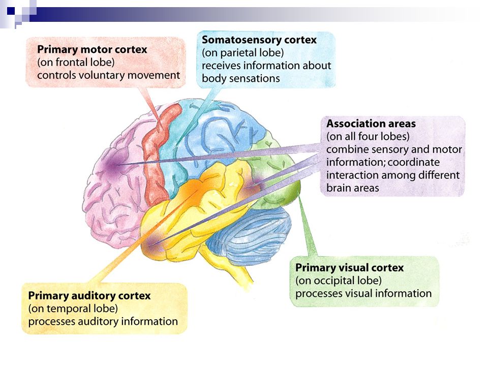
The Cerebral Cortex Frontal Lobes Parietal Lobes Occipital Lobes
involved in speaking and muscle movements and in making plans and judgments Parietal Lobes include the sensory cortex & processes somatic information Occipital Lobes include the visual areas, which receive visual information from the opposite visual field Temporal Lobes include the auditory areas

82
The Cerebral Cortex Motor Cortex Sensory Cortex
area at the rear of the frontal lobes that controls voluntary movements Sensory Cortex area at the front of the parietal lobes that registers and processes body sensations

85
The Cerebral Cortex

87
The Cerebral Cortex Functional MRI .......fMRI.........
scan shows the visual cortex activated as the subject looks at faces

88
Visual and Auditory Cortex

89
Association Areas More intelligent animals have increased “uncommitted” or association areas of the cortex

90
The Cerebral Cortex Aphasia Broca’s Area (Disrupts speaking)
impairment of language, usually caused by left hemisphere damage either to Broca’s area (impairing speaking) or to Wernicke’s area (impairing understanding) Broca’s Area (Disrupts speaking) an area of the left frontal lobe that directs the muscle movements involved in speech Wernicke’s Area (Disrupts understanding) an area of the left temporal lobe involved in language comprehension and expression
 or to Wernicke’s area (impairing understanding) Broca’s Area (Disrupts speaking) an area of the left frontal lobe that directs the muscle movements involved in speech. Wernicke’s Area (Disrupts understanding) an area of the left temporal lobe involved in language comprehension and expression.")
91
Specialization and Integration

92
Specialization and Integration
Brain activity when hearing, seeing, and speaking words

93
Brain Reorganization Plasticity
the brain’s capacity for modification, as evident in brain reorganization following damage (especially in children) and in experiments on the effects of experience on brain development
 and in experiments on the effects of experience on brain development.")
94
Brain Reorganization Hemispherectomy
The surgical removal of an entire cerebral hemisphere

95
Our Divided Brain Corpus Callosum large band of neural fibers
connects the two brain hemispheres carries messages between the hemispheres

96
Our Divided Brain The information highway from the eye to the brain

97
Split Brain a condition in which the two hemispheres of the brain are isolated by cutting the connecting fibers (mainly those of the corpus callosum) between them
 between them.")
98
Split Brain “What word did you see?” or “Point with your left
hand to the word you saw.” “Look at the dot.” Two words separated by a dot are momentarily projected.

99
Discovering PSY 2e p74 table 2.2

100
Disappearing Southpaws
The percentage of left-handers decreases sharply in samples of older people (adapted from Coren, 1993). The percentage of lefties sharply declines with age Age in years 14% 12 10 8 6 4 2 Percentage of left-handedness
. The percentage of. lefties sharply. declines with age Age in years. 14% Percentage of. left-handedness.")
101
Brain Structures and their Functions

102
The Endocrine System Endocrine System
the body’s “slow” chemical communication system a set of glands that secrete hormones into the bloodstream

103
Neural and Hormonal Systems
Hormones chemical messengers, mostly those manufactured by the endocrine glands, that are produced in one tissue and affect another Pituitary Gland under the influence of the hypothalamus, the pituitary regulates growth and controls other endocrine glands

104
The effects of the pituitary are clearly shown here
The effects of the pituitary are clearly shown here. Entertainer David Frost stands between the world’s tallest and smallest man. The tallest man in history was 8 feet 11 inches tall. He died at the age of 22, partly as a result of this defect. The shortest known person was 23 inches tall when she died at the age of 19. Today’s medicines can handle most of these problems if caught earlier enough, but these cases show what happen if the pituitary gland goes awry.

105
Neural and Hormonal Systems
Oxytocin– stimulates contractions of the uterus during labor and secretion of milk during nursing. Growth Hormone– stimulates the physical development of bones and muscles.

106
Neural and Hormonal Systems
Adrenal [ah-DREEN-el] Glands a pair of endocrine glands just above the kidneys secrete the hormones epinephrine (adrenaline) and norepinephrine (noradrenaline), which help to arouse the body in times of stress Cortisol– regulates metabolism and response to stress.
 and norepinephrine (noradrenaline), which help to arouse the body in times of stress. Cortisol– regulates metabolism and response to stress.")
107
Neural and Hormonal Systems
Pancreas Hormones Insulin– decreases blood sugar Glucagon– Increases blood sugar

108
Neural and Hormonal Systems
Thyroid Hormone Thyroxin– regulates metabolism and growth

109
Neural and Hormonal Systems
Sex Glands (Gonads) Female Sex Hormone– Estrogen (Ovary) Male Sex Hormone– Androgen (Testis)
 Female Sex Hormone– Estrogen (Ovary) Male Sex Hormone– Androgen (Testis)")
Similar presentations


>")
>")


>")


