Download presentation
Presentation is loading. Please wait.

1
3/25 Collect Onion Mitosis Lab Nervous system Homework – Chapter 48 and 49.2 Nervous System online due tomorrow (long) – We’re done!!! Review Manual Topics 8 and 9 – all relevant reading and questions due Wednesday Plant and Animal Systems Test Thursday and FRQ Friday – Big Idea Powerpoint is up on the website!!! Do first third tonight Next Practice AP Exam dates are here Saturday 4/13 10am-2pm and Saturday 4/20 12pm-4pm

2
Goals for Nervous System Know the function of the nervous system Include a picture of the system Know the anatomy of a neuron, and the functions of sensory, inter-, and motor neurons, and what a nerve is Know the mechanisms of impulse transmission in a neuron Know the process that leads to release of neurotransmitter, and what happens at the synapse Know the organization and function of the major divisions of the nervous system – central, peripheral, somatic, autonomic, sympathetic, parasympathetic Know the trends in nervous system evolution over animal phyla Know the components of a reflex arc and how they work Know one function for each major brain region Know gray vs. white matter, cerebrospinal fluid, and the types of glial cells

3
Nervous System Functions? (think about three main roles and types of neurons) What is a nerve? Ganglion? Nucleus?

4
Copyright © 2002 Pearson Education, Inc., publishing as Benjamin Cummings Fig. 48.1

5
Neuron – let’s label

6
Copyright © 2002 Pearson Education, Inc., publishing as Benjamin Cummings Fig. 48.2

7
Nervous System Activities http://session.masteringbiology.com/myct/ assignmentHome?assignmentID=2019767http://session.masteringbiology.com/myct/ assignmentHome?assignmentID=2019767 1 for neuron/action potential 2 synapse tutorials

8
Neural Impulse Electrochemical = Electric through neuron Chemical between neurons

9
At rest the sodium ions (Na + ) build up outside the cell and K + build up inside the cell.
 build up outside the cell and K + build up inside the cell.")
10
http://www.biology4all. com/resources_library/s ource/63.swf Review of the nerve impulse – THIS LINK WORKS!!!

11
Types of gated ions. –Chemically-gated ion channels open or close in response to a chemical stimulus. –Voltage-gated ion channels open or close in response to a change in membrane potential. Copyright © 2002 Pearson Education, Inc., publishing as Benjamin Cummings

12
Graded Potentials: Hyperpolarization and Depolarization –Graded potentials are changes in membrane potential Copyright © 2002 Pearson Education, Inc., publishing as Benjamin Cummings

13
Hyperpolarization. –Gated K + channels open K + diffuses out of the cell the membrane potential becomes more negative. Copyright © 2002 Pearson Education, Inc., publishing as Benjamin Cummings Fig. 48.8a Graded potentials are changes in membrane potential

14
Depolarization. –Gated Na + channels open Na + diffuses into the cell the membrane potential becomes less negative. Copyright © 2002 Pearson Education, Inc., publishing as Benjamin Cummings Fig. 48.8b

15
The Action Potential: All or Nothing Depolarization. –If graded potentials sum to -55mV a threshold potential is achieved. This triggers an action potential. –Axons only. Copyright © 2002 Pearson Education, Inc., publishing as Benjamin Cummings Fig. 48.8c

16
5 Steps of Action Potential Copyright © 2002 Pearson Education, Inc., publishing as Benjamin Cummings Fig. 48.9

17
During the undershoot both the Na + channel’s activation and inactivation gates are closed. –At this time the neuron cannot depolarize in response to another stimulus: refractory period. Copyright © 2002 Pearson Education, Inc., publishing as Benjamin Cummings

18
Saltatory conduction. –In myelinated neurons only unmyelinated regions of the axon depolarize. Thus, the impulse moves faster than in unmyelinated neurons. Copyright © 2002 Pearson Education, Inc., publishing as Benjamin Cummings Fig. 48.11

19
The small gap between the axon of one neuron and the dendrite of another neuron is called a synapse. Nervous System An action potential is carried across these gaps by neurotransmitters. The Synapse 33.1 Structure of the Nervous System Chapter 33 The neuron before the synapse is called the presynaptic neuron – what do you think the neuron after the synapse is called?

20
1. axon 2. synaptic knob 6. dendrite 5. synaptic gap 3. synaptic vesicles 4. cell membrane Structure of the synapse

21
The neurotransmitter diffuses across the synapse. Binds to receptors on the dendrite of a neuron. More nerve impulses are generated (or muscle or gland stimulated). The neurotransmitter is broken down by enzymes. Fast forward to end in this animation.
. The neurotransmitter is broken down by enzymes. Fast forward to end in this animation..")
22
Nervous System Chapter 33

23
Order these events of the neural impulse Neurotransmitters bind to next neuron’s dendrite, starting new impulse Threshold reached, action potential generated Calcium channels open Vesicles fuse with plasma membrane Neurotransmitters released into synaptic gap Action potential reaches end of axon

24
Excitatory postsynaptic potentials (EPSP) depolarize the postsynaptic neuron. –The binding of neurotransmitter to postsynaptic receptors open gated channels that allow Na + to diffuse into and K + to diffuse out of the cell. Inhibitory postsynaptic potential (IPSP) hyperpolarize the postsynaptic neuron. –The binding of neurotransmitter to postsynaptic receptors open gated channels that allow K+ to diffuse out of the cell and/or Cl- to diffuse into the cell. Neural integration occurs at the cellular level
 hyperpolarize the postsynaptic neuron. –The binding of neurotransmitter to postsynaptic receptors open gated channels that allow K+ to diffuse out of the cell and/or Cl- to diffuse into the cell. Neural integration occurs at the cellular level.")
25
Summation: graded potentials (EPSPs and IPSPs) are summed to either depolarize or hyperpolarize a postsynaptic neuron. Copyright © 2002 Pearson Education, Inc., publishing as Benjamin Cummings Fig. 48.14

26
Acetylcholine. –Excitatory to skeletal muscle. –Inhibitory to cardiac muscle. –Secreted by the CNS, PNS, and at vertebrate neuromuscular junctions. The same neurotransmitter can produce different effects on different types of cells Copyright © 2002 Pearson Education, Inc., publishing as Benjamin Cummings

27
Biogenic Amines. –Epinephrine and norepinephrine. Can have excitatory or inhibitory effects. Secreted by the CNS and PNS. Secreted by the adrenal glands. Copyright © 2002 Pearson Education, Inc., publishing as Benjamin Cummings

28
Dopamine –Generally excitatory; may be inhibitory at some sites. Widespread in the brain. Affects sleep, mood, attention, and learning. –Secreted by the CNS and PNS. –A lack of dopamine in the brain is associated with Parkinson’s disease. –Excessive dopamine is linked to schizophrenia. Copyright © 2002 Pearson Education, Inc., publishing as Benjamin Cummings

29
Serotonin. –Generally inhibitory. Widespread in the brain. Affects sleep, mood, attention, and learning –Secreted by the CNS. Copyright © 2002 Pearson Education, Inc., publishing as Benjamin Cummings

30
Amino Acids –Gamma aminobutyric acid (GABA). Inhibitory. Secreted by the CNS and at invertebrate neuromuscular junctions. –Also glycine, glutamate, aspartate Met-enkephalin (an endorphin). Neuropeptides. –Substance P. Gases that act as local regulators. –Nitric oxide. –Carbon monoxide. Copyright © 2002 Pearson Education, Inc., publishing as Benjamin Cummings
. Neuropeptides. –Substance P. Gases that act as local regulators. –Nitric oxide. –Carbon monoxide. Copyright © 2002 Pearson Education, Inc., publishing as Benjamin Cummings.")
31
3/26 Finish Nervous System Start Big Idea Powerpoint Homework – Review Manual Topics 8 and 9 – all relevant reading and questions due tomorrow Plant and Animal Systems Test Thursday and FRQ Friday – Big Idea Powerpoint is up on the website!!! Do second third tonight Next Practice AP Exam dates are here Saturday 4/13 10am-2pm and Saturday 4/20 12pm-4pm

32
A Simple Nerve Circuit – the Reflex Arc. –A reflex is an autonomic response. Copyright © 2002 Pearson Education, Inc., publishing as Benjamin Cummings Fig. 48.3

33
Neurons differ in terms of both function and shape. Copyright © 2002 Pearson Education, Inc., publishing as Benjamin Cummings Fig. 48.4

34
Types of Nerve Circuits. –Single presynaptic neuron several postsynaptic neurons. –Several presynaptic neurons single postsynaptic neuron. –Circular paths. Copyright © 2002 Pearson Education, Inc., publishing as Benjamin Cummings

35
Supporting Cells (Glia). –There are several types of glia. –Astrocytes are found within the CNS. –Structural and metabolic support. –By inducing the formation of tight junctions between capillary cells astrocytes help form the blood-brain barrier. –Like neurons, astrocytes communicate with one another via chemical signals. Oligodendrocytes are found within the CNS. –Form a myelin sheath by insulating axons. Copyright © 2002 Pearson Education, Inc., publishing as Benjamin Cummings

36
Schwann cells are found within the PNS. –Form a myelin sheath by insulating axons. Copyright © 2002 Pearson Education, Inc., publishing as Benjamin Cummings Fig. 48.5

37
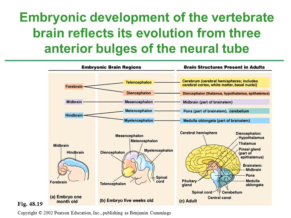
Embryonic development of the vertebrate brain reflects its evolution from three anterior bulges of the neural tube Copyright © 2002 Pearson Education, Inc., publishing as Benjamin Cummings Fig. 48.19
38
Copyright © 2002 Pearson Education, Inc., publishing as Benjamin Cummings Fig. 48.20 Let’s label functions – use p. 270

39
Self - Check Which part of the brain regulates thirst? Which part of the brain would be highly developed in an animal that is extremely coordinated like a monkey or cat? Which part of the brain do all your conscious thoughts come from?

40
The Reticular System, Arousal, and Sleep. –The reticular activating system (RAS) of the reticular formation. Regulates sleep and arousal. Acts as a sensory filter. Copyright © 2002 Pearson Education, Inc., publishing as Benjamin Cummings Fig. 48.21
 of the reticular formation. Regulates sleep and arousal. Acts as a sensory filter. Copyright © 2002 Pearson Education, Inc., publishing as Benjamin Cummings Fig")
41
–Sleep and wakefulness produces patterns of electrical activity in the brain that can be recorded as an electroencephalogram (EEG). Most dreaming occurs during REM (rapid eye movement) sleep. Copyright © 2002 Pearson Education, Inc., publishing as Benjamin Cummings Fig. 48.22b-d
 sleep. Copyright © 2002 Pearson Education, Inc., publishing as Benjamin Cummings Fig b-d.")
42
The cerebrum is derived from the embryonic telencephalon. The cerebrum is the most highly evolved structure of the mammalian brain Copyright © 2002 Pearson Education, Inc., publishing as Benjamin Cummings Fig. 48.24a

43
The cerebrum is divided into left and right cerebrum hemispheres. –The corpus callosum is the major connection between the two hemispheres. –The left hemisphere is primarily responsible for the right side of the body. –The right hemisphere is primarily responsible for the left side of the body. Cerebral cortex: outer covering of gray matter. –Neocortex: region unique to mammals. The more convoluted the surface of the neocortex the more surface area the more neurons. Basal nuclei: internal clusters of nuclei. Copyright © 2002 Pearson Education, Inc., publishing as Benjamin Cummings

44
The cerebrum is divided into frontal, temporal, occipital, and parietal lobes. Regions of the cerebrum are specialized for different functions Copyright © 2002 Pearson Education, Inc., publishing as Benjamin Cummings Fig. 48.24b

45
Copyright © 2002 Pearson Education, Inc., publishing as Benjamin Cummings Fig. 48.25

46
Lateralization of Brain Function. –The left hemisphere. Specializes in language, math, logic operations, and the processing of serial sequences of information, and visual and auditory details. Specializes in detailed activities required for motor control. –The right hemisphere. Specializes in pattern recognition, spatial relationships, nonverbal ideation, emotional processing, and the parallel processing of information. Copyright © 2002 Pearson Education, Inc., publishing as Benjamin Cummings

47
Language and Speech. –Broca’s area. Usually located in the left hemisphere’s frontal lobe Responsible for speech production. –Wernicke’s area. Usually located in the right hemisphere’s temporal lobe Responsible for the comprehension of speech. –Other speech areas are involved generating verbs to match nouns, grouping together related words, etc. Copyright © 2002 Pearson Education, Inc., publishing as Benjamin Cummings

48
Emotions. –In mammals, the limbic system is composed of the hippocampus, olfactory cortex, inner portions of the cortex’s lobes, and parts of the thalamus and hypothalamus. Mediates basic emotions (fear, anger), involved in emotional bonding, establishes emotional memory –For example, the amygdala is involved in recognizing the emotional content of facial expression. Copyright © 2002 Pearson Education, Inc., publishing as Benjamin Cummings Fig. 48.27
, involved in emotional bonding, establishes emotional memory –For example, the amygdala is involved in recognizing the emotional content of facial expression. Copyright © 2002 Pearson Education, Inc., publishing as Benjamin Cummings Fig")
49
Memory and Learning. –Short-term memory stored in the frontal lobes. –The establishment of long-term memory involves the hippocampus. The transfer of information from short-term to long-term memory. –Is enhanced by repetition (remember that when you are preparing for an exam). –Influenced by emotional states mediated by the amygdala. –Influenced by association with previously stored information. Copyright © 2002 Pearson Education, Inc., publishing as Benjamin Cummings
. –Influenced by emotional states mediated by the amygdala. –Influenced by association with previously stored information. Copyright © 2002 Pearson Education, Inc., publishing as Benjamin Cummings.")
50
The mammalian PNS has the ability to repair itself, the CNS does not. –Research on nerve cell development and neural stem cells may be the future of treatment for damage to the CNS. Research on neuron development and neural stem cells may lead to new approaches for treating CNS injuries and diseases Copyright © 2002 Pearson Education, Inc., publishing as Benjamin Cummings

51
3/27 Finish Big Idea Powerpoint I check review manual Homework – Plant and Animal Systems Test tomorrow and FRQ Friday – Big Idea Powerpoint is up on the website!!! Do last third tonight Next Practice AP Exam dates are here Saturday 4/13 10am-2pm and Saturday 4/20 12pm-4pm

52
3/28 Test

53
3/29 FRQ Spring Break homework – SparkNote diagnostic and review manual – intro chapters and diagnostic test – get familiar with layout of book, final lab report due week after Spring Break

Similar presentations


. Brain and spinal cord. Both contain fluid-filled spaces which contain cerebrospinal fluid (CSF). The central canal of the.>")

 CNS (brain and spinal cord) PNS (peripheral nervous system) PNS (peripheral nervous system)>")


