Download presentation
Presentation is loading. Please wait.

1
Nuclear Medicine

2
The History Henri Becquerel 1896-Discovered mysterious “rays”. 1903-Nobel Prize Marie Curie 1897- named mysterious rays “radioactivity” 1903-Nobel Prize Herman Blumgart 1925- First use of radioactive tracers for diagnosis. Hal Anger 1958- presented his first scintillation camera which led to cardiology.

3
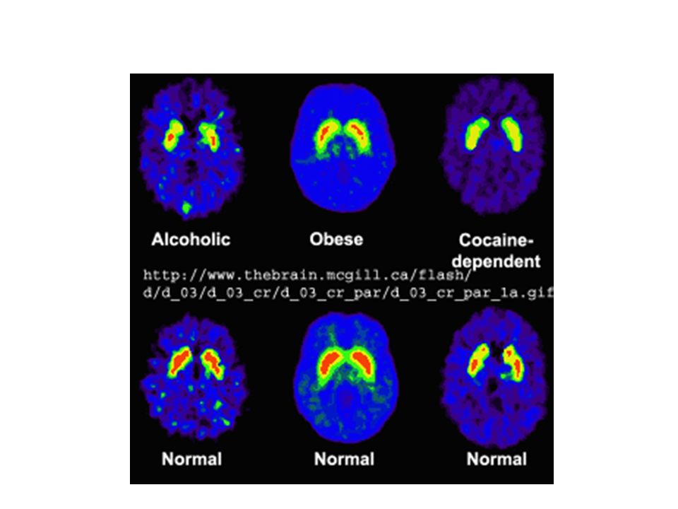
Diagnostic Imaging that involves the acquisition of physiologic images based on the detection of radiation from the emission of positrons. Best known for role in cancer imaging.

4
Does not produce an anatomical map of the body. Creates image of the spatial distribution of radiopharmaceuticals introduced into the body. – Based on fact that most pathological conditions are initiated by the biochemistry in the tissues. Cell swelling, tumor, ulcers, etc

5
Detects early indicators of disease by imaging the uptake and biodistribution of radioactive compounds introduce in the body. – Inhalation into lungs, injection in bloodstream, or oral administration – These sites are termed “hot spots”

6
Radiopharmaceuticals (radiotracers) are compounds consisting of a chemical substrate linked to a radioactive element. Abnormal tissue distribution or an increase/decrease in the rate a which the radiopharmaceutical accumulates in a tissue is a strong indicator of disease. Radiation (gamma rays) from the radioactive decay of the radiopharmaceutical is detected by a gamma cammera.
 from the radioactive decay of the radiopharmaceutical is detected by a gamma cammera..")
7
Nuclear Medicine Setup

8
Radioactivity Based particular isotopes of elements that have unstable nuclei. 4 radionuclides: – Neutron capture – Nuclear fission – Charged-particle bombardment – Radionuclide generators Most important = Mo – Commonly used method = on-site generator Radionuclides are “milked” from the generator daily

9
So what about these radionuclides in the body? Radioactive Decay! – Gamma ray decay Technetium provides gamma ray decay – Stable in body – Long half life (6.02 hours)
.")
10
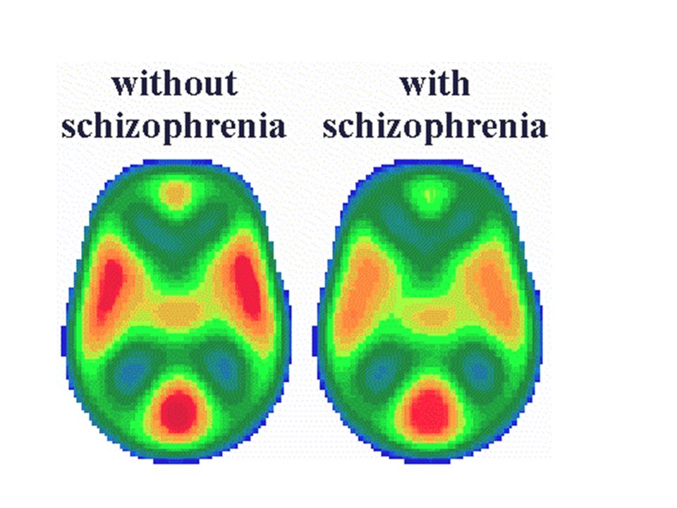
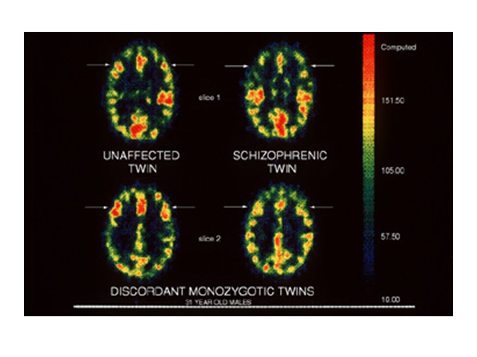
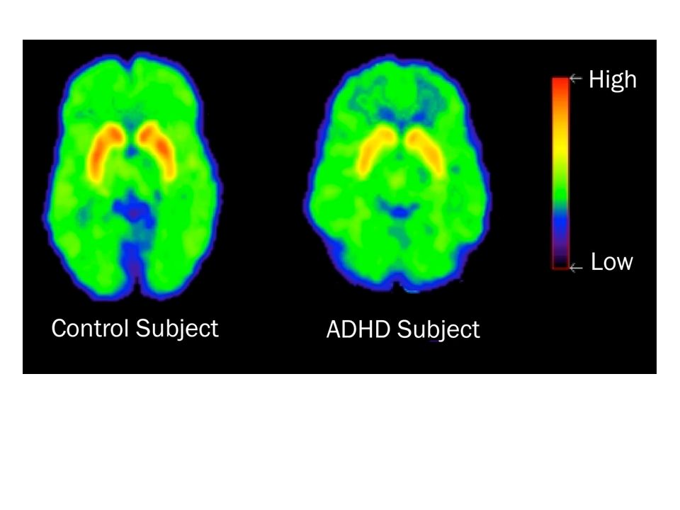
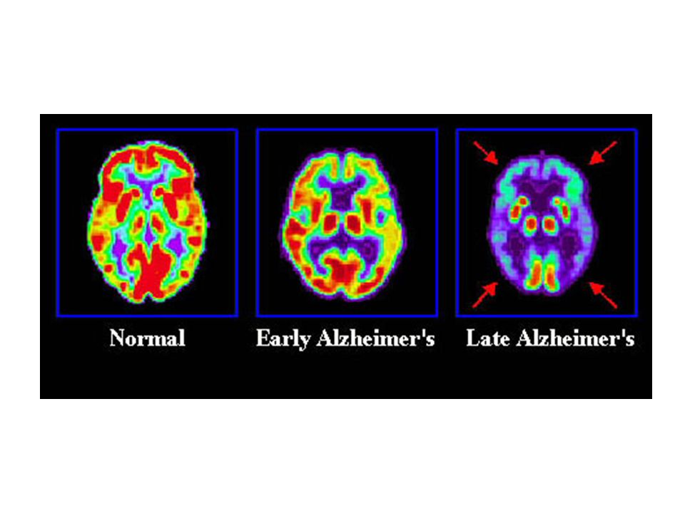
How Nuclear Medicine is Used Detect cancer and examine effects of cancer Determine blood flow in hear Signals coronary artery disease Evaluate memory disorders

11
SPECT Single Photon Emission Computed Tomography – Nuclear scans to get “slices” – Looks at actual anatomy of things (cancer, tumors, etc)
")
12
Gallium (Ga) – Used on Hodgkin’s disease, long cancer, leukemia, lymphoma, malignant melanoma
 – Used on Hodgkin’s disease, long cancer, leukemia, lymphoma, malignant melanoma")
13
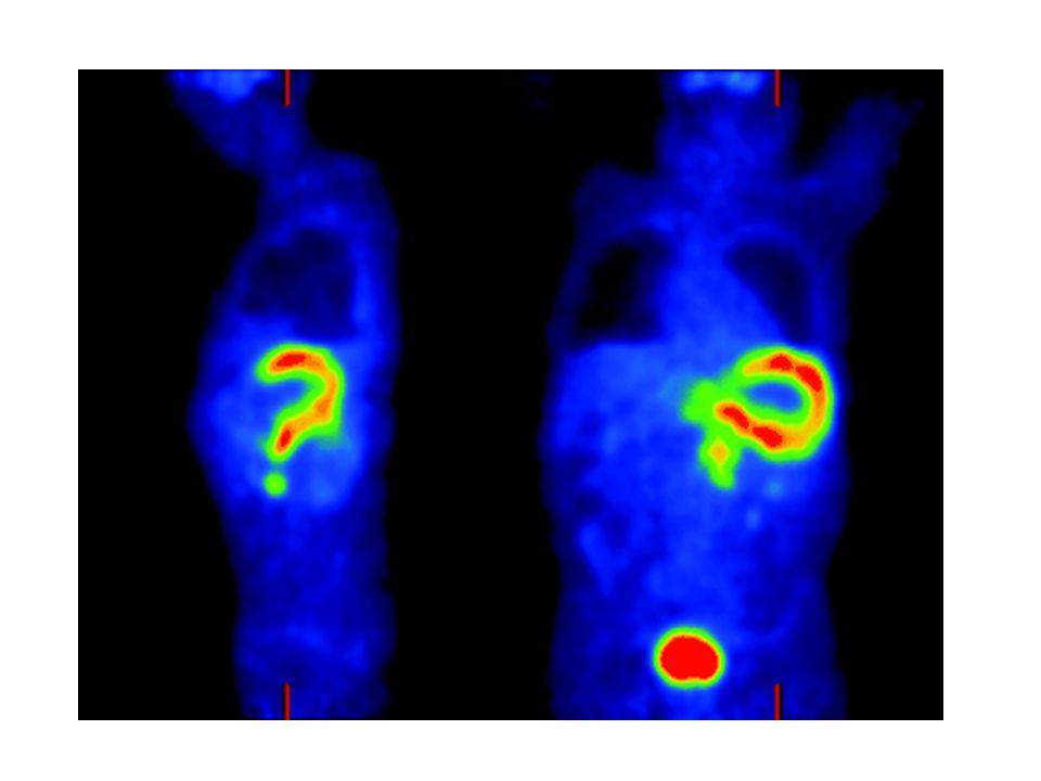
PET Positron emission tomography – Measures physiology and function Distribution, extent of uptake, rate of uptake, and rate of washout – Typically $1.5 to 2.5 million per system – Isotopes: C, O, F, and N (much shorter half life, so need on-site cyclotron generator) Undergo radioactive decay by emitting a positron (positively charged electron) Positron will annihilate with an electron = two gamma rays 180 degrees apart
 Undergo radioactive decay by emitting a positron (positively charged electron) Positron will annihilate with an electron = two gamma rays 180 degrees apart")
Similar presentations

>")


