Download presentation
Presentation is loading. Please wait.

1
Lecture 2 1.5 The Historical Roots of Microbiology
1.6 Microbial Diversity and the Advent of Molecular Microbiology 2.1 Elements of Cell and Viral Structure 2.2 Arrangement of DNA in Microbial Cells 2.3 The Tree of Life

2
Also refer to Table 1.1

3
Where do the microorganisms come from? Spontaneous generation?
Louis Pasteur ~1860 Where do the microorganisms come from? Spontaneous generation? Figure: 01-11a Caption: Pasteur’s experiment with the swan-necked flask. (a) Sterilizing the contents of the flask. (Madigan et al., Fig. 1.11) Heat was used to kill the microorganisms in the liquid
 Sterilizing the contents of the flask. (Madigan et al., Fig. 1.11) Heat was used to kill the microorganisms in the liquid.")
4
Figure: 01-11b Caption: Pasteur’s experiment with the swan-necked flask. (b) If the flask remained upright, no microbial growth occurred. (Madigan et al., Fig. 1.11) When dust was prevented from reaching the sterilized liquid, no microorganisms grew in the liquid
 When dust was prevented from reaching the sterilized liquid, no microorganisms grew in the liquid.")
5
Contact with dust resulted in growth of microorganisms in the liquid
Figure: 01-11c Caption: Pasteur’s experiment with the swan-necked flask. (c) If microorganisms trapped in the neck reached the sterile liquid, microbial growth ensued. (Madigan et al., Fig. 1.11) Contact with dust resulted in growth of microorganisms in the liquid → disproved spontaneous generation
 If microorganisms trapped in the neck reached the sterile liquid, microbial growth ensued. (Madigan et al., Fig. 1.11) Contact with dust resulted in growth of microorganisms in the liquid. → disproved spontaneous generation.")
7
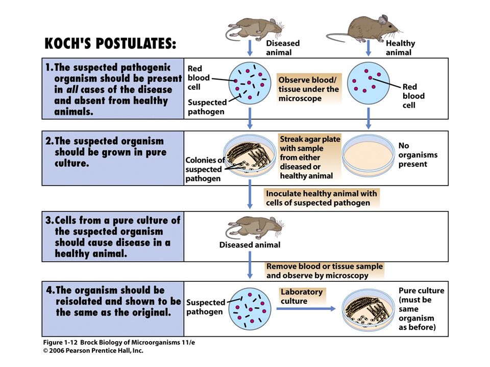
Robert Koch, 1870s: Proof that microorganisms can cause disease
-“germ theory of disease” (Madigan et al., Fig. 1.12) Anthrax, caused by Bacillus anthracis Organism present in the blood of all diseased animals - cause or result of the disease?
 Anthrax, caused by Bacillus anthracis. Organism present in the blood of all diseased animals. - cause or result of the disease")
8
Pure culture (Madigan et al., Fig. 1.12)
")
9
(Madigan et al., Fig. 1.12)
")
10
Conclusion - specific organisms cause specific disease
(Madigan et al., Fig. 1.12) Conclusion - specific organisms cause specific disease Koch’s postulates can be extended beyond disease-causing organisms
 Conclusion - specific organisms cause specific disease. Koch’s postulates can be extended beyond disease-causing organisms.")
11
comparative structure of prokaryotic and eukaryotic cells:
Figure: 02-01a-b Caption: Internal structure of microbial cells. (a) Diagram of a prokaryote. (b) Diagram of a eukaryote. (Madigan et al., Fig 2.1) prokaryotic: nucleoid no organelles eukaryotic: nucleus organelles
 Diagram of a prokaryote. (b) Diagram of a eukaryote. (Madigan et al., Fig 2.1) prokaryotic: nucleoid. no organelles. eukaryotic: nucleus. organelles.")
12
bacterial cell, 1 x 3 μm (Heliobacterium modesticaldum)
Figure: 02-02a Caption: (a) Heliobacterium modesticaldum (Bacteria); the cell measures 1 x 3 µm. (Madigan et al., Fig. 2.2)
 Heliobacterium modesticaldum (Bacteria); the cell measures 1 x 3 µm. (Madigan et al., Fig. 2.2)")
13
(Saccharomyces cerevisiae)
yeast cell, 8 μm dia (Saccharomyces cerevisiae) Figure: 02-02c Caption: (c) Saccharomyces cerevisiae (Eukarya); the cell measures 8 µm in diameter.
 Figure: 02-02c. Caption: (c) Saccharomyces cerevisiae (Eukarya); the cell measures 8 µm in diameter.")
14
viruses: very small microorganisms (10s of nm dia), but not cells
not dynamic open systems do not take nutrients or expel wastes static structure; behave as more-or-less as particles, except when infecting host possess genes but no biosynthetic machinery rely on host machinery to reproduce viruses known to infect all cells viruses of bacteria = bacteriophages see Madigan et al., Fig. 2.3a, b

15
relative sizes of different microorganisms:
Figure: 02-03c Caption: (c) The size of the viruses shown in (a) and (b) in comparison to a bacterial and eukaryotic cell.
 The size of the viruses shown in (a) and (b) in comparison to a bacterial and eukaryotic cell.")
16
ribosomal RNA (rRNA) gene sequencing and phylogeny:
(Madigan et al., Fig. 2.6) Figure: 02-06a-e Caption: Ribosomal RNA gene sequencing and phylogeny. (a) Cells, either from a pure culture or from an environmental sample, are broken open; (b) the gene-encoding ribosomal RNA is isolated, and many identical copies made by the technique called the polymerase chain reaction (PCR); f Section (c) The gene is sequenced (f Section 10.13), and (d) the sequences obtained are aligned in a computer. An algorithm makes pair-wise comparisons and generates a tree (e) that depicts the differences in ribosomal RNA sequence between the organisms analyzed. If an environmental sample is used, the isolated ribosomal RNA genes from the different microorganisms in the sample must first be sorted out (cloned) before copies are made and sequencing done. For further discussion of these methods, see Sections 11.5 and 18.5. all organisms possess ribosomes → rRNAs useful molecules for assessing relationships between organisms rRNA genes isolated gene sequences determined and compared phylogenetic tree depicts differences between organisms analyzed
 Figure: 02-06a-e. Caption: Ribosomal RNA gene sequencing and phylogeny. (a) Cells, either from a pure culture or from an environmental sample, are broken open; (b) the gene-encoding ribosomal RNA is isolated, and many identical copies made by the technique called the polymerase chain reaction (PCR); f Section (c) The gene is sequenced (f Section 10.13), and (d) the sequences obtained are aligned in a computer. An algorithm makes pair-wise comparisons and generates a tree (e) that depicts the differences in ribosomal RNA sequence between the organisms analyzed. If an environmental sample is used, the isolated ribosomal RNA genes from the different microorganisms in the sample must first be sorted out (cloned) before copies are made and sequencing done. For further discussion of these methods, see Sections 11.5 and all organisms possess ribosomes → rRNAs useful molecules for assessing relationships between organisms. rRNA genes isolated. gene sequences determined and compared. phylogenetic tree depicts differences between organisms analyzed.")
17
The “Five Kingdoms” of Life
Plants Animals Fungi Monera (prokaryotes) Protists (slime molds, flagellates, Giardia) human-centric organization
 Protists (slime molds, flagellates, Giardia) human-centric organization.")
18
The Three Domains of Life
Figure: 02-07 Caption: The phylogenetic tree of life as defined by comparative ribosomal RNA sequencing. The tree consists of three domains of organisms: the Bacteria and the Archaea, cells of which are prokaryotic, and the Eukarya (eukaryotes). Only a few of the groups of organisms within each domain are shown. See more detailed trees of each domain in Figures 2.9, 2.18, and 2.22, and the phylogenetic trees in Chapters 11–14. Hyperthermophiles are prokaryotes that grow best at temperatures of 80˚C or higher. The group shaded in red are macroorganisms. All other organisms on the tree of life are microorganisms.
. Only a few of the groups of organisms within each domain are shown. See more detailed trees of each domain in Figures 2.9, 2.18, and 2.22, and the phylogenetic trees in Chapters 11–14. Hyperthermophiles are prokaryotes that grow best at temperatures of 80˚C or higher. The group shaded in red are macroorganisms. All other organisms on the tree of life are microorganisms.")
21
Nomenclature Bacteria are named using the binomial system used for other living things whereby each species is given two names The first name is the Genus name (equivalent to your surname) and the second name is the species name (equivalent to your Christian name) Bacteria belong to the one species if they have 90% similarity of all observed characteristics A group of similar species that have 80% similarity is called a Genus
 and the second name is the species name (equivalent to your Christian name) Bacteria belong to the one species if they have 90% similarity of all observed characteristics. A group of similar species that have 80% similarity is called a Genus.")
22
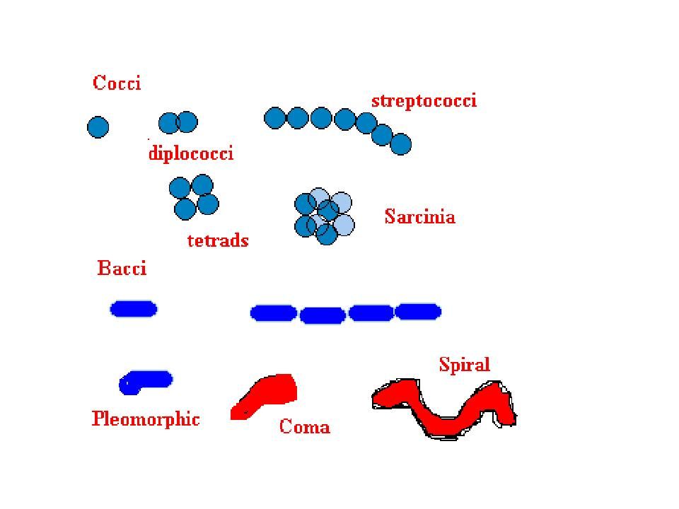
Names and morphology The genus name always start with a capital letter and the species name is in lower case and in singular e.g. Staphylococcus aureus Such binomial species names are always underlined or written in Italics e.g. not all streptococci are Streptococcus in fact some streptococci are Leuconostoc And not all staphylococci are Staphylococcus in fact some staphylococci are Micrococcus not all bacilli are Bacillus in fact some bacilli are Chlostridium etc etc.

23
Criteria for Classification of Prokaryotes
Cultural Morphology Microscopic Morphology Cellular Components Growth Characteristics Metabolic Pathways Molecular Genetics Location in Broth Cell Shape Cell Wall Atmospheric requirements Carbon requirements DNA base ratio Colony Appearance Cell Size Gram Stain pH tolerance Nitrogen requirements DNA sequence Pigmentation Arrangement Capsule Temperature requirements Sulfur RNA Internal Structures Symbiotic lifestyle Fermentation Probes Accessory Structures Antibiotic sensitivity Respiration PCR End Products

Similar presentations

 – Made up of at least one cell – Has DNA – Needs energy and.>")


>")


 Lecture 1 Introduction, History and Microscopy (Text Chapters: 1.1-1.8; 4.1-4.3)>")


