Download presentation
Presentation is loading. Please wait.

1
SPINE IMAGING

20
OD LM B TP

21
OD SP

22
AAC1 AAC2

23
SP B TP AP TF

24
PA TF AA OD

45
B L SP P R CVJ CTJ

47
SP B PM

48
ZJ

51
IVS B SP

52
MPR

53
F SIJ

54
Pathologies

55
Ankylosing spondylitis is a type of arthritis that affects the spine. Spondylitis may cause pain and stiffness from the neck down to the lower back. The bones of the spine, called vertebrae, may grow or fuse together, resulting in a rigid spine. These changes may be mild or severe, and may lead to a stooped-over posture.

56
Spondylolisthesis Description The most common X-ray identified cause of low back pain in adolescent athletes is a stress fracture in one of the bones (vertebrae) that make up the spinal column. Technically, this condition is called spondylolysis (spon-dee-low-lye-sis). It usually affects the fifth lumbar vertebra in the lower back, and much less commonly, the fourth lumbar vertebra. If the stress fracture weakens the bone so much that it is unable to maintain its proper position, the vertebra can start to shift out of place. This condition is called spondylolisthesis (spon-dee-low- lis-thee-sis). If too much slippage occurs, the bones may begin to press on nerves and surgery may be necessary to correct the condition
. It usually affects the fifth lumbar vertebra in the lower back, and much less commonly, the fourth lumbar vertebra. If the stress fracture weakens the bone so much that it is unable to maintain its proper position, the vertebra can start to shift out of place. This condition is called spondylolisthesis (spon-dee-low- lis-thee-sis). If too much slippage occurs, the bones may begin to press on nerves and surgery may be necessary to correct the condition.")
58
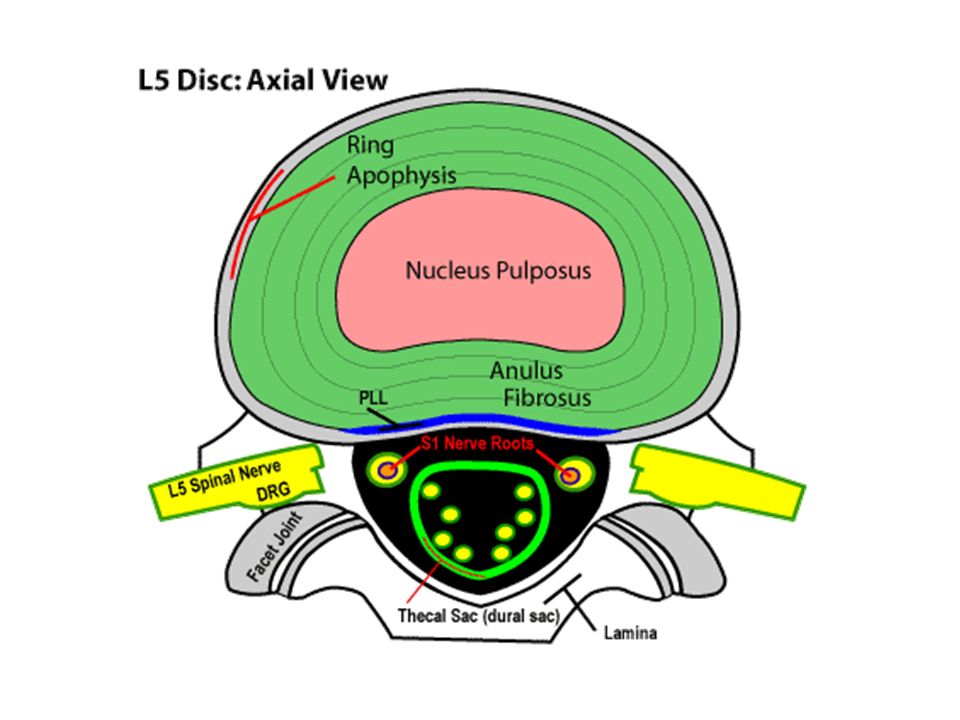
HNP-herniated nucleus pulposus

59
Many types of tumors start in the central nervous system (CNS) (brain and spinal cord). If you have one of these tumors, your symptoms, outlook for survival (prognosis), and treatment depend on your age, the tumor type, and the precise location of the tumor within the CNS Astrocytoma: Most tumors that arise within the brain itself start in brain cells called astrocytes. These tumors are called astrocytomas. About 35% of brain tumors are astrocytomas. Most astrocytomas cannot be cured because they spread widely throughout the surrounding normal brain tissue. Sometimes astrocytomas spread along the cerebrospinal fluid pathways. With only rare exceptions, astrocytomas, however, do not spread outside of the brain or spinal cord
, and treatment depend on your age, the tumor type, and the precise location of the tumor within the CNS Astrocytoma: Most tumors that arise within the brain itself start in brain cells called astrocytes. These tumors are called astrocytomas. About 35% of brain tumors are astrocytomas. Most astrocytomas cannot be cured because they spread widely throughout the surrounding normal brain tissue. Sometimes astrocytomas spread along the cerebrospinal fluid pathways. With only rare exceptions, astrocytomas, however, do not spread outside of the brain or spinal cord.")
60
Astrocytoma

61
Definition Lumbar Spinal Stenosis is derived from the word stenosis meaning narrowing. Imagine the spinal canal is a circle. The circle can be average, big or small. Since the spinal nerves travel in the circle at this level of the spine, any narrowing of the circle could put pressure on the spinal nerves. Unless the individual is born with a small spinal canal (congenital stenosis), spinal narrowing occurs most commonly from progressive degenerative changes (acquired spinal stenosis).
, spinal narrowing occurs most commonly from progressive degenerative changes (acquired spinal stenosis)..")
62
Spine stenosis

63
Fractures- Jefferson’s A Jefferson fracture consists of a fracture of the C1 ring. This results from an axial loading injury to the head with compression force to C1 (typically from diving).
..")
64
Hangman’s fracture- C2-C3 Unstable hangman's type fracture of the C2 body and posterior elements extending into the left foramen transversarium.

65
COMPRESSION FRACTURE

66
BURST FRACTURE Burst fractures are comminuted fractures of the vertebral bodies often associated with bone fragments in the canal

67
VACUUM “GAS” PHENOMENON Vacuum" phenomena relate to the accumulation of gas, principally nitrogen, in crevices within the intervertebral disk or vertebra.

68
Protocols

69
C-SPINE

70
SCOUT: LAT LANDMARK: XIPHOID SLICE PLANE: OML I.V. CONTRAST: FOR EVALUATION OF DEGENERATIVE DISK DISEASE, DIFFERENTIATION OF THE DISK FROM THE SURGICAL SCAR TISSUE BREATH HOLD: QUIET RESPIRATION SLICE THICKNESS: 2-4 MM ( IF ONE DISK TO SCAN- 2MM) INDEX: CONTIGUOUS SLICES START LOCATION: PEDICLE OF C3 END LOCATION: THROUGH C7 FILMING: SOFT TISSUE AND BONE + MPR RECONSTRUCTION (IF SPIRAL)
 INDEX: CONTIGUOUS SLICES START LOCATION: PEDICLE OF C3 END LOCATION: THROUGH C7 FILMING: SOFT TISSUE AND BONE + MPR RECONSTRUCTION (IF SPIRAL).")
71
T-SPINE

72
SCOUT: LAT LANDMARK: STERNAL NOTCH SLICE PLANE: SPIRAL I.V. CONTRAST: FOR EVALUATION OF DEGENERATIVE DISK DISEASE, DIFFERENTIATION OF THE DISK FROM THE SURGICAL SCAR TISSUE BREATH HOLD: QUIET RESPIRATION SLICE THICKNESS: 3-5 MM INDEX: CONTIGUOUS SLICES IF ONE VERTEBRAE START PEDICLE ABOVE END LOCATION: PEDICLE BELOW FILMING: SOFT TISSUE AND BONE + MPR RECONSTRUCTION (IF SPIRAL)
.")
73
L-SPINE

74
SCOUT: LAT LANDMARK: XIPHOID SLICE PLANE: Angle the gantry so the slices will be parallel to the intervertebral disk spaces. I.V. CONTRAST: FOR EVALUATION OF DEGENERATIVE DISK DISEASE, DIFFERENTIATION OF THE DISK FROM THE SURGICAL SCAR TISSUE BREATH HOLD: QUIET RESPIRATION SLICE THICKNESS: 3-5 MM INDEX: CONTIGUOUS START LOCATION: PEDICLE OF L3 END LOCATION: S1 FILMING: SOFT TISSUE AND BONE

78
SCOUT: LAT LANDMARK: XIPHOID SLICE PLANE: AXIAL OR SPIRAL I.V. CONTRAST: FOR EVALUATION OF DEGENERATIVE DISK DISEASE, DIFFERENTIATION OF THE DISK FROM THE SURGICAL SCAR TISSUE BREATH HOLD: QUIET RESPIRATION SLICE THICKNESS: 3-5 MM INDEX: 3-5 MM START LOCATION: PEDICLE OF L3 END LOCATION: S1 FILMING: SOFT TISSUE AND BONE + MPR RECONSTRUCTION 3-D RECON: 50% OVERLAP

79
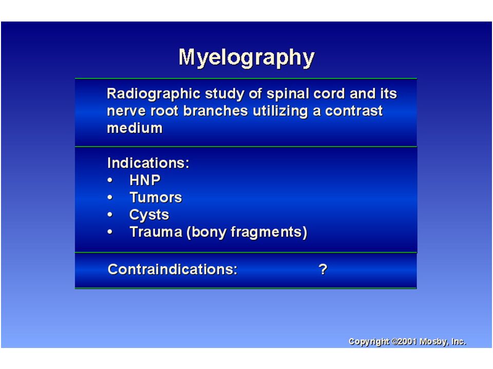
MYELOGRAPHY

82
CT MYELOGRAM

83
PURPOSE OF CT MYELOGRAM TO DETECT: HNP TUMOR INVADING CANAL BONY FRAGMENTS IN THE CANAL CYSTS

84
IV CONTRAST USED IN CT MYELOGRAM TO DIAGNOSE DEGENERATIVE DISK DISEASE (EPIDURAL SPACE WILL ENHANCE) DIFFERENTIATION OF THE DISKS FROM THE SURGICAL SCAR TISSUE
 DIFFERENTIATION OF THE DISKS FROM THE SURGICAL SCAR TISSUE")
85
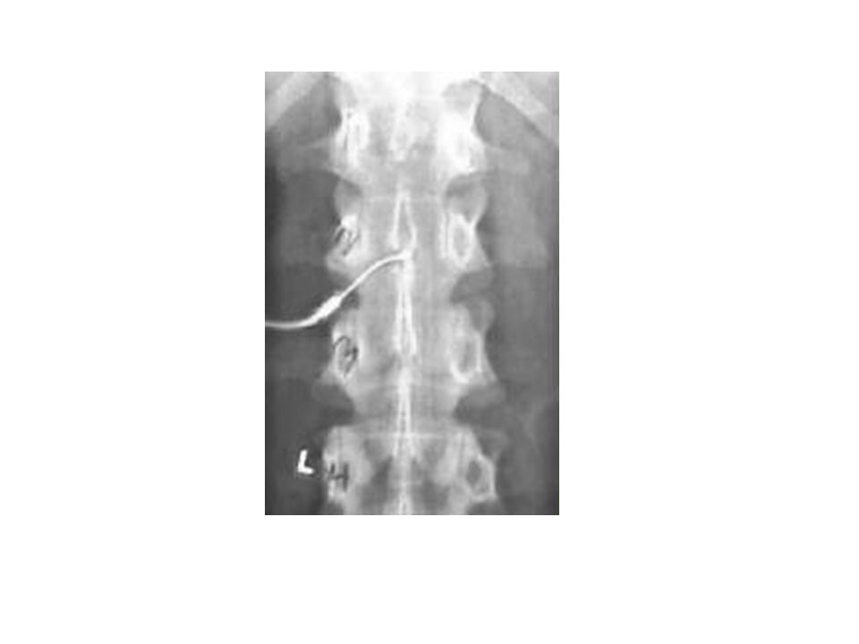
CONTRAST – INTRATHECAL INJECTION

87
SCANNING 1-4 HOURS AFTER THE CONTRAST INSTILLED THE DELAY ALLOWS FOR CONTRAST DILUTION SO THE INTRADURAL SPACES ARE CLEARLY VISUALIZED ROLLING OF THE PATIENT BEFORE THE SCAN PREVENTS LAYERING OF THE CONTRAST PRONE POSITION TO PREVENT POOLING OF THE CONTRAST

88
CERVICAL MYELOGRAM

89
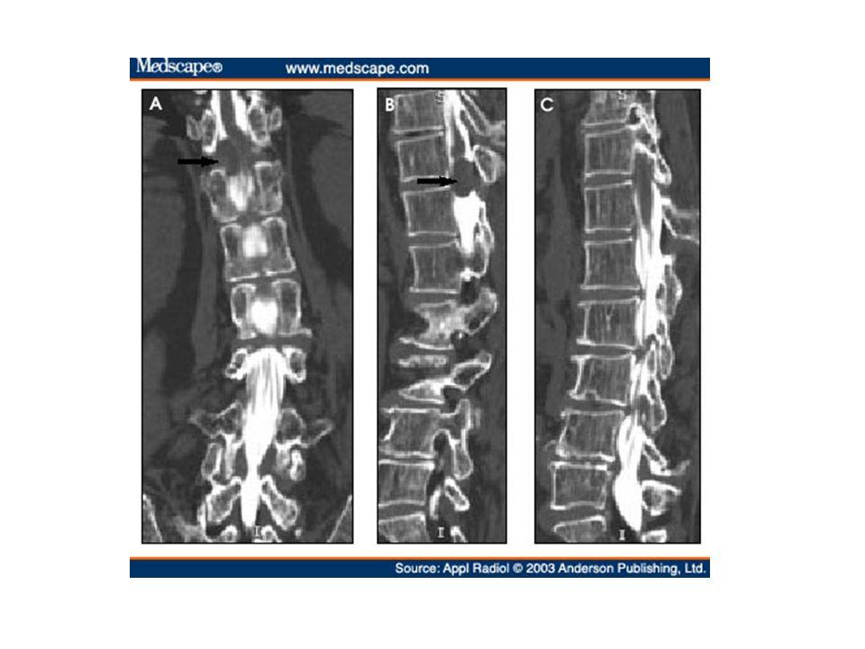
LUMBAR MYELOGRAM

90
IMAGES

Similar presentations


 by bony structures (vertebral body, facets, pedicles) as well as soft tissue structures (ligamentum.>")


 10.1 A 10.1 B 10.1 C Precontrast sagittal T1 wtd. MRI of.>")


