Download presentation
Presentation is loading. Please wait.

1
A tour of the cell

2
Cells All organisms are made of cells
The cell is the simplest collection of matter that can be alive Cell structure is correlated to cellular function All cells are related by their descent from earlier cells

3
Eukaryotic Cells – membranes to separate function

4
What is a cell? Basic features of all cells Plasma membrane
Semifluid substance called cytosol Chromosomes (carry genes) Ribosomes (make proteins)
 Ribosomes (make proteins)")
5
Prokaryotes

6
Eukaryotic vs prokaryotic
No nucleus Nucleoid Membrane-less organelles Eukaryotic Nucleus Nuclear envelope Organelles with membranes

7
Plasma membrane Cell wall Capsule Flagella
Figure 6.5 Fimbriae Nucleoid Ribosomes Plasma membrane Bacterial chromosome Cell wall Capsule 0.5 m Flagella (a) A typical rod-shaped bacterium (b) A thin section through the bacterium Bacillus coagulans (TEM) Figure 6.5 A prokaryotic cell.
 A typical rod-shaped bacterium. (b) A thin section through the bacterium Bacillus coagulans (TEM) Figure 6.5 A prokaryotic cell.")
8
Eukaryotic Cells Have plasma membranes selective barrier
TEM of a plasma membrane Eukaryotic Cells Outside of cell Have plasma membranes selective barrier double layer of phospholipids Inside of cell 0.1 m Carbohydrate side chains Hydrophilic region Hydrophobic region Hydrophilic region Phospholipid Proteins (b) Structure of the plasma membrane
 Structure of the plasma membrane.")
9
Surface area increases while total volume remains constant
5 1 1 Total surface area [sum of the surface areas (height width) of all box sides number of boxes] 6 150 750 Figure 6.7 Geometric relationships between surface area and volume. Total volume [height width length number of boxes] 1 125 125 Surface-to-volume (S-to-V) ratio [surface area volume] 6 1.2 6
 of all box sides number of boxes] Figure 6.7 Geometric relationships between surface area and volume. Total volume [height width length number of boxes] Surface-to-volume (S-to-V) ratio [surface area volume]")
10
Animal Cell ENDOPLASMIC RETICULUM (ER) Nuclear envelope Rough ER
Figure 6.8a Animal Cell ENDOPLASMIC RETICULUM (ER) Nuclear envelope Rough ER Smooth ER Flagellum NUCLEUS Nucleolus Chromatin Centrosome Plasma membrane CYTOSKELETON: Microfilaments Intermediate filaments Microtubules Ribosomes Figure 6.8 Exploring: Eukaryotic Cells Microvilli Golgi apparatus Peroxisome Mitochondrion Lysosome
 Nuclear envelope. Rough ER. Smooth ER. Flagellum. NUCLEUS. Nucleolus. Chromatin. Centrosome. Plasma membrane. CYTOSKELETON: Microfilaments. Intermediate filaments. Microtubules. Ribosomes. Figure 6.8 Exploring: Eukaryotic Cells. Microvilli. Golgi apparatus. Peroxisome. Mitochondrion. Lysosome.")
11
Animal Cells Fungal Cells 1 m Parent cell 10 m Cell wall Buds
Figure 6.8b Animal Cells Fungal Cells 1 m Parent cell 10 m Cell wall Buds Vacuole Cell 5 m Nucleus Nucleus Nucleolus Mitochondrion Human cells from lining of uterus (colorized TEM) Yeast cells budding (colorized SEM) A single yeast cell (colorized TEM) Figure 6.8 Exploring: Eukaryotic Cells
 Yeast cells budding (colorized SEM) A single yeast cell (colorized TEM) Figure 6.8 Exploring: Eukaryotic Cells.")
12
Rough endoplasmic reticulum
Figure 6.8c Nuclear envelope Rough endoplasmic reticulum Plant Cell NUCLEUS Smooth endoplasmic reticulum Nucleolus Chromatin Ribosomes Central vacuole Golgi apparatus Microfilaments Intermediate filaments CYTOSKELETON Microtubules Figure 6.8 Exploring: Eukaryotic Cells Mitochondrion Peroxisome Chloroplast Plasma membrane Cell wall Plasmodesmata Wall of adjacent cell

13
Plant Cells Protistan Cells Figure 6.8d Chlamydomonas (colorized SEM)
Flagella Cell 1 m 5 m 8 m Cell wall Nucleus Chloroplast Nucleolus Mitochondrion Vacuole Nucleus Nucleolus Chloroplast Chlamydomonas (colorized SEM) Cells from duckweed (colorized TEM) Cell wall Chlamydomonas (colorized TEM) Figure 6.8 Exploring: Eukaryotic Cells
 Cells from duckweed (colorized TEM) Cell wall. Chlamydomonas (colorized TEM) Figure 6.8 Exploring: Eukaryotic Cells.")
14
The Nucleus

15
The Nucleus: Home of Genetic Instructions
The nucleus contains most of the DNA in a eukaryotic cell Ribosomes use the information from the DNA to make proteins

16
Nuclear Envelope: Separation of Nucleus and cytoplasm
Double membrane of lipid bilayer Surrounds nucleus Tightly controlled

17
Close-up of nuclear envelope Chromatin
Figure 6.9a Nucleus Nucleolus Chromatin Nuclear envelope: Inner membrane Outer membrane Nuclear pore Rough ER Pore complex Ribosome Figure 6.9 The nucleus and its envelope. Close-up of nuclear envelope Chromatin

18
Surface of nuclear envelope
Figure 6.9b 1 m Nuclear envelope: Inner membrane Outer membrane Nuclear pore Figure 6.9 The nucleus and its envelope. Surface of nuclear envelope

19
Pore complexes regulate entry and exit of nucleus
Pore complexes (TEM) Nuclear lamina are a matrix of proteins that line the interior of the nuclear membrane Provide support to the envelope 1 m Figure 6.9 The nucleus and its envelope. Nuclear lamina (TEM)
 Nuclear lamina are a matrix of proteins that line the interior of the nuclear membrane. Provide support to the envelope. 1 m. Figure 6.9 The nucleus and its envelope. Nuclear lamina (TEM)")
20
Chromatin to chromosomes
DNA is organized into discrete units called chromosomes Each chromosome composed of a single DNA molecule associated with proteins The DNA and proteins of chromosomes together called chromatin The nucleolus located within the nucleus and is the site of ribosomal RNA (rRNA) synthesis
 synthesis.")
21
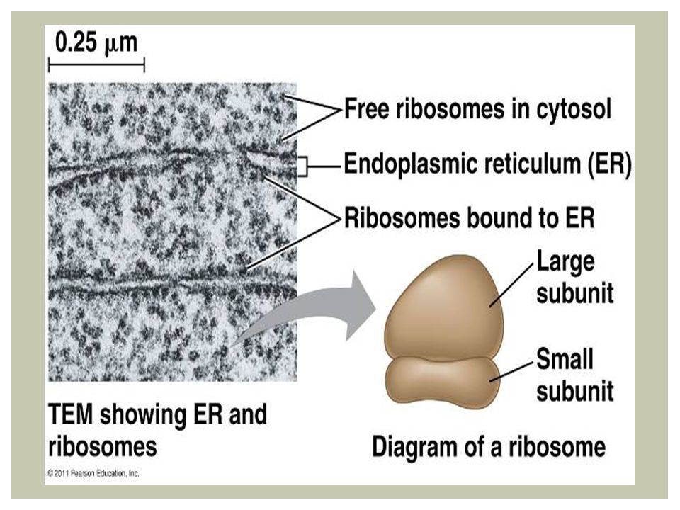
Ribosomes Made of ribosomal RNA and protein
Carry out protein synthesis in two locations

23
Endomembrane system

24
endomembrane regulates protein traffic and performs metabolic functions in the cell continuous or connected via transfer by vesicles Nuclear envelope Endoplasmic reticulum Golgi apparatus Lysosomes Vacuoles Plasma membrane

25
Smooth ER Rough ER Nuclear envelope ER lumen Cisternae Transitional ER Ribosomes Transport vesicle

26
Collectively account for more than ½ the membrane in a cell
ER Collectively account for more than ½ the membrane in a cell Membranous cisternae Lumen = internal space Nuclear Envelope continuous with lumen

27
200 nm Smooth ER Rough ER

28
Smooth vs Rough ER Synthesizes lipids Metabolizes carbohydrates
Smooth ER Rough ER Synthesizes lipids Metabolizes carbohydrates Detoxifies drugs and poisons Stores calcium ions Has bound ribosomes, which secrete glycoproteins Distributes transport vesicles Is a membrane factory for the cell

29
Ribosome Protein Lumen Endoplasmic Reticulum

30
Golgi Apparatus Made of flattened stacks = cisternae
2 distinct “sides” cis and trans Modify molecules as they move through Synthesizes macromolecules

31
Golgi Apparatus cis face (“receiving” side of Golgi apparatus) 0.1 m
Cisternae trans face (“shipping” side of Golgi apparatus) TEM of Golgi apparatus
 TEM of Golgi apparatus.")
32
Lysosomes Membranous sac of enzymes Used to digest macromolecules
Contents and membranes of lysozymes synthesized in ER ER protected by 3D structure

33
Intracellular digestion
Phagocytosis – engulf “food” Lysosomes fuse with food vacuole Create nutrients for cell Autophagy – recycling cell’s own materials Damage organelle tagged Surrounded by membrane Lysosome fuses

34
Vesicle containing two damaged organelles
Nucleus 1 m Mitochondrion fragment Lysosome Peroxisome fragment Digestive enzymes Lysosome Lysosome Peroxisome Plasma membrane Digestion Food vacuole Mitochondrion Digestion Vesicle (a) Phagocytosis (b) Autophagy
 Phagocytosis. (b) Autophagy.")
35
Animation: Lysosome Formation Right-click slide / select “Play”
© 2011 Pearson Education, Inc.

36
Vacuole diverse maintenance chambers
Central vacuole Membranous vesicle from ER Many functions: food, contractile, storage Function varies based on cell structure Cytosol Central vacuole Nucleus Cell wall Chloroplast 5 m

37
Nucleus Rough ER Smooth ER Plasma membrane Figure 6.15-1
Figure 6.15 Review: relationships among organelles of the endomembrane system. Plasma membrane

38
Nucleus Rough ER Smooth ER cis Golgi Plasma membrane trans Golgi
Figure Nucleus Rough ER Smooth ER cis Golgi Figure 6.15 Review: relationships among organelles of the endomembrane system. Plasma membrane trans Golgi

39
Nucleus Rough ER Smooth ER cis Golgi Plasma membrane trans Golgi
Figure Nucleus Rough ER Smooth ER cis Golgi Figure 6.15 Review: relationships among organelles of the endomembrane system. Plasma membrane trans Golgi

40
Mitochondria and chroloplasts

41
Mitochondria Mitochondria are the sites of cellular respiration
Turn O2 into ATP Have own DNA Double membrane Contain free ribosomes Grow independently of rest of cell

42
Free ribosomes in the mitochondrial matrix membrane
Figure 6.17a Intermembrane space Outer membrane DNA Inner Free ribosomes in the mitochondrial matrix membrane Cristae Figure 6.17 The mitochondrion, site of cellular respiration. Matrix 0.1 m (a) Diagram and TEM of mitochondrion
 Diagram and TEM of mitochondrion.")
43
Outer membrane Inner membrane Cristae Matrix 0.1 m
Figure 6.17aa Outer membrane Inner membrane Figure 6.17 The mitochondrion, site of cellular respiration. Cristae Matrix 0.1 m

44
Network of mitochondria in a protist cell (LM)
Figure 6.17b 10 m Mitochondria Mitochondrial DNA Figure 6.17 The mitochondrion, site of cellular respiration. Nuclear DNA (b) Network of mitochondria in a protist cell (LM)
 Network of mitochondria in a protist cell (LM)")
45
Chloroplasts Chloroplasts are the sites of photosynthesis
Thylakoids are membranous sacs stacked into granum Contain chlorophyll and other enzymes necessary for photosynthesis

46
(a) Diagram and TEM of chloroplast
Figure 6.18a Ribosomes Stroma Inner and outer membranes Granum DNA Figure 6.18 The chloroplast, site of photosynthesis. Thylakoid Intermembrane space 1 m (a) Diagram and TEM of chloroplast
 Diagram and TEM of chloroplast.")
47
Stroma Inner and outer membranes Granum 1 m Figure 6.18aa
Figure 6.18 The chloroplast, site of photosynthesis. 1 m

48
(b) Chloroplasts in an algal cell
Figure 6.18b 50 m Chloroplasts (red) Figure 6.18 The chloroplast, site of photosynthesis. (b) Chloroplasts in an algal cell
 Figure 6.18 The chloroplast, site of photosynthesis. (b) Chloroplasts in an algal cell.")
49
Endosymbiont Theory Endoplasmic reticulum Nucleus
Engulfing of oxygen- using nonphotosynthetic prokaryote, which becomes a mitochondrion Nuclear envelope Ancestor of eukaryotic cells (host cell) Mitochondrion Engulfing of photosynthetic prokaryote At least one cell Chloroplast Nonphotosynthetic eukaryote Mitochondrion Photosynthetic eukaryote
 Mitochondrion. Engulfing of photosynthetic prokaryote. At least one cell. Chloroplast. Nonphotosynthetic eukaryote. Mitochondrion. Photosynthetic eukaryote.")
50
peroxisome Peroxisomes are specialized metabolic compartments bounded by a single membrane Peroxisomes produce hydrogen peroxide and convert it to water How peroxisomes are related to other organelles is still unknown

51
1 m Chloroplast Peroxisome Mitochondrion Figure 6.19
Figure 6.19 A peroxisome.

52
Cell Separation

53
Centrifuged at 1,000 g (1,000 times the force of gravity) for 10 min
Figure 6.4 TECHNIQUE Homogenization Tissue cells Homogenate Centrifuged at 1,000 g (1,000 times the force of gravity) for 10 min Centrifugation Supernatant poured into next tube Differential centrifugation 20,000 g 20 min 80,000 g 60 min Pellet rich in nuclei and cellular debris Figure 6.4 Research Method: Cell Fractionation 150,000 g 3 hr Pellet rich in mitochondria (and chloro- plasts if cells are from a plant) Pellet rich in “microsomes” (pieces of plasma membranes and cells’ internal membranes) Pellet rich in ribosomes
 for 10 min. Centrifugation. Supernatant poured into next tube. Differential centrifugation. 20,000 g. 20 min. 80,000 g. 60 min. Pellet rich in nuclei and cellular debris. Figure 6.4 Research Method: Cell Fractionation. 150,000 g. 3 hr. Pellet rich in mitochondria (and chloro- plasts if cells are from a plant) Pellet rich in microsomes (pieces of plasma membranes and cells’ internal membranes) Pellet rich in ribosomes.")
54
Homogenization Tissue cells Homogenate Centrifugation TECHNIQUE
Figure 6.4a TECHNIQUE Homogenization Tissue cells Figure 6.4 Research Method: Cell Fractionation Homogenate Centrifugation

55
Centrifuged at 1,000 g (1,000 times the force of gravity) for 10 min
Figure 6.4b TECHNIQUE (cont.) Centrifuged at 1,000 g (1,000 times the force of gravity) for 10 min Supernatant poured into next tube Differential centrifugation 20,000 g 20 min 80,000 g 60 min Pellet rich in nuclei and cellular debris 150,000 g 3 hr Figure 6.4 Research Method: Cell Fractionation Pellet rich in mitochondria (and chloro- plasts if cells are from a plant) Pellet rich in “microsomes” Pellet rich in ribosomes
 Centrifuged at 1,000 g (1,000 times the force of gravity) for 10 min. Supernatant poured into next tube. Differential centrifugation. 20,000 g. 20 min. 80,000 g. 60 min. Pellet rich in nuclei and cellular debris. 150,000 g. 3 hr. Figure 6.4 Research Method: Cell Fractionation. Pellet rich in mitochondria (and chloro- plasts if cells are from a plant) Pellet rich in microsomes Pellet rich in ribosomes.")
Similar presentations


 date = Wednesday November 12 Test Date= Friday November 14.>")
, visible light passes through a specimen and then through glass lenses, which magnify the image The quality of an.>")
 Robert Hooke -- 1665: examined thinly sliced cork and coined term “cell”>")


 you have a test soon!.>")

>")


 (b) Structure of the plasma membrane Outside of cell Inside of cell 0.1 µm Hydrophilic region Hydrophobic region.>")
