Download presentation
Presentation is loading. Please wait.

1
Essentials of Human Anatomy & Physiology Copyright © 2003 Pearson Education, Inc. publishing as Benjamin Cummings Seventh Edition Elaine N. Marieb Chapter 12 The Lymphatic System and Body Defenses

2
Lymph Nodes Slide 12.6b Copyright © 2003 Pearson Education, Inc. publishing as Benjamin Cummings Figure 12.3

3
The Lymphatic System Slide 12.1 Copyright © 2003 Pearson Education, Inc. publishing as Benjamin Cummings Two parts Lymphatic vessels Lymphoid tissues and organs Functions Transport fluids back to the blood Essential roles in body defense and resistance to disease Absorb digested fat at the intestinal villi

4
Lymphatic Characteristics Slide 12.2 Copyright © 2003 Pearson Education, Inc. publishing as Benjamin Cummings Lymph – excess tissue fluid carried by lymphatic vessels Properties of lymphatic vessels One way system toward the heart No pump Lymph moves toward the heart Milking action of skeletal muscle Rhythmic contraction of smooth muscle in vessel walls

5
Lymphatic Vessels Slide 12.3b Copyright © 2003 Pearson Education, Inc. publishing as Benjamin Cummings Figure 12.1

6
Lymphatic Vessels Slide 12.4a Copyright © 2003 Pearson Education, Inc. publishing as Benjamin Cummings Lymphatic collecting vessels Collects lymph from lymph capillaries Carries lymph to and away from lymph nodes Figure 12.2

7
Lymphatic Vessels Slide 12.4b Copyright © 2003 Pearson Education, Inc. publishing as Benjamin Cummings Lymphatic collecting vessels (continued) Returns fluid to circulatory veins near the heart Right lymphatic duct Thoracic duct Figure 12.2
 Returns fluid to circulatory veins near the heart Right lymphatic duct Thoracic duct Figure")
8
Lymph Slide 12.5a Copyright © 2003 Pearson Education, Inc. publishing as Benjamin Cummings Materials returned to the blood Water Blood cells Proteins

9
Lymph Slide 12.5b Copyright © 2003 Pearson Education, Inc. publishing as Benjamin Cummings Harmful materials that enter lymph vessels Bacteria Viruses Cancer cells Cell debris

10
Lymph Nodes Slide 12.6a Copyright © 2003 Pearson Education, Inc. publishing as Benjamin Cummings Filter lymph before it is returned to the blood Defense cells within lymph nodes Macrophages – engulf and destroy foreign substances Lymphocytes – provide immune response to antigens

11
Lymph Nodes Slide 12.6b Copyright © 2003 Pearson Education, Inc. publishing as Benjamin Cummings Figure 12.3

12
Lymph Node Structure Slide 12.7b Copyright © 2003 Pearson Education, Inc. publishing as Benjamin Cummings Figure 12.4

13
Other Lymphoid Organs Slide 12.9 Copyright © 2003 Pearson Education, Inc. publishing as Benjamin Cummings Several other organs contribute to lymphatic function Spleen Thymus Tonsils Peyer’s patches Figure 12.5

14
The Spleen Slide 12.10 Copyright © 2003 Pearson Education, Inc. publishing as Benjamin Cummings Located on the left side of the abdomen Filters blood Destroys worn out blood cells Forms blood cells in the fetus Acts as a blood reservoir

15
The Thymus Slide 12.11 Copyright © 2003 Pearson Education, Inc. publishing as Benjamin Cummings Located low in the throat, overlying the heart Functions at peak levels only during childhood Produces hormones (like thymosin) to program lymphocytes
 to program lymphocytes.")
16
Tonsils Slide 12.12 Copyright © 2003 Pearson Education, Inc. publishing as Benjamin Cummings Small masses of lymphoid tissue around the pharynx Trap and remove bacteria and other foreign materials Tonsillitis is caused by congestion with bacteria

18
Peyer’s Patches Slide 12.13 Copyright © 2003 Pearson Education, Inc. publishing as Benjamin Cummings Found in the wall of the small intestine Resemble tonsils in structure Capture and destroy bacteria in the intestine

19
Mucosa-Associated Lymphatic Tissue (MALT) Slide 12.14 Copyright © 2003 Pearson Education, Inc. publishing as Benjamin Cummings Includes: Peyer’s patches Tonsils Other small accumulations of lymphoid tissue Acts as a guard to protect respiratory and digestive tracts

20
Body Defenses Slide 12.15a Copyright © 2003 Pearson Education, Inc. publishing as Benjamin Cummings The body is constantly in contact with bacteria, fungi, and viruses (pathogens) The body has two defense systems for foreign materials 1.Nonspecific defense system Mechanisms protect against a variety of invaders Responds immediately to protect body from foreign materials
 The body has two defense systems for foreign materials 1.Nonspecific defense system Mechanisms protect against a variety of invaders Responds immediately to protect body from foreign materials.")
21
Body Defenses Slide 12.15b Copyright © 2003 Pearson Education, Inc. publishing as Benjamin Cummings 2.Specific defense system Specific defense is required for each type of invader Also known as the immune system

22
Nonspecific Body Defenses Slide 12.16 Copyright © 2003 Pearson Education, Inc. publishing as Benjamin Cummings Body surface coverings Intact skin Mucous membranes Specialized human cells Chemicals produced by body Fever

23
Surface Membrane Barriers – First Line of Defense Slide 12.17a Copyright © 2003 Pearson Education, Inc. publishing as Benjamin Cummings The skin Physical barrier Acidic to inhibit bacterial growth Sebum is toxic to bacteria Vaginal secretions are very acidic

24
Surface Membrane Barriers – First Line of Defense Slide 12.17b Copyright © 2003 Pearson Education, Inc. publishing as Benjamin Cummings Stomach mucosa Secretes hydrochloric acid Has protein-digesting enzymes Saliva and lacrimal fluid contain lysozyme Mucus traps microogranisms in digestive and respiratory pathways

25
Defensive Cells Slide 12.18a Copyright © 2003 Pearson Education, Inc. publishing as Benjamin Cummings 1.Phagocytes (neutrophils and macrophages) Engulfs foreign material into a vacuole Enzymes from lysosomes digest the material Figure 12.6b
 Engulfs foreign material into a vacuole Enzymes from lysosomes digest the material Figure 12.6b.")
26
Phagocyte

27
Defensive Cells Slide 12.18b Copyright © 2003 Pearson Education, Inc. publishing as Benjamin Cummings 2.Natural killer cells Can lyse and kill cancer cells Can destroy virus- infected cells Figure 12.6b

28
Inflammatory Response - Second Line of Defense Slide 12.19 Copyright © 2003 Pearson Education, Inc. publishing as Benjamin Cummings Triggered when body tissues are injured Produces four cardinal signs Redness Heat Swelling Pain Results in a chain of events leading to protection and healing

29
Functions of the Inflammatory Response Slide 12.20 Copyright © 2003 Pearson Education, Inc. publishing as Benjamin Cummings Prevents spread of damaging agents Disposes of cell debris and pathogens Sets the stage for repair

30
Steps in the Inflammatory Response Slide 12.21 Copyright © 2003 Pearson Education, Inc. publishing as Benjamin Cummings Figure 12.7

31
Antimicrobial Chemicals Slide 12.22a Copyright © 2003 Pearson Education, Inc. publishing as Benjamin Cummings 1.Complement A group of at least 20 plasma proteins Activated when they encounter and attach to cells (complement fixation) Figure 12.8
 Figure")
32
Antimicrobial Chemicals Slide 12.22b Copyright © 2003 Pearson Education, Inc. publishing as Benjamin Cummings Complement (continued) Damage foreign cell surfaces Will rupture or lyse the foreign cell membrane Figure 12.8
 Damage foreign cell surfaces Will rupture or lyse the foreign cell membrane Figure")
34
Antimicrobial Chemicals Slide 12.22c Copyright © 2003 Pearson Education, Inc. publishing as Benjamin Cummings 2.Interferon Secreted proteins of virus-infected cells Bind to healthy cell surfaces to inhibit viruses binding

35
Fever Slide 12.23 Copyright © 2003 Pearson Education, Inc. publishing as Benjamin Cummings Abnormally high body temperature High temperatures inhibit the release of iron and zinc from liver and spleen needed by bacteria Increases the speed of tissue repair

36
Specific Defense: The Immune System – Third Line of Defense Slide 12.24 Copyright © 2003 Pearson Education, Inc. publishing as Benjamin Cummings Antigen specific – recognizes and acts against particular foreign substances Systemic – not restricted to the initial infection site Has memory – recognizes and mounts a stronger attack on previously encountered pathogens

37
Types of Immunity Slide 12.25 Copyright © 2003 Pearson Education, Inc. publishing as Benjamin Cummings Humoral immunity Antibody-mediated immunity Cells produce chemicals for defense Cellular immunity Cell-mediated immunity Cells target virus infected cells

38
Antigens (Nonself) Slide 12.26 Copyright © 2003 Pearson Education, Inc. publishing as Benjamin Cummings Any substance capable of exciting the immune system and provoking an immune response Examples of common antigens Foreign proteins Nucleic acids Large carbohydrates Some lipids Pollen grains Microorganisms

39
Self-Antigens Slide 12.27 Copyright © 2003 Pearson Education, Inc. publishing as Benjamin Cummings Human cells have many surface proteins Our immune cells do not attack our own proteins Our cells in another person’s body can trigger an immune response because they are foreign Restricts donors for transplants

40
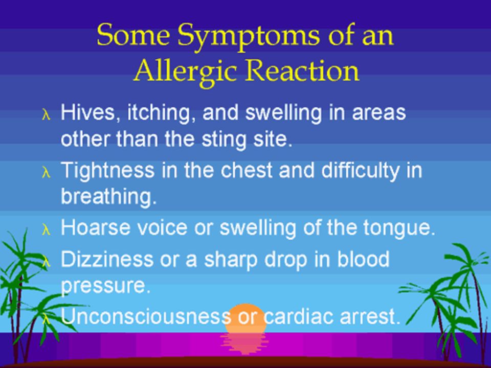
Allergies Slide 12.28 Copyright © 2003 Pearson Education, Inc. publishing as Benjamin Cummings Many small molecules (called haptens or incomplete antigens) are not antigenic, but link up with our own proteins The immune system may recognize and respond to a protein-hapten combination The immune response is harmful rather than protective because it attacks our own cells
 are not antigenic, but link up with our own proteins The immune system may recognize and respond to a protein-hapten combination The immune response is harmful rather than protective because it attacks our own cells.")
43
Cells of the Immune System Slide 12.29 Copyright © 2003 Pearson Education, Inc. publishing as Benjamin Cummings Lymphocytes B lymphocytes become immunocompetent in the bone marrow T lymphocytes become immunocompetent in the thymus Macrophages Arise from monocytes Become widely distributed in lymphoid organs

44
Activation of Lymphocytes Slide 12.30 Copyright © 2003 Pearson Education, Inc. publishing as Benjamin Cummings Figure 12.9

45
Humoral (Antibody-Mediated) Immune Response Slide 12.31a Copyright © 2003 Pearson Education, Inc. publishing as Benjamin Cummings B lymphocytes with specific receptors bind to a specific antigen The binding event activates the lymphocyte to undergo clonal selection A large number of clones are produced (primary humoral response)
.")
46
Humoral (Antibody Mediated) Immune Response Slide 12.31b Copyright © 2003 Pearson Education, Inc. publishing as Benjamin Cummings Most B cells become plasma cells Produce antibodies to destroy antigens Activity lasts for four or five days Some B cells become long-lived memory cells (secondary humoral response)
.")
47
Humoral Immune Response Slide 12.32 Copyright © 2003 Pearson Education, Inc. publishing as Benjamin Cummings Figure 12.10

48
Active Immunity Slide 12.34 Copyright © 2003 Pearson Education, Inc. publishing as Benjamin Cummings Your B cells encounter antigens and produce antibodies Active immunity can be naturally or artificially acquired Figure 12.12

49
Passive Immunity Slide 12.35 Copyright © 2003 Pearson Education, Inc. publishing as Benjamin Cummings Antibodies obtained from someone else Naturally from a mother to her fetus Artificially from immune serum Immunological memory does not occur Protection provided by “borrowed antibodies”

50
Antibodies (Immunoglobulins) (Igs) Slide 12.37 Copyright © 2003 Pearson Education, Inc. publishing as Benjamin Cummings Soluble proteins secreted by B cells (plasma cells) Carried in blood plasma Capable of binding specifically to an antigen
 Carried in blood plasma Capable of binding specifically to an antigen.")
51
Antibody Classes Slide 12.39 Copyright © 2003 Pearson Education, Inc. publishing as Benjamin Cummings Antibodies of each class have slightly different roles Five major immunoglobulin classes – (Do Not Need to know!) IgM – can fix complement IgA – found mainly in mucus IgD – important in activation of B cell IgG – can cross the placental barrier IgE – involved in allergies
 IgM – can fix complement IgA – found mainly in mucus IgD – important in activation of B cell IgG – can cross the placental barrier IgE – involved in allergies.")
52
Cellular (Cell-Mediated) Immune Response Slide 12.42 Copyright © 2003 Pearson Education, Inc. publishing as Benjamin Cummings Antigens must be presented by macrophages to an immunocompetent T cell (antigen presentation) T cells must recognize nonself and self (double recognition) After antigen binding, clones form as with B cells, but different classes of cells are produced
 T cells must recognize nonself and self (double recognition) After antigen binding, clones form as with B cells, but different classes of cells are produced.")
53
Cellular (Cell-Mediated) Immune Response Slide 12.43 Copyright © 2003 Pearson Education, Inc. publishing as Benjamin Cummings Figure 12.15

54
T Cell Clones Slide 12.44a Copyright © 2003 Pearson Education, Inc. publishing as Benjamin Cummings Cytotoxic T cells Specialize in killing infected cells Insert a toxic chemical (perforin) Helper T cells Recruit other cells to fight the invaders Interact directly with B cells
 Helper T cells Recruit other cells to fight the invaders Interact directly with B cells.")
56
T Cell Clones Slide 12.44b Copyright © 2003 Pearson Education, Inc. publishing as Benjamin Cummings Suppressor T cells Release chemicals to suppress the activity of T and B cells Stop the immune response to prevent uncontrolled activity A few members of each clone are memory cells

57
Summary of the Immune Response Slide 12.45 Copyright © 2003 Pearson Education, Inc. publishing as Benjamin Cummings Figure 12.16

58
Organ Transplants and Rejection Slide 12.46a Copyright © 2003 Pearson Education, Inc. publishing as Benjamin Cummings Major types of grafts Autografts – tissue transplanted from one site to another on the same person (ideal) Isografts – tissue grafts from an identical person (identical twin) (ideal) Allografts – tissue taken from an unrelated person (close tissue match) Xenografts – tissue taken from a different animal species (not successful)
 Isografts – tissue grafts from an identical person (identical twin) (ideal) Allografts – tissue taken from an unrelated person (close tissue match) Xenografts – tissue taken from a different animal species (not successful).")
59
Disorders of Immunity: Autoimmune Diseases Slide 12.50a Copyright © 2003 Pearson Education, Inc. publishing as Benjamin Cummings The immune system does not distinguish between self and nonself The body produces antibodies and sensitized T lymphocytes that attack its own tissues

60
Disorders of Immunity: Autoimmune Diseases Slide 12.50b Copyright © 2003 Pearson Education, Inc. publishing as Benjamin Cummings Multiple sclerosis – white matter of brain and spinal cord are destroyed Juvenile diabetes – destroys pancreatic beta cells that produce insulin Rheumatoid arthritis – destroys joints Systemic lupus erythematosus (SLE) – affects joints, kidney, heart, lung and skin Glomerulonephritis – impairment of renal function
 – affects joints, kidney, heart, lung and skin Glomerulonephritis – impairment of renal function.")
61
Disorders of Immunity: Immunodeficiencies Slide 12.49 Copyright © 2003 Pearson Education, Inc. publishing as Benjamin Cummings Production or function of immune cells or complement is abnormal May be congenital or acquired Includes AIDS – Acquired Immune Deficiency Syndrome

62
HIV targets cells Retrovirus attaches to CD4 receptors of T helper cells –Transmission: Body fluids, i.e., blood, semen, breast milk, vaginal secretions Immune Deficiency: AIDS

63
The Structure of HIV Figure 9.19

64
Time Course of the Progression of AIDS after HIV Infection Figure 9.21

65
AIDS progression: –Phase I: few weeks to a few years; flu like symptoms, swollen lymph nodes, chills, fever, fatigue, body aches. Virus is multiplying, antibodies are made but ineffective for complete virus removal –Phase II: within six months to 10 years; opportunistic infections present, Helper T cells affected, 5% may not progress to next phase –Phase III: Helper T cells fall below 200 per cubic millimeter of blood AND the person has an opportunistic infection or type of cancer. Person is now termed as having “AIDS” May include pneumonia, meningitis, tuberculosis, encephalitis, Kaposi’s sarcoma, and non-Hodgkin’s lumphoma….

66
More than 36 million infected with HIV worldwide Most infections in sub-Sahara of Africa Increasing spread in Asia and India Most often spread by heterosexual contact outside U.S. AIDS Pandemic

Similar presentations