Download presentation
Presentation is loading. Please wait.

1
Flexible Fiberoptic Bronchoscopy Chapter 16

2
Endoscopy Procedures that look into the body’s tubes and cavities – Colonoscopy – Esophagoscopy/Gastroscopy – Bronchoscopy Used to diagnose various diseases and explain conditions

3
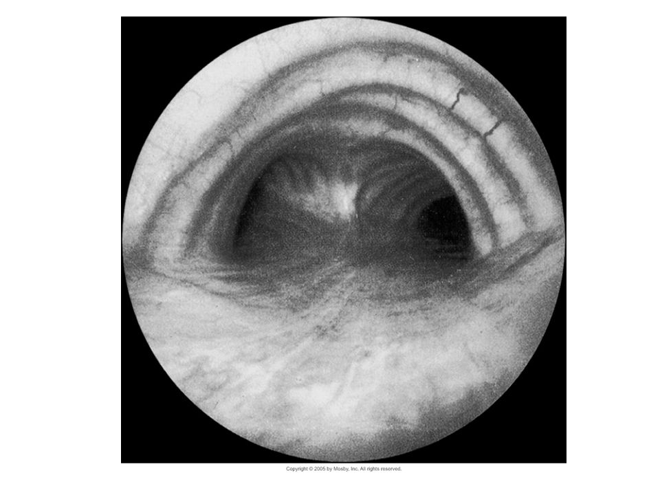
Bronchoscopy Allows visualization of the airways (tracheobronchial tree) Performed to diagnose problems with the airway or treat problems such as an object or growth in the airway
 Performed to diagnose problems with the airway or treat problems such as an object or growth in the airway")
4
Scopes Rigid bronchoscope Flexible Fiberoptic Scopes

5
Figure 4-4 Flexible fiberoptic bronchoscope. The four channels consist of two that provide a light source, one vision channel, and one open channel that accommodates instruments or allows administration of an anesthetic or oxygen.

7
Indications Abnormal CXR Excessive bronchial secretions Acute smoke inhalation injuries Hemoptysis Pneumonia Unexplained Cough Tracheal disease, stridor and localized wheezing Intubation damage Atelectasis Laser excision Removal of foreign bodies Lung lavage Difficult intubations Suctioning of excessive secretions, mucus plugs Selective lavage Management of life threatening hemoptysis

8
Classifications Direct visualization of the tracheobronchial tree for abnormalities (e.g., tumors, inflammation, strictures) Biopsy of tissue from observed lesions Aspiration of “deep” sputum for culture and sensitivity and for cytologic determinations Direct visualization of the larynx for identification of vocal cord paralysis, if present. With pronunciation of “eeee” the cords should move toward the midline. Aspiration of retained secretions in patients with airway obstruction or postoperative atelectasis Control of bleeding within the bronchus Removal of foreign bodies that have been aspirated Brachytherapy, which is endobronchial radiation therapy using an iridium wire placed via the bronchoscope Palliative laser obliteration of bronchial neoplastic

9
Biopsy Biting forceps Grasping forceps Shielded brushes Unshielded brushes Sheathed needles Sampling catheters

10
Foreign Body Retrieval Grasping forceps Snares

11
Flexible bronchoscopic view of a large foreign body (a Lite-Brite peg) lodged in the right main bronchus of a 7-year-old boy (left, A) Swanson K. L. et.al. Chest 2002;121:1695-1700 ©2002 by American College of Chest Physicians

12
BAL Tip of the scope is wedged into the bronchus Aliquots of sterile saline are instilled in to flood the alveoli A little more than half of the lavage is suctioned back to into a collection chamber Fluid contains cellular debris, microorganisms used for diagnosis

13
Interventional Bronchoscopy Laser Therapy – Thermal tissue damage to destroy obstructing lesions – Saline lavage to clean debris Cryotherapy – Tissue destruction via intracellular freezing – Bronchogenic carcinomas Stents – Tracheobronchial prostheses – May require opening the airway with other techniques prior to placement

14
Fluoroscopic Guidance Real time moving images of internal structures Allows precision in locating areas of interest Use with caution for both patient and health care providers

15
Role of the RCP Know the type of procedure being performed Preparing the patient Explain your role “Prep” the upper airway Prepare the equipment and workspace Establish monitoring Procedural sedation Observe safety protocols

16
Patient Preparation Explain the procedure to the patient. Allay any fears and allow the patient to verbalize any concerns. Obtain informed consent for this procedure. Keep the patient on nothing by mouth (NPO) status for 4 to 8 hours before the test to reduce the risk of aspiration. Instruct the patient to perform good mouth care to minimize the risk of introducing bacteria into the lungs during the procedure. Remove and safely store the patient's dentures, glasses, or contact lenses before administering the preprocedural medications. Administer the preprocedural medications as ordered. Atropine may be used to prevent vagal-induced bradycardia and to minimize secretions. Meperidine may be used to sedate the patient and relieve anxiety. Reassure the patient that he or she will be able to breathe during this procedure. Instruct the patient not to swallow the local anesthetic sprayed into the throat. Provide a basin for expectoration of the lidocaine.
 status for 4 to 8 hours before the test to reduce the risk of aspiration. Instruct the patient to perform good mouth care to minimize the risk of introducing bacteria into the lungs during the procedure. Remove and safely store the patient s dentures, glasses, or contact lenses before administering the preprocedural medications. Administer the preprocedural medications as ordered. Atropine may be used to prevent vagal-induced bradycardia and to minimize secretions. Meperidine may be used to sedate the patient and relieve anxiety. Reassure the patient that he or she will be able to breathe during this procedure. Instruct the patient not to swallow the local anesthetic sprayed into the throat. Provide a basin for expectoration of the lidocaine..")
17
Procedure – The patient's nasopharynx and oropharynx are anesthetized topically with lidocaine spray before the insertion of the bronchoscope. A bite block may be used. – The patient is placed in the sitting or supine position, and the scope is inserted through the nose or mouth and into the pharynx. – After the scope passes into the larynx and through the glottis, more lidocaine is sprayed into the trachea to prevent the cough reflex. – The scope is passed farther, well into the trachea, bronchi, and the first- and second-generation bronchioles, for systematic examination of the bronchial tree. – Biopsy specimens and washings are taken if a pathologic condition is suspected. – If bronchoscopy is performed for pulmonary toilet (removal of mucus), each bronchus is aspirated until clear. – Monitor the patient's oxygen saturation to be sure that the patient is well oxygenated. These patients often have pulmonary diseases that already compromise their oxygenation. When a scope is placed, breathing may be further impaired.
, each bronchus is aspirated until clear. – Monitor the patient s oxygen saturation to be sure that the patient is well oxygenated. These patients often have pulmonary diseases that already compromise their oxygenation. When a scope is placed, breathing may be further impaired..")
18
Post Procedure Instruct the patient not to eat or drink anything until the tracheobronchial anesthesia has worn off and the gag reflex has returned, usually in approximately 2 hours. Observe the patient's sputum for hemorrhage if biopsy specimens were removed. A small amount of blood streaking may be expected and is normal for several hours. Large amounts of bleeding can cause a chemical pneumonitis. Observe the patient closely for evidence of impaired respiration or laryngospasm. The vocal cords may go into spasms after intubation. Emergency resuscitation equipment should be readily available. Inform the patient that postbronchoscopy fever often develops within the first 24 hours. If a tumor is suspected, collect a postbronchoscopy sputum sample for a cytologic determination. Inform the patient that warm saline gargles and lozenges may be helpful if a sore throat develops. Note that a chest x-ray film may be ordered to identify a pneumothorax if a deep biopsy was obtained.

19
Potential Complications Fever Bronchospasm Hemorrhage (after biopsy) Hypoxemia Pneumothorax Infection Laryngospasm Aspiration Cardiac arrest – arrhythmias Respiratory depression hypotension Age-Related Concerns Children have a smaller bronchus. The bronchoscope can significantly decrease the available space for them to breathe. They are at higher risk of hypoxemia than adults.

Similar presentations


 into the atmosphere Filter, moisten,>")


>")


>")

 that is secreted into the airways of the respiratory tract(lungs,bronchi, trachea)>")
