Download presentation
Presentation is loading. Please wait.

1
Breast Pathology Seminar CASE PRESENTATION PART 1 Elba Torres Matundan MD FCAP Victor Carlo Vargas MD FCAP

2
What is your diagnosis?

3
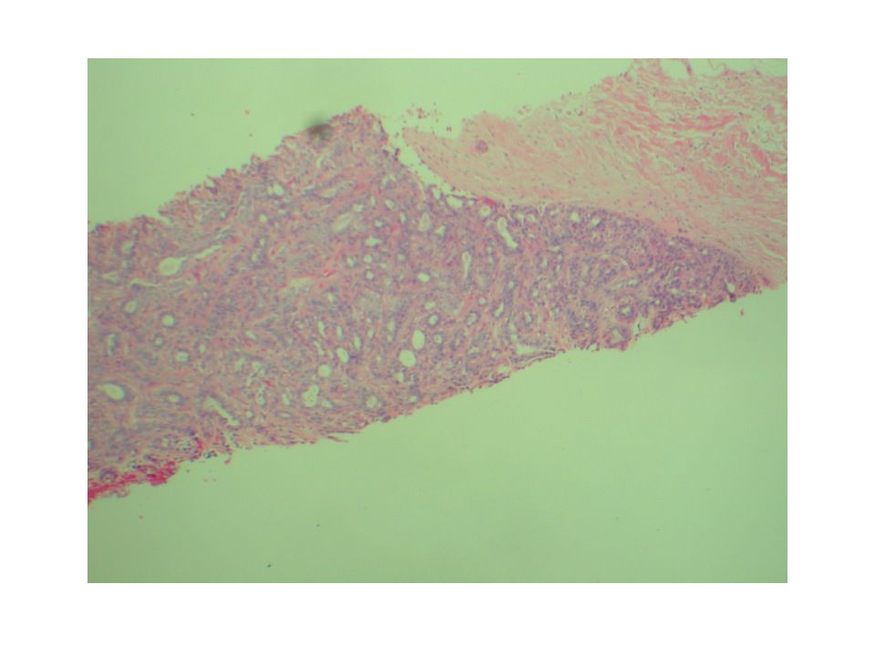
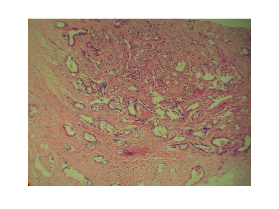
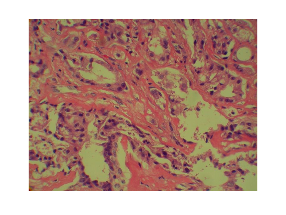
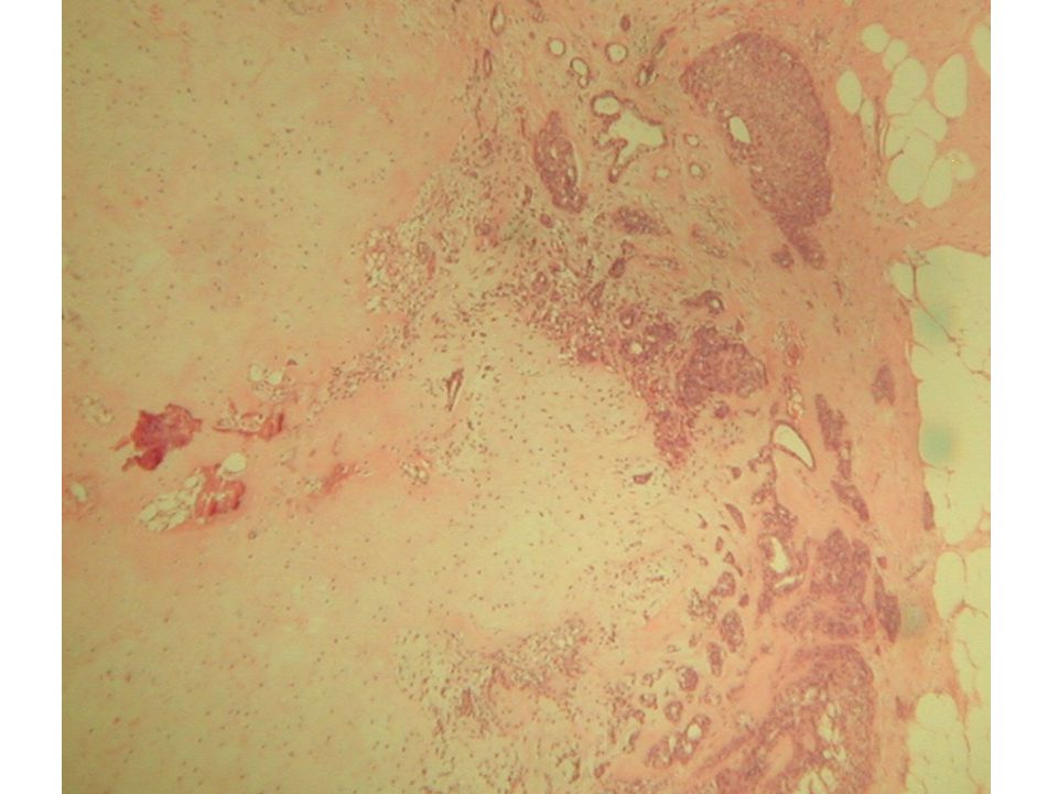
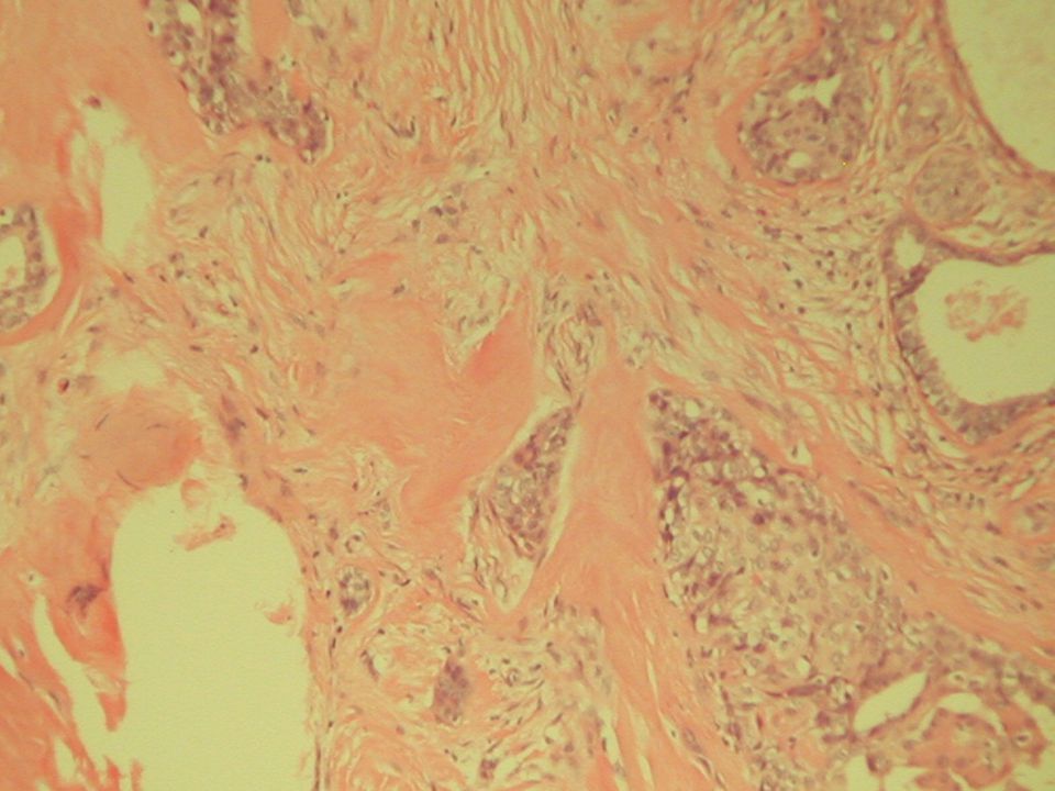
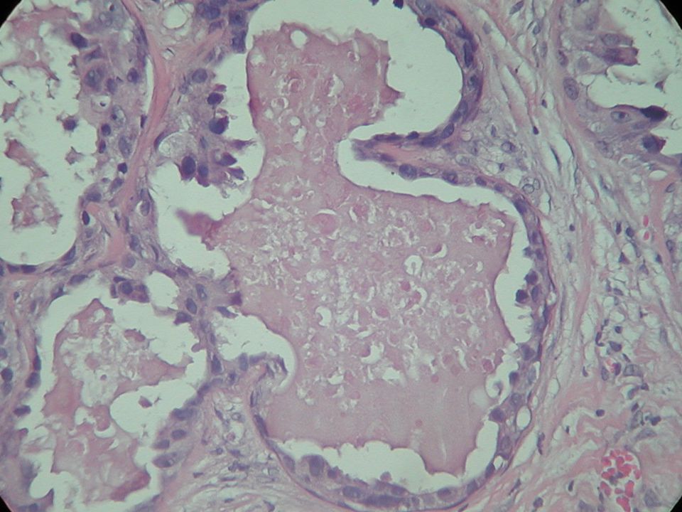
CASE 1 A 48 year old female was found to have a lobular mass on routine mammogram.

6
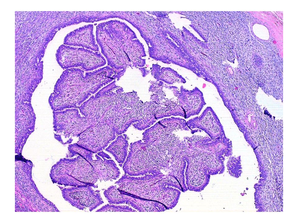
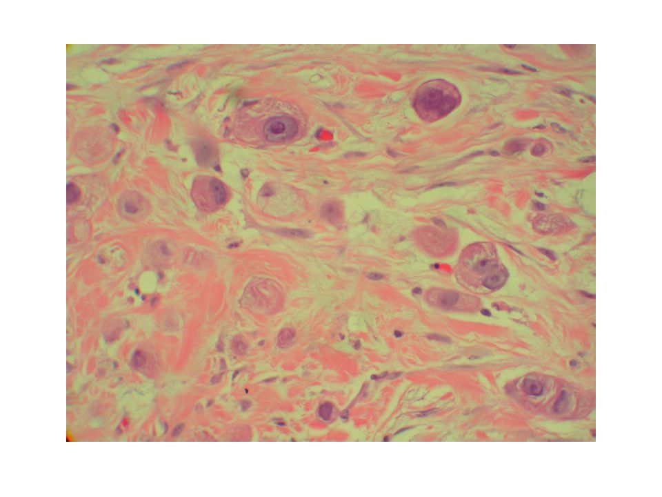
CASE 2 A 20 year old female with clinical history of cardiac myxoma and melanotic schwannomas was found to have a right breast mass.

8
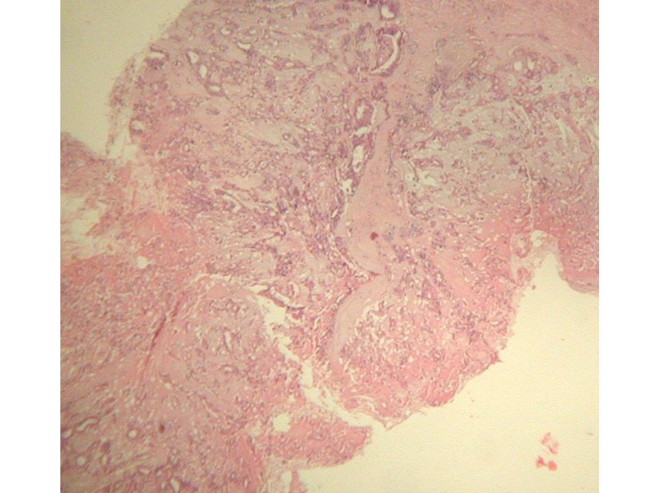
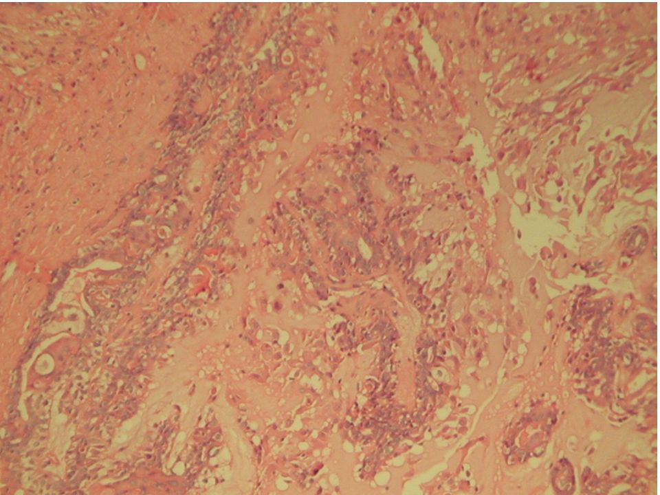
CASE 3 A 62 year old woman was found to have spiculated mass with microcalcifications on mammogram.

10
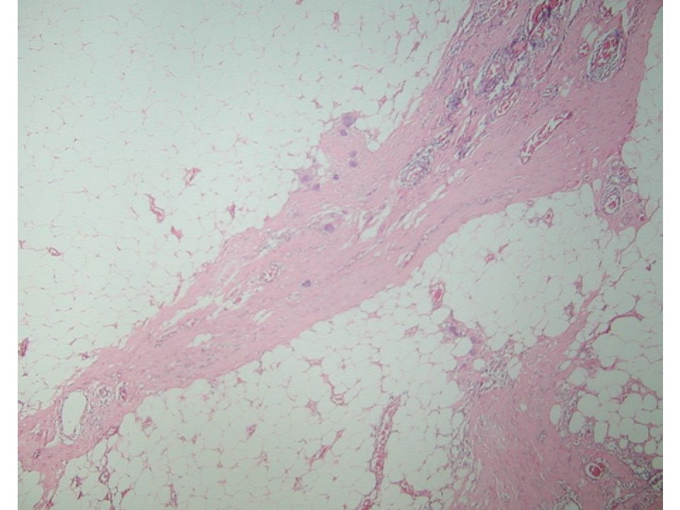
CASE 4 A 16 year old female with a palpable nodular lesion.

13
CASE 5 A 40 year old female was found to have a mobile, circumscribed, painless mass.

16
CASE 6 A 45 year old female with firm nodular mass.

19
CASE 7 A 12 year old female with a palpable nodular mass.

22
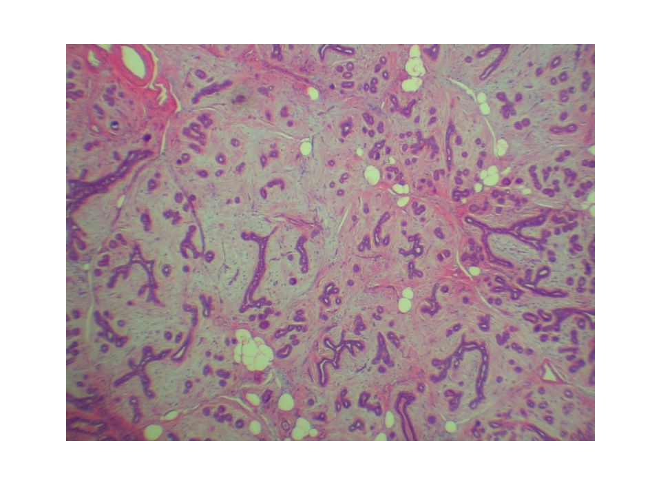
CASE 8 A 53 year old woman with history of invasive ductal carcinoma on the left breast. Now present with nodule on the same breast.

25
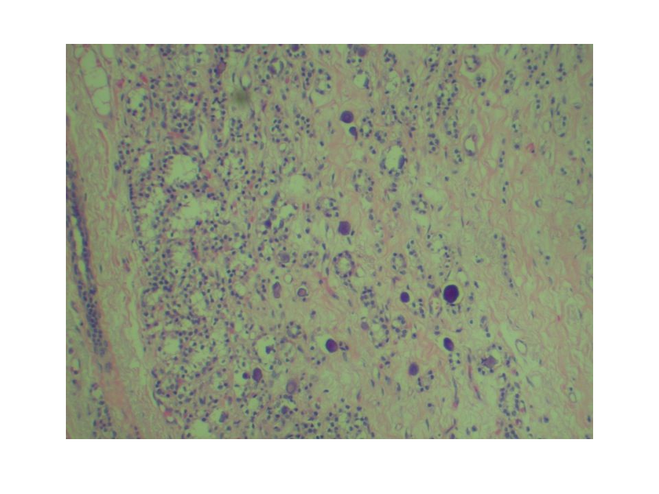
CASE 9 A 50 year old female with firm to hard palpable mass.

28
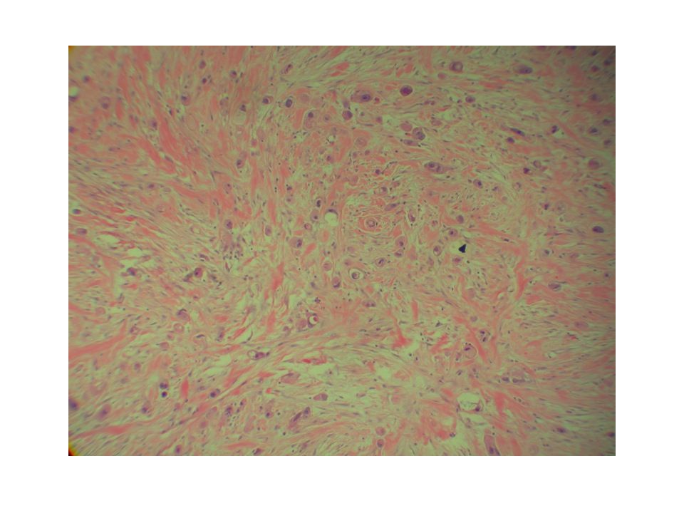
CASE 10 A 17 year old female presents with a clinically palpable mass

30
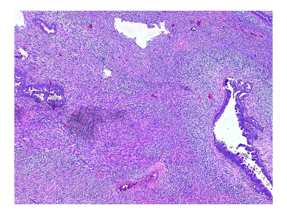
CASE 11 A 32 year old female with nodular palpable mass.

32
CASE 12 A 42 year old female with a history of two left prior breast biopsies for fibroadenomas over the past five years developed a large mass in the same breast.

36
CASE 13 A 53 year old female with calcifications on routine mammogram.

39
CASE 14 A 30 year old female with positive family history of breast cancer was found to have an ill-defined mass.

42
CASE 15 A 33 year old female was found to have a circumscribed nodular mass.

43
biopsy

45
excision

47
CASE 16 A 64 year old female with left breast invasive ductal carcinoma presents a right breast spiculated retroareolar lesion on mammogram.

49
CASE 17 A 25 year old female with positive family history of breast cancer with a nodular mass on the left breast.

51
CASE 18 A 47 year old female with history of invasive ductal carcinoma presents with a nodular lesion.

54
Breast Pathology Seminar CASE PRESENTATION PART 2 Elba Torres Matundan MD FCAP Victor Carlo Vargas MD FCAP

55
What is your diagnosis?

56
CASE 1 50 year old woman with breast density; What is your diagnosis? 1.Metaplastic carcinoma 2.Adenomyoepithelioma 3.Invasive carcinoma with apocrine features 4.Papilloma

62
Case 2 45 year old woman with retroareolar papilloma; What is the most relevant clinical history in order to make a diagnosis? 1.Is the mass ulcerated? 2.History of chemotherapy 3.History of radiation 4.History of prior core needle biopsy

68
Case 3 50 year old with palpable, well defined breast mass. What is your diagnosis? 1.Mixed tumor (pleomorphic adenoma) 2.Metaplastic carcinoma with heterologous elements 3.Invasive ductal carcinoma 4.Papillary carcinoma
 2.Metaplastic carcinoma with heterologous elements 3.Invasive ductal carcinoma 4.Papillary carcinoma.")
75
CASE 3 50 year old woman with calcifications; What is the greatest diagnostic pitfall (incorrect) with this kind of lesion seen on core biopsy? 1.Call it columnar cell change with atypia 2.Columnar cell change without atypia 3.Columnar cell hyperplasia with atypia 4.High grade DCIS, clinging pattern

78
THE END

Similar presentations