Download presentation
Presentation is loading. Please wait.

7
The patient is a 65 year old man with a history of hypertension and valvular heart disease who presented with spontaneous hemorrhage of the right lower extremity soft tissue. He had neither constitutional nor ocular symptoms and no evidence of muscular weakness. A chest x-ray showed an anterior mediastinal mass confirmed by CT scan. The mass was contiguous with the inferior thymus. CT-guided biopsy of the mass was performed and cytologic analysis of the aspirate showed keratin- and vimentin-positive spindle cells. The anterior mediastinal mass was surgically resected

12
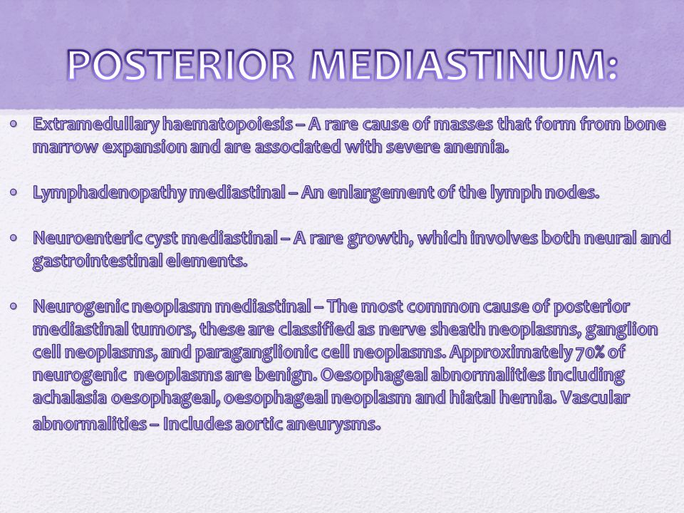
CAUSES The cause of mediastinal tumors is often unknown. Although the cause may be unknown, certain kinds of mediastinal tumors may be associated with other conditions. For example, thymoma can be associated with other conditions, such as myasthenia gravis, polymyositis, lupus erythematosus, rheumatoid arthritis, and thyroiditis.

13
SIGNS AND SYMPTOMS

14
COMPLICATIONS Complications of mediastinal tumors include: Spinal cord compression Spread to nearby structures such as the heart, lining around the heart (pericardium), and great vessels (aorta and vena cava) Radiation, surgery, and chemotherapy can all have serious complications.
, and great vessels (aorta and vena cava) Radiation, surgery, and chemotherapy can all have serious complications.")
15
DIAGNOSIS

16
Chest x-ray Computed tomography (CT) scan of the chest or CT-guided needle biopsy Magnetic resonance imaging (MRI) of the chest Mediastinoscopy with biopsy (Performed under general anesthesia, this examination of the chest cavity uses a lighted tube inserted through a small incision under the chest bone; a sample of tissue is taken to determine if cancer is present. Mediastinoscopy with biopsy allows doctors to accurately diagnose 80 to 90% of mediastinal tumors, and 95 to 100% of anterior mediastinal tumors.)
.")
17
TREATMENT The treatment used for mediastinal tumors depends on the type of tumor and its location: Thymic cancers require surgery, followed by radiation or chemotherapy. Types of surgery include thoracoscopy (a minimally invasive approach), mediastinoscopy (minimally invasive) and thoracotomy (a procedure performed through an incision in the chest). Lymphomas are recommended to be treated with chemotherapy followed by radiation. Neurogenic tumors found in the posterior (back) mediastinum are treated surgically.
, mediastinoscopy (minimally invasive) and thoracotomy (a procedure performed through an incision in the chest). Lymphomas are recommended to be treated with chemotherapy followed by radiation. Neurogenic tumors found in the posterior (back) mediastinum are treated surgically..")
18
CASE REPORT A 20 year old Caucasian man with no significant past medical history presented to his primary care physician for chest discomfort and cough. Two months prior to presentation, he reported having an unremarkable viral syndrome which resolved with no medical intervention. His primary care physician prescribed a short course of antibiotics for empiric treatment of pneumonia with some initial improvement in symptoms. His chest discomfort returned and he developed progressive dyspnea on exertion which led to a chest radiograph

20
CASE REPORT CONTD. An abnormality was noted in the left mediastinum which prompted his physician to order a computed tomography (CT) of the chest and to refer him to a pulmonary specialist. This CT scan revealed a rounded, well-demarcated mass in the superoanterior mediastinal compartment. The largest diameter measured 6.8 x 4.8cm
 of the chest and to refer him to a pulmonary specialist. This CT scan revealed a rounded, well-demarcated mass in the superoanterior mediastinal compartment. The largest diameter measured 6.8 x 4.8cm.")
22
CASE REPORT CONTD. Further review of systems revealed subjective fever/chills along with night sweats. He denied wheezing, hemoptysis, weight loss, scrotal mass, palpable lymph nodes or any neurologic deficits.He was a non-smoker and did not take any medications or use illicit drugs

23
PHYSICAL EXAM The patient was in no acute distress. Vital signs were unremarkable. Cardiac exam demonstrated regular rate and rhythm with no murmur, gallop or rub. Lungs were clear to auscultation bilaterally without wheezes or rales. Abdomen was soft with no hepato/splenomegaly. There was no palpable cervical, supraclavicular or axillary lymphadenopathy. Genitourinary exam was negative for testicular masses. Neurologic exam showed no focal deficits. Cranial nerves appeared intact.

24
http://path.upmc.edu/cases/case131/images/micro3.jpg http://www.thoracic.org/clinical/ats-clinical-cases/pages/12-12.php http://clinicaldepartments.musc.edu/anesthesia/intranet/educatio n/resident%20research/files/Catherine%20Tobin.pdfhttp://clinicaldepartments.musc.edu/anesthesia/intranet/educatio n/resident%20research/files/Catherine%20Tobin.pdf http://www.nlm.nih.gov/medlineplus/ency/article/001086.htm http://lungcancer.about.com/od/glossary/g/mediastinum.htm http://www.meddean.luc.edu/lumen/MedEd/radio/curriculum/Pul monary/mediastinum_teach.htmhttp://www.meddean.luc.edu/lumen/MedEd/radio/curriculum/Pul monary/mediastinum_teach.htm

Similar presentations


>")


 or a.>")
