Download presentation
Presentation is loading. Please wait.

1
Purpura and Vasculitis

2
Objectives To differentiate between different types of purpura.
To know the difference between inflammatory and non inflammatory purpura. To have an approach to diagnose purpuric lesions. To be familiar with serious and non serious conditions and when to refer to a specialist.

3
Purpura Reddish-purplish skin lesions from extravasation of RBCs into the skin. Non palpable purpura: classified according to size to Petechiae less than 2 mm, and Ecchymosis or bruises for larger lesions. Palpable purpura is the inflammatory type of different sizes, the main sign of vasculitis.

4
Purpura Petechiae Ecchymosis or bruises

5
Purpura causes Vascular damage from trauma, ageing, nutritional or vasculitis. Decreased platelets numbers or function. Coagulopathy like DIC, drug induced bleeding like heparin and warfarin. Or other clotting factors deficiency.

6
Vasculitis Large vessels: Giant cell arteritis, Takayasu's arteritis.
Medium sized vessels: polyarteritis nodosa, Kawasaki disease. Small vessels: Wegener's granulomatosis, Churg-Strauss syndrome, Microscopic polyangiits, Essential cryoglobulinemia vasculitis, Henoch-Schönlein purpura, Cutaneous small-vessel vasculitis (CSVV), and Urticarial vasculitis.
, and Urticarial vasculitis.")
7
Cutaneous small-vessel vasculitis (CSVV)
Also called Leukocytoclastic vasculitis. characterized by neutrophilic infiltration into the peripheral small dermal blood vessels. Purpura, urticaria, erythema-multiforme-like erythema, papules, nodules, pustules, blistering, erosion and ulceration occur, mainly in the lower extremities. An immune complex of an antigen (e.g., bacterium, virus, drug) and the antibody against that antigen deposit on the arteriolovenular walls. These activate the immune system and cause vasculitis (type III allergic reaction). Penicillin, sulfa drugs, thiazides, NSAIDs and other drugs, chemical substances, hemolytic streptococcus bacteria, or viruses may be the foreign antigen. Collagen diseases and antibodies against malignant tumors can also be causes.
 and the antibody against that antigen deposit on the arteriolovenular walls. These activate the immune system and cause vasculitis (type III allergic reaction). Penicillin, sulfa drugs, thiazides, NSAIDs and other drugs, chemical substances, hemolytic streptococcus bacteria, or viruses may be the foreign antigen. Collagen diseases and antibodies against malignant tumors can also be causes.")
8
Cutaneous small-vessel vasculitis (CSVV)
")
9
Pathology In the upper and middle dermal layer, fragments of nuclear debris and leakage of erythrocytes are found in the peripheral arteriola. Neutrophilic infiltration occurs in the arteriolovenous small blood vessels and capillaries. Thickening of the blood vessel walls and fibrinoid necrosis.

10
Investigations CBC, urea, creatinine. ESR ( usually raised)
Complements (decreased) Urinalysis ( for protein and hematuria if kidneys involved) Occult blood in stool ANA, ANCA Cryoglobulins , hepatitis B, C, HIV. CRP, ASO titer and throat swab culture for streptococcal infection Skin biopsy
 Urinalysis ( for protein and hematuria if kidneys involved) Occult blood in stool. ANA, ANCA. Cryoglobulins , hepatitis B, C, HIV. CRP, ASO titer and throat swab culture for streptococcal infection. Skin biopsy.")
11
Management When the cause is a drug or infection, those should be removed. For a lesion in the lower extremities, the legs should be raised and kept warm and the patient should get bed rest. Oral NSAIDs and dapsone is effective in relieving symptoms. Systemic corticosteroid therapy and immunosuppressant are useful for severe cases with systemic symptoms.

13
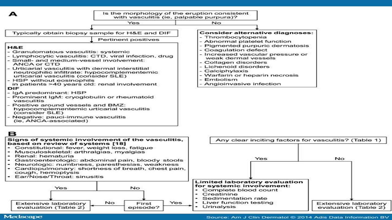
Table 5. Summary of key points for cutaneous small-vessel vasculitis.
Clinical presentation Classic: symmetric, partially blanchable palpable purpura on dependent areas (e.g., lower extremities) Variants: urticarial, ulcerative, pustular, vesicular, nodular, targetoid Histopathology Classic: polymorphonuclear cells (e.g., neutrophils) around postcapillary venules, with vessel wall fibrinoid deposits, endothelial swelling, and red blood cell extravasation Variants: granulomatous, lymphocytic, eosinophilic Differential diagnoses Thrombocytopenia, disorders of coagulation, embolic phenomena, pigmented purpuric dermatoses, microvascular occlusion Evaluation 1. Confirm diagnosis of vasculitis through clinicopathologic correlation (i.e., clinical morphology, skin biopsy) 2. Assess for an underlying cause of vasculitis (i.e., infection, medication, autoimmune connective tissue disease, inflammatory condition, malignancy) 3. Exclude systemic (extracutaneous) involvement of vasculitis Management 1. Treatment of underlying cause of vasculitis (if identified) 2. Other treatment options depend upon whether the vasculitis is isolated or recurrent; widespread or symptomatic; or associated with systemic involvement
 Variants: urticarial, ulcerative, pustular, vesicular, nodular, targetoid. Histopathology. Classic: polymorphonuclear cells (e.g., neutrophils) around postcapillary venules, with vessel wall fibrinoid deposits, endothelial swelling, and red blood cell extravasation. Variants: granulomatous, lymphocytic, eosinophilic. Differential diagnoses. Thrombocytopenia, disorders of coagulation, embolic phenomena, pigmented purpuric dermatoses, microvascular occlusion. Evaluation. 1. Confirm diagnosis of vasculitis through clinicopathologic correlation (i.e., clinical morphology, skin biopsy) 2. Assess for an underlying cause of vasculitis (i.e., infection, medication, autoimmune connective tissue disease, inflammatory condition, malignancy) 3. Exclude systemic (extracutaneous) involvement of vasculitis. Management. 1. Treatment of underlying cause of vasculitis (if identified) 2. Other treatment options depend upon whether the vasculitis is isolated or recurrent; widespread or symptomatic; or associated with systemic involvement.")
14
Henoch-Schönlein purpura (HSP)
A specific type of cutaneous small-vessel vasculitis. Mostly affects children. May be preceded by symptoms of upper respiratory tract infection. Presents as multiple palpable purpura occur mostly in the lower legs, buttocks and to lesser extent on forearms. could be accompanied by arthralgia, abdominal pain, nausea, vomiting, melena and kidney involvement with hematuria , proteinuria, or acute nephritis.

15
Henoch-Schönlein purpura (HSP)
")
16
Henoch-Schönlein purpura (HSP)
In children, the onset is mostly after upper respiratory infection; association with hemolytic streptococcus has been pointed out. Drugs (penicillin, aspirin) These antigens combine with antibodies (mainly IgA) in the body, and the immunocomplex deposits on the vascular walls. Immunoreaction is induced to cause vasculitis and purpura. Pathology shows leukocytoclastic vasculitis with IgA deposition is observed by direct immunofluorescence. The histology of the kidney in HSP patients often shows crescentic glomerulonephritis.
 These antigens combine with antibodies (mainly IgA) in the body, and the immunocomplex deposits on the vascular walls. Immunoreaction is induced to cause vasculitis and purpura. Pathology shows leukocytoclastic vasculitis with IgA deposition is observed by direct immunofluorescence. The histology of the kidney in HSP patients often shows crescentic glomerulonephritis.")
17
Henoch-Schönlein purpura (HSP)
Treatment by bed rest, pain relief and antibiotics if strep throat infection is present. Systemic steroids for severe cases with systemic involvement. HSP generally has a good prognosis and resolves within several weeks in most cases; however, it may recur. Serious complications may occur in other organs, such as nephritis with IgA deposition in the mesangium area, enterorrhagia, intussusception, intestinal perforation, or cerebral hemorrhage. Adults may have prolonged kidney involvement.

18
Urticarial vasculitis
Presents with urticarial weals that lasts more than 24 hours unlike urticaria. And usually leave hyperpigmentation after resolution. Could be associated with fever, arthralgia, abdominal pain and angioedema especially in hypocomplementaemic urticarial vasculitis that is associated with SLE. Causes include connective tissue diseases, viral and bacterial infections, drugs like ACE inhibitors, penicillin, sulfonamides, fluoxetine and thiazides. Also leukemia could cause it. Most cases are idiopathic and improve spontaneously after few months. Treatment is to remove the offending agents if present, treat the underlying diseases. Symptomatic therapy with antihistamines and NSAIDs. Dapsone, colchicine, hydroxycholorquine, steroids, azathioprine, and cyclosporine.

19
Urticarial vasculitis

20
Cutaneous polyarteritis nodosa
a rare form of vasculitis which involves small and medium sized arteries of dermis and subcutaneous tissue. tender subcutaneous nodules and livedo reticularis that may ulcerate on legs and feet. Pathogenesis is unknown. Systemic involvement is uncommon except for peripheral neuropathy, and progression to classical polyarteritis nodosa is an exception. may have neuromuscular involvement in the form of peripheral neuropathy that presents with tingling, numbness, sensory disturbances, weakness, and absent reflexes.

21
Cutaneous polyarteritis nodosa
In cutaneous PAN, histopathological examination shows features of nodular arteritis with polymorphonuclear infiltrates involving medium sized arteries in deep reticular dermis. There is extensive fibrinoid necrosis. This is in contrast to classical PAN which rarely shows nodular arteritis and the picture is of small vessel leukocytoclastic. Cutaneous PAN runs a chronic course lasting months to years, and has a waxing and waning phenomenon. Patients are generally treated with non-steroidal anti-inflammatory drugs and oral steroids. Immunosuppressive drugs can also be used in low doses in more severe kinds of cutaneous PAN and as steroid-sparing drugs.

22
Cutaneous polyarteritis nodosa

23
pigmented purpuric dermatoses
a group of chronic diseases of mostly unknown etiology characterized by extravasation of erythrocytes in the skin with marked hemosiderin deposition. Mostly affect males. Venous hypertension, exercise, and gravitational dependency are important cofactors that appear to influence disease presentation. Histologically, a perivascular T-cell lymphocytic infiltrate is centered on the superficial small blood vessels of the skin, which show signs of endothelial cell swelling and narrowing of the lumen. Extravasation of red blood cells with marked hemosiderin deposition in macrophages is also found but no vasculitis. The lesions are chronic and persist for years. With time, many of the lesions tend to extend and may become darker brown in color. No effective treatment available. It is a cosmetic problem mostly.

24
pigmented purpuric dermatoses

Similar presentations


 Dr. Raid Jastania. Vasculitis Inflammation of the walls of the vessels Causes of inflammation: –Infectious, physical, chemical,>")


 Ulcerative colitis is an inflammatory bowel disease (IBD) that causes chronic inflammation of the digestive tract It is.>")

 Aneurysms & Dissections Veins & Lymphatics Tumors.>")


 is the sudden onset of: – Haematuria (macroscopic/microscopic)>")

 Caused by renal diseases that increase the permeability across the glomerular filtration.>")