Download presentation
Presentation is loading. Please wait.

1
Blood Vessels Human Anatomy Chapter 19

2
The blood vessels of the body form a closed circulatory system
The blood vessels of the body form a closed circulatory system. Blood is pumped from the heart to the body and returned to the heart. Blood vessels have the ability to constrict or dilate and increase or decrease pressure. Blood vessels may divide and grow as the body requires. Arteries carry blood away from the heart and veins bring blood towards the heart. I. Structure of blood vessels walls Blood vessels have three layers or tunics in a circular fashion that create a cavity called the lumen: a. Tunica Intima- contains endothelium (simple squamous epithelium). In vessels with a small diameter the endothelium is surrounded by a thin layer of loose connective tissue called subendothelial layer.
. In vessels with a small diameter the endothelium is surrounded by a thin layer of loose connective tissue called subendothelial layer.")
3
b. Tunica Media- this layer is the thickest layer composed of smooth muscle arranged in circular sheets and connective tissue. The sympathetic NS controls the smooth muscle activity. Vasoconstriction occurs when the smooth muscle contracts, this decreases the flow of blood. Vasodilation occurs when the smooth muscle relaxes, increases the diameter and thus the blood flow.

4
c. Tunica externa- Outer most layer, composed of connective tissue
c. Tunica externa- Outer most layer, composed of connective tissue. It is designed to protect, stretches and anchors the blood vessels. d. Lumen- cavity in the middle of the blood vessel that is filled with blood. The diameter of the lumen changes with vasoconstriction or vasodilation. It can also be influenced by plaque build up.

5
Artery & vein cross section

6
Artery Wall- notice thick muscular layer.

7
Vein Wall Capillary walls

8
II. Types of blood vessels
All three vessel types are found in systemic and pulmonary circulation. A. arteries- The closer the artery is to the heart the larger its diameter and the thicker its walls. As the artery branches it becomes smaller in diameter and thinner until it becomes a capillary. Because arteries have a thick muscle layer they have the ability to change blood pressure. 1. Elastic arteries- (2.5-1 cm) largest, closer to the heart, have thick sheets of elastin in the tunica media, also called conducting arteries, able to withstand large fluctuations in blood pressure. 2. Muscular arteries- (1cm-0.33 mm) distal to elastic arteries, they reach the organs, their tunica media is thicker in diameter than the lumen. Also the tunica media is lined with layer of connective tissue of both sides (internal elastic lamina and external elastic lamina). The muscular layer constricts the vessel as needed by each individual organ. 3. Arterioles- (0.3mm-10 mm) They have the smallest diameter, the tunicas are considerably thinner but are still innervated to induce vasoconstriction or vasodilation. They connect to capillaries.
 largest, closer to the heart, have thick sheets of elastin in the tunica media, also called conducting arteries, able to withstand large fluctuations in blood pressure. 2. Muscular arteries- (1cm-0.33 mm) distal to elastic arteries, they reach the organs, their tunica media is thicker in diameter than the lumen. Also the tunica media is lined with layer of connective tissue of both sides (internal elastic lamina and external elastic lamina). The muscular layer constricts the vessel as needed by each individual organ. 3. Arterioles- (0.3mm-10 mm) They have the smallest diameter, the tunicas are considerably thinner but are still innervated to induce vasoconstriction or vasodilation. They connect to capillaries.")
9
Cross-section of elastic artery

10
B. Capillaries- these are the smallest blood vessels (8-10mm) forcing RBC to pass by in one after the other in line. They are thin enough to allow for chemical exchange, only composed of a single layer or endothelial cells lined by basal lamina. This is also the site through which WBC exit the blood stream.
 forcing RBC to pass by in one after the other in line. They are thin enough to allow for chemical exchange, only composed of a single layer or endothelial cells lined by basal lamina. This is also the site through which WBC exit the blood stream..")
11
1. Capillary beds- this is a network or capillaries that is connected to a terminal arteriole and postcapillary venule. The terminal arteriole branches into the metarteriole (intermediate between arteriole and true capillary) that further branches into the capillary bed. Throughout the metarteriole there are precapillary sphincters (smooth muscle) that control the flow of blood into the true capillaries. The capillaries connect into the thoroughfare channel (intermediate between capillaries and venule) that connects the venule.
 that further branches into the capillary bed. Throughout the metarteriole there are precapillary sphincters (smooth muscle) that control the flow of blood into the true capillaries. The capillaries connect into the thoroughfare channel (intermediate between capillaries and venule) that connects the venule..")
12
2. Capillary permeability- small molecules enter and leave the capillaries through intercellular clefts. To increase permeability capillaries containing pores, called fenestrated capillaries, are located in areas of high exchange. All other capillaries are called continuous, there are no pores. Permeability happens in different forms: a. direct diffusion- as with oxygen and carbon dioxide b. intercellular cleft- as with WBC, most common c. cytoplasmic vesicles (caveolae)- as when large molecules like proteins and carbohydrates are transferred from the intestine into the blood. d. fenestrations- in areas of large exchange as when water exits blood to make synovial fluid within the synovial joints. The capillaries that have the lowest permeability are those of the blood brain barrier. They are only permeable to lipid soluble substances and components such as oxygen and carbon dioxide.
- as when large molecules like proteins and carbohydrates are transferred from the intestine into the blood. d. fenestrations- in areas of large exchange as when water exits blood to make synovial fluid within the synovial joints. The capillaries that have the lowest permeability are those of the blood. brain barrier. They are only permeable to lipid soluble substances and. components such as oxygen and carbon dioxide.")
13
Continous Capillary Fenestrated Capillary
Sinusoidal Capillary

15
3. Sinusoids- wide leaky fenestrated capillaries in areas of extensive change and crossing or large materials. These may be sites in which cells move into the blood like it happens in bone marrow and spleen.

16
C. Veins-blood vessels that carry blood from the capillaries to the heart. As they move away from the capillaries thay increase in diameter. The blood pressure in veins is less than in arteries and their walls are also thinner than arteries. 1. Venules- (8-100 mm) they are the smallest and thinnest, specially as they are closer to the capillaries. As they approach the veins they become thicker. 2. Veins- lumens vary in size but compared to arteries they are larger. Their tunica externa is thicker than tunica media. The largest veins are the vena cavae the tunica externa also has bands of smooth muscle. 3. Vein valves- these assist in the transport of blood back to the heart. They prevent back flow. Similar to the valves of the heart, these valve have cusps formed by the endothelial cells of the tunica intima. In areas of the body that blood flow is directly against gravity there are more valves. There are no valves in veins of the thoracic and abdominal cavities. 4. Returning blood to the heart- normal body movements produce muscle contractions that bring blood back to the heart. Skeletal muscle contractions force the valves to open and propel blood towards the heart.
 they are the smallest and thinnest, specially as they are closer to the capillaries. As they approach the veins they become thicker. 2. Veins- lumens vary in size but compared to arteries they are larger. Their tunica externa is thicker than tunica media. The largest veins are the vena cavae the tunica externa also has bands of smooth muscle. 3. Vein valves- these assist in the transport of blood back to the heart. They prevent back flow. Similar to the valves of the heart, these valve have cusps formed by the endothelial cells of the tunica intima. In areas of the body that blood flow is directly against gravity there are more valves. There are no valves in veins of the thoracic and abdominal cavities. 4. Returning blood to the heart- normal body movements produce muscle contractions that bring blood back to the heart. Skeletal muscle contractions force the valves to open and propel blood towards the heart.")
18
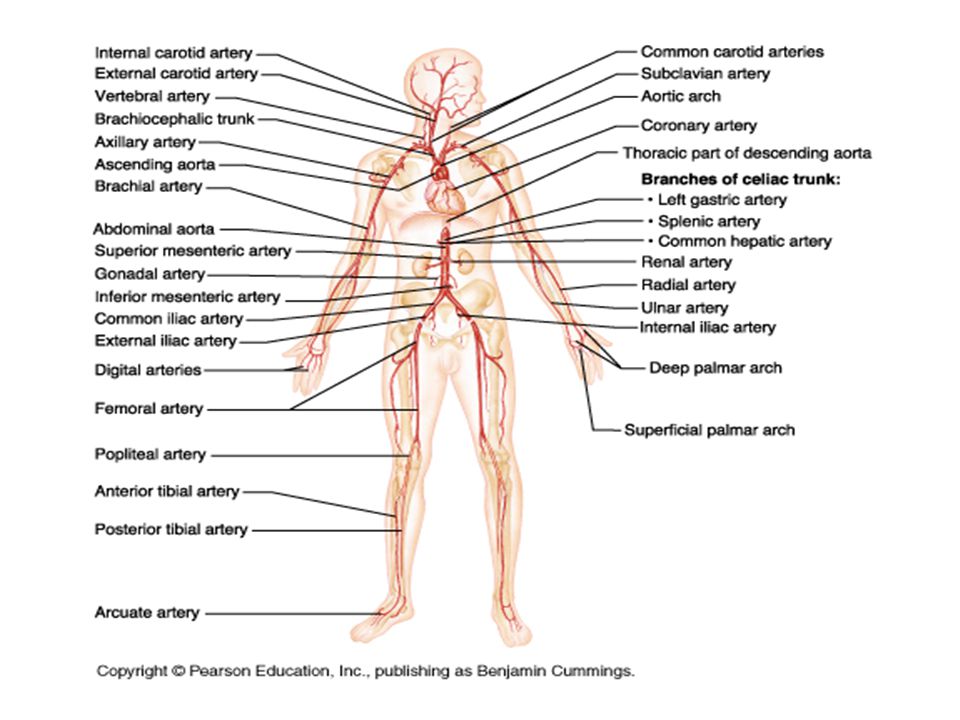
D. Vascular Anastomoses- areas where several blood vessels unite or interconnect, they may be arteries or veins. Anastomoses may provide alternative routes for blood flow. They occur around organs like the heart, brain, or abdominal organs or around joints. Blood flow blockage of areas where there are no arterial anastomoses may result in severe tissue damage. E. Vasa vasorum- tiny arteries, capillaries, and veins in larger blood vessels to provide their own blood supply and nourishment for the outer half of the wall layers. These tiny blood vessels may arise from the same blood vessels it nourishes or those near by. Part 2: Blood vessels of the body- Focus on knowing the names of the blood vessel given to you in the practicum list. Also study the diagrams through out the book so you can visualize the pattern the blood vessels take throughout the body.

19
I. The pulmonary circulation- blood vessels that travel from the heart to the lungs, within the lungs, and back to the heart. Includes all types of vessels. Arteries carry deoxygenated blood and veins carry oxygenated blood.

20
II. The systemic circulation-blood vessels that travel from the heart throughout the body and back to the heart. Arteries carry oxygenated blood and veins carry deoxygenated blood. A. Systemic arteries 1. aorta 2. arteries of the head and neck 3. arteries of the upper limbs 4. arteries of the thorax 5. arteries of the abdomen 6. arteries of the pelvis and lower limbs B. Systemic veins 1. venae cavae and their major tribuatires 2-6 same as those for arteries 7. Portal-systemic anastomoses

23
Blood Vessels entering or leaving the heart

29
View of iliac and femoral arteries

38
Vascular system within the liver

41
III. Disorders of the blood vessles-
a. Arthrosclerosis- hardening of artery due to fatty deposits. The artery loses its flexibility. b. Aneurysm- widening or out pocketing or an artery or vein increasing the changes of the vessel rupturing. It may result from weaken walls c. Deep vein thrombosis of the lower limb- formation of clots in lower legs. The clot can detach, travel through the body and cause a embolism or stroke. d. Venous disease- inadequate drainage of lower limbs due to failure of valves, it can possibly causing ulceration. e. Microangiopathy of diabetes- common long term diabetes mellitus complication- patient had thicken but leaky capillaries slowing tissue fluid flow affecting delicate organs like the kidneys, and retina, and nerves. f. Arteriovenous malformation- congenital condition in which capillaries do not form and arteries connect directly into veins. Usually occurs in cerebrum. The vein becomes weaken an aneurysm can occur. Fetal Circulation- study figure on page 555

Similar presentations


 Structure of Blood Vessels 1. Tunica Intima – simple squamous epithelium 2. Tunica Media – sheets of smooth muscle Contraction.>")


