Download presentation
Presentation is loading. Please wait.

1
Trachea diseases and esophagus diseases The Otolaryngology Faculty of Shanghai Jiao Tong University School of Medicine Xiang Mingliang

2
Anatomy of trachea and bronchus

3
Trachea: in the middle of neck and thoracic cavity. 16-20 trachea rings in total, and 7- 8 ring in the neck.

4
Bronchus tree. The angle between the trachea and the bronchus: left45 o, right25 o

5
carina of trachea: the end of trachea. A important mark in the bronchoscopy

6
Anatomy of esophagus

7
Muscular organ. Pipe in shape Inner layer: mucosa Middle layer: submucosal tissue external layer: muscle

8
Physiological stenosis in esophagus 1 ) from upper incisor 16cm 2 ) from upper incisor 23cm 3 ) from upper incisor 27cm 4 ) from upper incisor 36cm
 from upper incisor 16cm 2 ) from upper incisor 23cm 3 ) from upper incisor 27cm 4 ) from upper incisor 36cm")
9
foreign body in trachea and bronchus Source : external 、 internal Classification of foreign body : plant : peanut 、 seeds. animal : fishbone 、 bone pieces mineral : iron nail synthetic product : dental prosthesis

10
90% cases ﹤ 5 years, 80% cases ﹤ 3 years causes : 1 ) poorly chew function, poorly pharyngeal reflex 2 ) unconscious , aspiration 3) unhealthy habit: foreign body in mouth 4 ) medical:
 poorly chew function, poorly pharyngeal reflex 2 ) unconscious , aspiration 3) unhealthy habit: foreign body in mouth 4 ) medical:")
11
Factors associated with clinic presentation foreign body sorts : peanut , chemical bronchitis Size: Shape: Stay time: Stay site Infection or not:

12
presentation –Inhaled period : cough , dyspnea –Quiet period : no any symptom –Infection period : cough , dyspnea, fever –Complication period : atelectasis 、 emphysema 、 lung infection

13
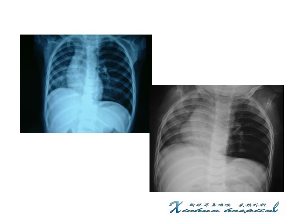
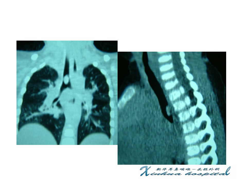
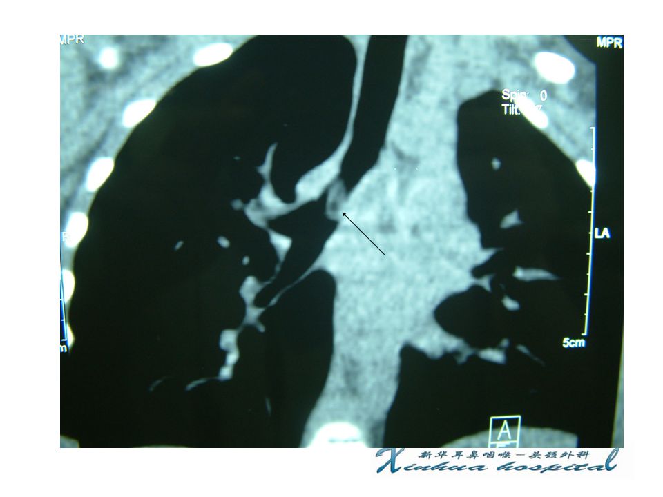
diagnosis : history of foreign body , dyspnea 、 cough , disorder sign in X-ray X-ray : 1 ) obstructive emphysema 2 ) obstructive atelectasis CT scan : three-dimensional rebuild treatment : bronchoscopy
 obstructive emphysema 2 ) obstructive atelectasis CT scan : three-dimensional rebuild treatment : bronchoscopy")
17
Foreign body in bronchus

18
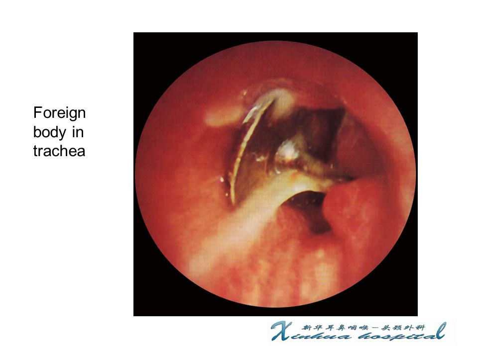
Foreign body in trachea

20
Foreign body in bronchus

21
Foreign body in esophagus cause More cases in old: poorly chew function, disesthesia of mouth, dental prosthesis More cases in children : like to put toy in his mouth Eating too quick Stenosis or lump in esophagus suicide

22
Sorts of foreign body animal : fishbone,bone pieces, meat lump metals : coin synthetic product : dental prosthesis plant : kernel

23
Location of foreign body The first stenosis: most seen The second stenosis The third stenosis

24
Clinic symptoms dysphagia odynophagia dyspnea : large foreign body

25
X-ray examination nonopaque : positive in barium x-ray study opaque : x-ray flat. entopic and lateral examination of esophagus

26
complication Esophagus perforation Infection of periesophagus organ : neck,mediastinum Large artery disruption Tracheal-esophageal fistula

27
diagnosis : history of foreign body , d ysphagia,odynophagia , disorder sign in X- ray treatment : esophagoscopy Electronic gastroscopy open : foreign body in out of esophagus, injury of large artery

28
Foreign body in esophagus

30
Thanks

Similar presentations


 –Partial.>")

>")


 into the atmosphere Filter, moisten,>")


