Download presentation
Presentation is loading. Please wait.

1
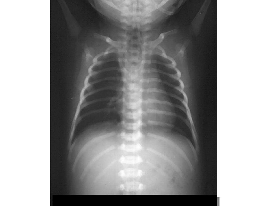
Pneumothorax & pneumopericardium

2
Pneumothorax, pneumopericardium pneumoperitoneum, subcut. emphysema

3
Pulmonary interstitial emphysema

4
CATHETER AND TUBE POSITION
1. ENDOTRACHEAL TUBE : 1 cm below vocal cord & 2 cm above carina 2. NASOGASTRIC TUBE : In stomach 3. UVC : in IVC or RA proper 4. UAC : in thoracic aorta below level of DA ( T4 ) and above origin of celiac artery ( T11 )
 and above origin of celiac artery ( T11 )")
5
UAC at T3- 4 level

6
UAC tip is at T11

7
UVC tip is in portal vein

8
RESPIRATORY DISTRESS IN NEWBORN SURGICAL CAUSES
DIAPHRAGMATIC HERNIA Common on left side Multiple lucencies or cysts ( bowel loops ) in the chest Shifting of heart and mediastinum to the other side Decreased bowel gas or gasless abdomen
 in the chest. Shifting of heart and mediastinum to the other side. Decreased bowel gas or gasless abdomen.")
11
FOREIGN BODY ASPIRATION
Common in children years old Tracheal obstruction Bronchial obstruction Incomplete - check valve : Air trapping Complete : Atelectasis S/S : Dyspnea, hyperpnea, cough, +/- Hx of FB aspiration

12
F 11 mo , หายใจหอบหลังกินถั่วติดคอมา 1อาทิตย์

13
IiInInspiration film

14
Expiration film , more air trapping in right lung

15
Scope : peanut in Rt.main bronchus
Post F.B. removal film

16
F 4mo, cough, หายใจหอบหลังสำลักเม็ดส้มมา 1 wk

17
Post F.B.removal

18
Alimentary Tract Disease in Children

19
ALIMENTARY TRACT OBSTRUCTION IN NEWBORN
Normal abdominal bowel gas pattern : ‘Polygonal pattern’ Stomach : immediate after birth Small bowel : within 3 hours Colon : within 5 hours Rectum : within 6-8 hours Prone film to demonstrate air in rectum ( R/O obstruction ) Upright or left lateral decubitus film to demonstrate free air
 Upright or left lateral decubitus film to demonstrate free air.")
20
Normal bowel pattern = polygonal

21
ESOPHAGEAL ATRESIA ( EA )
MC = proximal eso. atresia with distal TEF High incidence of associated anomalies : VACTERL ( V-vertebral, A-anorectal , C-cardiovascular , TE-tracheoesophageal ,R-renal , L-limb )
")
24
GASTRIC OUTLET OBSTRUCTION IDIOPATHIC HYPERTROPHIC PYLORIC STENOSIS
S/S : Non-billous vomitting Onset ~ 1 month old Minimal upper abdominal distension +/-palpable mass ( olive ) at epigastrium
 at epigastrium.")
25
IDIOPATHIC HYPERTROPHIC PYLORIC STENOSIS ( IHPS )
X-RAY FINDINGS : Plain film : Gastric dilatation with large amount of air/fluid content , Decreased small bowel gas US : Elongation with thickening of muscle wall of pyloric canal UGI : Narrowing and elongation of pyloric canal ‘String sign’ Muscle indentation on lumen of stomach and duodenum ‘shoulder sign’

26
M 5 mo , vomiting for 5 d.

Similar presentations


 –Partial.>")


