Download presentation
Presentation is loading. Please wait.

1
PROTOZOA Chapter 12

2
Introduction Protozoans are unicellular, eukaryotic chemoheterotrophic organisms. Most protozoa have two stages Trophozoite – the feeding and growing stage Some protozoa will produce a protective capsule called a cyst. A cyst allows the parasite to exist outside of the host and be the infective stage allowing the parasite to get to another host.

3
Reproduction Protozoa reproduce asexually by: Fission (mitosis)
Budding Schizogony a multiple fission – nucleus undergoes multiple divisions before the cell divides Protozoa reproduce sexually by: Conjugation Gamete formation

4
FIGURE 13-3 Paramecium undergoing transverse fission (X264).
Fission of a Ciliate FIGURE 13-3 Paramecium undergoing transverse fission (X264).
.")
5
Reproduction Definitive Host harbors the sexually reproducing stage of parasite Intermediate Host harbors asexually reproducing portion of the parasite’s life cycle.

6
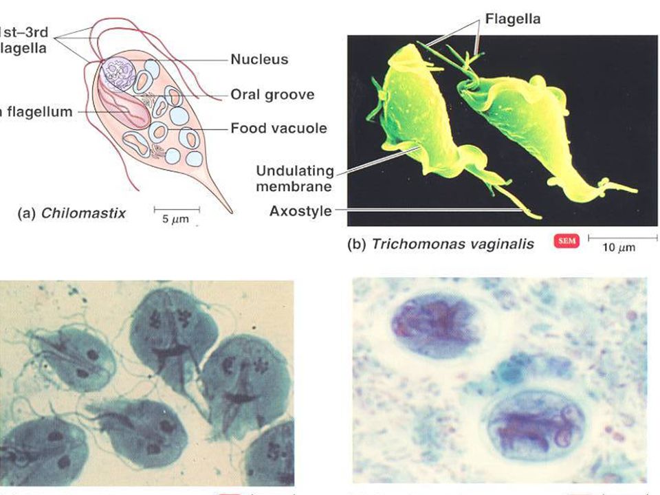
Representative Parasitic Protozoans
Flagellated Forms Giardia lamblia – enteritis – fecal contamination of drinking water. Common in campers and hikers. Resistant to 1-2ppm Cl2 use chemicals plus filtration Trichomonas vaginalis – urethritis vaginitis - sexually transmited or contact with vaginal urethral discharges

8
Trypanosoma species do include nonflagellated forms in lifecycle
Trypanosoma species do include nonflagellated forms in lifecycle. Hemoflagellates cause two distinctly different forms of disease: T. gambiense African sleeping sickness vector = Tsetse fly. Some animal reservoirs suspected specifically hoofed game animals. Enter wound created by fly bite enter blood and lymph, eventually invading central nervous system. Control breeding sites of Tsetse, insecticides, screens or netting, insect repellant. Insecticides have not been successful Female Tsetse flies mate once. Maybe irradiation of male flies to induce sterility.

9
T. cruzi – American trypanosomiasis Chagas disease
T. cruzi – American trypanosomiasis Chagas disease. Vector = reduvid bug (kissing bug) because they frequently bite humans around mouth (lips) feeding on tissue fluids and defecating in the wound. Parasites then migrate to cardiac muscle, liver, and brain. Many animals serve as reserviors for this parasite.
 because they frequently bite humans around mouth (lips) feeding on tissue fluids and defecating in the wound. Parasites then migrate to cardiac muscle, liver, and brain. Many animals serve as reserviors for this parasite.")
10
UM FIGURE 13-25 Trypanosoma brucei trypomastigotes in a blood smear (X1000). The nucleus and undulating membrane (UM) are visible.
 are visible.")
11
Leopard changing their spots
Antigenic shift No good vaccine

12
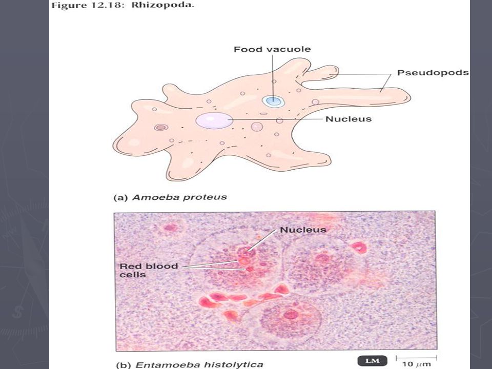
Amoeboid Forms Entamoeba histolytica - mobility is by pseudopod (false foot). Life cycle simple divided into two stages: trophozoite active feeding stage, and the cyst the quiescent resistant infective stage. The amoebic trophozoites remain actively motile, feeding on red blood cells, as long as environmental conditions are favorable. Dysentery and anemia. The cyst develops when environmental temperature or moisture drops. In diagnosis the cysts are the only forms recognized. Trophozoites only in fresh stool specimens. Ulcers in intestinal mucosa cause amoebic dysentary. May invade peritoneal cavity invasion of liver. Inflammation, hemorage, secondary bacterial infection. More than 5% americans asymptomatic carriers. Montezumas’ revenge. Drinking water and salads.
. Life cycle simple divided into two stages: trophozoite active feeding stage, and the cyst the quiescent resistant infective stage. The amoebic trophozoites remain actively motile, feeding on red blood cells, as long as environmental conditions are favorable. Dysentery and anemia. The cyst develops when environmental temperature or moisture drops. In diagnosis the cysts are the only forms recognized. Trophozoites only in fresh stool specimens. Ulcers in intestinal mucosa cause amoebic dysentary. May invade peritoneal cavity invasion of liver. Inflammation, hemorage, secondary bacterial infection. More than 5% americans asymptomatic carriers. Montezumas’ revenge. Drinking water and salads.")
14
FIGURE 13-6 Entamoeba histolytica trophozoite (X800, iron hematoxylin stain). Trophozoites range in size from 12 to 60 µm. Notice the small, central karyosome, the beaded chromatin at the nucleus’ margin, the ingested red blood cells and the finely granular cytoplasm. Compare with an Entamoeba coli trophozoite in Figure 13-8.

15
a b CB FIGURE 13-7 Entamoeba histolytica cysts. (a) Cysts are spherical with diameter of 10 to 20 µm. Two of the four nuclei are visible; other nuclear characteristics are as in the trophozoite. Compare with an Entamoeba coli cyst in Figure 13-9 (X1320, iron hematoxylin stain). (b) E. histolytica cyst (X1200, trichrome stain) with cytoplasmic chromatoidal bars (CB). These are found in approximately 10% of the cysts, have blunt ends and are composed of ribonucleoprotein.
 Cysts are spherical with diameter of 10 to 20 µm. Two of the four nuclei are visible; other nuclear characteristics are as in the trophozoite. Compare with an Entamoeba coli cyst in Figure 13-9 (X1320, iron hematoxylin stain). (b) E. histolytica cyst (X1200, trichrome stain) with cytoplasmic chromatoidal bars (CB). These are found in approximately 10% of the cysts, have blunt ends and are composed of ribonucleoprotein.")
16
Naeglaria fowleri – Meningoencephalitis
Naeglaria fowleri – Meningoencephalitis. Swimming in polluted water results in asymptomatic colonization of nasal passages. Invade cribiform plate through olfactory openings resulting in rapid fulminating fatal meningoencephalitis. Spinal fluid contains many erythrocytes and trophozoites. Post mortum diagnosis of trophozoites in the brain.

17
V FIGURE 13-14 Naegleria fowleri trophozoite from culture (X1000, iron hematoxylin stain). Trophozoites are between 10 and 35 µm in size. Notice the large karyosome within the nucleus and the lobed pseudopods. Vacuoles (V) are also visible in the cytoplasm.
. Trophozoites are between 10 and 35 µm in size. Notice the large karyosome within the nucleus and the lobed pseudopods. Vacuoles (V) are also visible in the cytoplasm.")
19
Ciliates Balantidium coli is the only member of the ciliate group that is pathogenic for humans. The organism is similar to amoebiasis in that the organisms cause tissue invasion and intestinal ulceration. Large trophozoite with large macronulcleus Swine and monkeys important reservoirs. Fecal oral route, swine feces contaminating local water supplies. Food handlers substandard hygienic conditions. Extraintestinal invasion of other organis is extremely rare in balantidiasis

20
C C Cy FIGURE 13-15 Balantidium coli trophozoite (X800). Trophozoites are oval in shape and have dimensions of 50 to 100 µm long by 40 to 70 µm wide. Cilia (C) cover the cell surface. Internally, the macronucleus is prominent; the adjacent micronucleus is not. An anterior cytostome (Cy) is usually visible.
. Trophozoites are oval in shape and have dimensions of 50 to 100 µm long by 40 to 70 µm wide. Cilia (C) cover the cell surface. Internally, the macronucleus is prominent; the adjacent micronucleus is not. An anterior cytostome (Cy) is usually visible.")
21
FIGURE 13-16 Balantidium coli cyst (X1000)
FIGURE 13-16 Balantidium coli cyst (X1000). Cysts are usually spherical and have a diameter in the range of 50 to 75 µm. There is a cyst wall and the cilia are absent. As in the trophozoite, the macronucleus is prominent, but the micronucleus may not be.
. Cysts are usually spherical and have a diameter in the range of 50 to 75 µm. There is a cyst wall and the cilia are absent. As in the trophozoite, the macronucleus is prominent, but the micronucleus may not be.")
22
Plasmodium species Sporozoan parasites that require two hosts for completion of their life cycle. The mosquito for the sexual reproductive stages. And the human or other animal (monkey) for the asexual stages. Human infection is initiated by the bite of an Anopheles mosquito which introduces the sporozoites into the blood stream via the mosquito saliva that acts as an anticoagulant. They go to the liver where schizogamy occurs for days.
 for the asexual stages. Human infection is initiated by the bite of an Anopheles mosquito which introduces the sporozoites into the blood stream via the mosquito saliva that acts as an anticoagulant. They go to the liver where schizogamy occurs for days.")
23
Plasmodium continued The hepatocytes rupture releasing merozoites that bind to red blood cells initiating the erythrocyte cycle of malaria (ring, trophozoite, and schizont stages). In the human (intermediate host) asexual reproduction occurs and results in the rupture of the erythrocyte and release of merozoites the infect more erythrocytes. Some merozoites also develop into male and female gametocytes. If a female mosquito ingests the gametocytes, the sexual reproduction of malaria can be initiated with the eventual production of sporozoites in the gut of the mosquitoand the sporozoites migrate to the salivary glands of the mosquito to be injected into the indefinate host.
. In the human (intermediate host) asexual reproduction occurs and results in the rupture of the erythrocyte and release of merozoites the infect more erythrocytes. Some merozoites also develop into male and female gametocytes. If a female mosquito ingests the gametocytes, the sexual reproduction of malaria can be initiated with the eventual production of sporozoites in the gut of the mosquitoand the sporozoites migrate to the salivary glands of the mosquito to be injected into the indefinate host.")
24
One disease selects for another
In Africa there has been a population of individuals that have a genetic hemaglobinopathy Sickle cell anemia. This disease is controlled by two codominant genes H = normal hemoglobin and h= abnormal hemoglobin or sickle anemia. A person that carries the h gene has fragile erythrocytes that will not support the Plasmodium parasite. Therefore, only HH children will succumb to malaria and the rest of the population will carry the sickle cell gene.

26
FIGURE 13-20 Giardia lamblia trophozoite (X1320, iron hematoxylin stain). Trophozoites have a long, tapering posterior end and range in size from 9 to 21 µm by 5 to 15 µm. There are two nuclei with small karyosomes. The two median bodies and the four pairs of flagella are not visible in this specimen.

27
M FIGURE 13-21 Giardia lamblia cyst (X1000, trichrome stain). Giardia cysts are smaller than trophozoites (8 to 12 µm by 7 to 10 µm), but the four nuclei with eccentric karyosomes and the median bodies (M) are still visible.
. Giardia cysts are smaller than trophozoites (8 to 12 µm by 7 to 10 µm), but the four nuclei with eccentric karyosomes and the median bodies (M) are still visible.")
28
FIGURE 13-27 Plasmodium life cycle.

29
FIGURE 13-28 Plasmodium falciparum ring stage in a red blood cell (X2640). The ring is the trophozoite. Note the chromatin dots in the nucleus.

30
FIGURE 13-30 Erythrocyte infected with Plasmodium vivax (X1000)
FIGURE 13-30 Erythrocyte infected with Plasmodium vivax (X1000). The parasite is in the ring stage, and the red cell exhibits characteristic Schüffner’s dots in the cytoplasm. Schüffner’s dots are also seen in red cells infected with P. ovale.
. The parasite is in the ring stage, and the red cell exhibits characteristic Schüffner’s dots in the cytoplasm. Schüffner’s dots are also seen in red cells infected with P. ovale.")
31
FIGURE 13-32 Plasmodium falciparum developing schizont in a red blood cell (X2640). These are usually not seen in peripheral blood smears since they reside in visceral capillaries.

32
FIGURE 13-34 Plasmodium malariae schizont with 8 merozoites in a distinctive rosette arrangement (X1200).
.")
33
FIGURE 13-33 A mature Plasmodium vivax schizont composed of approximately 16 merozoites (X1200). More than 12 merozoites distinguishes P. vivax from P. malariae and P. ovale, which both typically have eight, but up to 12. P. falciparum may have up to 24 merozoites, but they are not typically seen in peripheral blood smears and so are not confused with P. vivax.

34
FIGURE 13-35 Plasmodium falciparum gametocyte in an erythrocyte (X1200). Differentiation between microgametocytes and megagametocytes is difficult in this species.

35
Toxoplasmosis Toxoplasma gondii is an intracellular parasite that is found in a wide variety of animals including birds, mice, cats and humans. The essential reservoir is the common house cat That has eaten an infected rodent. The infective cysts are passed in cat feces where they can be ingested by mice and/or humans. If infection occurs during the first trimester of pregnancy spontaneous abortion, still birth or severe neurological disease. Disease is usually mild to asymptomatic in immunologically competent adults. Immunocomprimised patient AIDS result with severe neurological disease. Demostration of the banna shaped trophozoites in spinal fluid is one method of diagnosis. Newer monoclonal antibody serological methods will also diagnose the presence of the parasite.

37
FIGURE 13-36 Toxoplasma gondii trophozoites (X1000)
FIGURE 13-36 Toxoplasma gondii trophozoites (X1000). Notice the bow shaped cells with prominent nuclei.
. Notice the bow shaped cells with prominent nuclei.")
38
Cryptosporidium parvum
Newly recognized parasite in immune compromised humans, children, aged, AIDS Respiratory and gall bladder infections = major cause of death Transmitted through fecal oral route from cows, rodents and dogs and cats mainly water borne, but veterinary personnel, animal handlers, day care centers, and homosexuals are at high risk. Resistant to usually water purification methods –chlorination and ozone

39
FIGURE 13-38 Cryptosporidium parvum oocysts from a human fecal sample (X1000, modified acid-fast stain). Oocysts contain sporozoites (not visible) and are the infective stage. They are typically about 5µm in size.
 and are the infective stage. They are typically about 5µm in size..")
Similar presentations


 Drs. Babcock and Hopkins Spring 2009>")

 can be heterotrophic or autotrophic most live in water (though some live.>")
 Can move like animals and respond to changes in the environment.>")


>")