Download presentation
Presentation is loading. Please wait.

1
About Plant Biology Chapter 1

2
Why Study Plant Biology?
Show interrelationships between plants and other fields of study Prepare for careers in plant biology Gain fundamental knowledge for upper division plant biology courses Share expertise gained with nonbotanists

3
What is a Plant? An organism that is green and photosynthetic
Additional characteristics Cell wall composed of cellulose Multicellular body Can control water loss Have strengthening tissues Can reproduce by means of microscopic, drought-resistant spores

4
Ecologic Services Sources of food, fabric, shelter, medicine
Produce atmospheric oxygen and organic nitrogen Build new land Inhibit erosion Control atmospheric temperature Decompose and cycle essential mineral nutrients

5
Importance of Plants to Human Civilizations
Trees for lumber to make warships Fuel to smelt metals, cure pottery, generate power and heat Sources of wealth spices Sources of industrial products Rubber oil

6
Natural Plant Losses Plant losses occurring at a faster rate than ever before Factors include Agriculture Urbanization Overgrazing Pollution Extinction

7
Environmental Laws Described in 1961 by plant biologist Barry Commoner
Laws becoming more true every day Four “environmental laws” Everything is connected to everything else. Everything must go somewhere. Nature knows best. There is no such thing as a free lunch.

8
Scientific Method Codefined and promoted in 17th century by Rene Decartes and Francis Bacon Steps involved in scientific method Make observations Ask questions Make educated guesses about possible answers Base predictions on the guesses Devise ways to test predictions Draw conclusions

9
Scientific Method Hypothesis – “educated guess” based on observations and questioning Predicted result occurs – hypothesis is most likely correct Individuals using scientific method should be objective and unbiased

10
Scientific Method Original Hypothesis Devise method to test hypothesis
Analyze results Results support hypothesis Results support hypothesis but suggest minor refinements Results do not support original hypothesis but fall within range that could be expected if original hypothesis is slightly modified Results are so unexpected that they do not support original hypothesis and require a new hypothesis Retest using minor refinements of process Test new hypotheses Test using slightly modified hypothesis

11
Studying Plants From Different Perspectives
Plant genetics – study of plant heredity Plant systematics – study of plant evolution and classification Plant ecology – study of how the environment affects plant organisms Plant anatomy – study of a plant’s internal structure Plant morphology – study of how a plant develops from a single cell into its diverse tissues and organs

12
Study Plants from Taxonomic Classification
Microbiology – study of bacteria Mycology – study of fungi Phycology – study of algae Bryology – study of mosses

13
Interrelationships Among Several Plant Biology Disciplines
Genes ENVIRONMENT Genetics Evolution Taxonomy & Systematics Ecology Paleoecology Biogeography METABOLISM Physiology PLANT TAXONOMIC GROUPS STRUCTURE Phycology Microbiology Mycology Bryology Anatomy DEVELOPMENT Morphology

14
Plant Classification Taxonomy Linnaean system Easy to use
Based on idea that species never changed Grouped organisms according to arbitrary similarities Fails to meet needs of modern biologists

15
Linnaean Taxa Taxa Ending Kingdom Division -phyta Class -opsida Order
-ales Family -aceae Genus No standard ending Species

16
Plant Classification Whittaker’s Five Kingdoms
Developed in 1969 by Robert Whittaker Each kingdom assumed to be monophyletic group of species Molecular biology techniques Cladistics Show five kingdom system also does not recognize evolutionary groups

17
Whittaker’s Five Kingdoms
Description Monera Included bacteria Fungi Included molds, mildews, rusts, smuts, and mushrooms Protista Included simple organisms, some were photosynthetic, mostly aquatic organisms called algae Plantae Included more complex photosynthetic organisms that typically grew on land Animalia Included typically motile, multicellular, nonphotosynthetic organisms

18
Plant Classification Cladistics Based on evolutionary groups
Compare DNA base pair sequences of organisms to determine relatedness Obtain percent similarity between organisms

19
Plant Classification Clades – evolutionary groups
Cladogram = phylogenetic tree Branching diagram Emphasizes shared features from common ancestor Future discoveries may require modifications of cladogram

20
Plant Classification Domain Three domains based on cladistics
Neutral term Groups of organisms as large or larger than a kingdom Monophyletic Three domains based on cladistics Eukarya Bacteria Archaea

21
Domain Eukarya Made up of Whittaker’s plant, animal, and fungal kingdoms Eukaryotic cells Membrane-bounded organelles Linear chromosomes Protists Not monophyletic Controversy over where to place organisms

22
Domain Bacteria Organisms originally were placed in Whittaker’s Kingdom Monera Microscopic Prokaryotic cells No membrane-bounded organelles Circular chromosome Sexual reproduction unknown Found in every habitat on Earth

23
Domain Bacteria Beneficial aspects Decomposers
Some carry on photosynthesis Cyanobacteria or blue-green algae Nitrogen fixation Convert inorganic N2 into ammonium for plant use Cyanobacteria

24
Domain Bacteria Detrimental effects Pathogens – cause diseases
Human diseases Botulism, bubonic plague, cholera, syphilis, tetanus, tuberculosis Plant diseases

25
Domain Archaea Organisms originally were placed in Whittaker’s Kingdom Monera Prokaryotic Different cell structure and chemistry than organisms in Domain Bacteria

26
Domain Archaea Divided into three groups based on habitat
Bacteria of sulfur-rich anaerobic hot springs and deep ocean hydrothermal vents Bacteria of anaerobic swamps and termite intestines Bacteria of extremely saline waters Extreme halophiles Photosynthetic – pigment bacteriorhodopsin

27
Three Domains Domain Cell Type Description Eukarya Eukaryotic Archaea
Membrane bounded organelles, linear chromosomes Archaea Prokaryotic Found in extreme environments, cell structure and differ from members of Domain Bacteria Bacteria Ordinary bacteria, found in every habitat on earth, play major role as decomposers

28
Kingdom Fungi Eukaryotic cells Typically microscopic and filamentous
Rigid cell wall made of chitin Reproduce sexually in a variety of complex life cycles and spores Widely distributed throughout world – mainly terrestrial

29
Kingdom Fungi Economic importance Decomposers
Form associations with roots of plants Important foods for animals and humans Mushrooms, morels Decomposing action of yeast Flavored cheeses, leavened bread, alcoholic beverages

30
Kingdom Fungi Economic importance Production of antibiotics Pathogens
Penicillium Pathogens Invade both plant and animal tissue Cause illnesses Reduce crop yields

31
Kingdom Protista Eukaryotic cells
Reproduce both sexually and asexually Catch-all group Photosynthetic organisms – algae Nonphotosynthetic organisms – slime molds, foraminiferans, protozoans

32
Kingdom Protista Algae Arrangements Photosynthetic
Single cells, clusters, filaments, sheets, three-dimensional packets of cells Photosynthetic Float in uppermost layers of all oceans and lakes

33
Kingdom Protista Phytoplankton “grasses of the sea” Microscopic algae
Form base of natural food chain Produce 50% of all oxygen in atmosphere

34
Kingdom Plantae Included all organisms informally called plants
Bodies more complex than bacteria, fungi, or protists Eukaryotic

35
Kingdom Plantae Unique biochemical traits of plants
Cell walls composed of cellulose Accumulate starch as carbohydrate storage product Special types of chlorophylls and other pigments

36
Kingdom Plantae Ecologic and economic importance of plants
Form base of terrestrial food chains Principal human crops Provide building materials, clothing, cordage, medicines, and beverages

37
Challenge for 21st Century
While the human population increases, the major challenge of retaining natural biological diversity and developing a sustainable use of the world’s forests, grasslands, and cropland remains. As you study plant biology, think of the ways that you can contribute to this challenge.

38
Proteins take on a variety of shapes, which enables specific interactions (function) with other molecules. Fig Stages in the formation of a functioning protein

39
The Plant Cell and the Cell Cycle
Chapter 3

42
A colony of Volvox aureus, a green algae
A colony of Volvox aureus, a green algae. Yellow masses are daughter colonies.

43
Plant vascular tissue

44
Eucaryotic Cell structure
Rough endoplasmic reticulum-site of secreted protein synthesis Smooth ER-site of fatty acid synthesis Ribosomes-site of protein synthesis Golgi apparatus- site of modification and sorting of secreted proteins Lysosomes-recycling of polymers and organelles Nucleus-double membrane structure confining the chromosomes Nucleolus-site of ribosomal RNA synthesis and assembly of ribosomes Peroxisome-site of fatty acid and amino acid degradation Flagella/Cilia- involved in motility Mitochondria-site of oxidative phosphorylation Chloroplast-site of photosynthesis Intermediate filaments- involved in cytoskeleton structure

45
Plant vs Animal Cells Plant cells have chloroplasts and perform photosynthesis Outermost barrier in plant cells is the cell wall Outermost barrier in animal cells is the plasma membrane

49
Chloroplasts in leaf cells, in an electron micrograph, false color image of EM.

50
Cell Basic unit of plant structure and function Robert Hooke
Looked at cork tissue under microscope “little boxes or cells distinct from one another ….that perfectly enclosed air” Nehemiah Grew Recognized leaves as collections of cells filled with fluid and green inclusions

51
Cell Theory Statement Year Contributor
All plants and animals are composed of cells. 1838 Matthias Schleiden and Theodor Schwann Cells reproduce themselves. 1858 Rudolf Virchow All cells arise by reproduction from previous cells.

52
Basic Similarities of Cells
Cells possess basic characteristic of life Movement Metabolism Ability to reproduce Organelles “little organs” Carry out specialized functions within cells

53
Light Microscope View cells 20-200 µm in diameter
Can view living or stained specimens Resolution (resolving power) Ability to distinguish separate objects Limited by lenses and wavelengths of light used Smallest object that can be resolved is ~ 0.2 µm in diameter
 Ability to distinguish separate objects. Limited by lenses and wavelengths of light used. Smallest object that can be resolved is ~ 0.2 µm in diameter.")
54
Confocal Microscope Laser illumination
Detecting lens focuses on single point at a time Scans entire sample to assemble picture No reduction in contrast due to scattered light Can generate 3-D images

55
Transmission Electron Microscope
Responsible for discovery of most of smaller organelles in cell Greater resolution Uses beams of electrons rather than light Magnets for lenses Ultrathin section examined in vacuum View image on fluorescent plate or photographic film

56
Scanning Electron Microscope
Collected electrons used to form picture in television picture tube High resolution view of surface structures Requires vacuum Recent refinements Can operate in low vacuum Can view live plant cells and insects

57
Microscope Comparisons
Source for illumination Nature of lenses Condition of specimen Image formation Light microscope White light Glass Living or killed stained specimen View directly through microscope Confocal microscope Laser Killed stained specimens Image analyzed on digital computer screen Transmission electron microscope Electrons Magnets Ultrathin section of killed specimen contained within vacuum View on fluorescent plate or photographic film Scanning electron microscope Surface view of killed specimen contained within vacuum, with low vacuum can view living cells Television picture tube

58
Generalized Plant Cell

59
chloroplast vacuole nucleus cell wall mitochondrion
Figure 3.3: Plant cells, showing the major organelles. Leaf (b) and root (c) cells of timothy grass (Phleum pratense) as seen in a transmission electron microscope. Fig. 3-3 (b & c), p. 33
 and root (c) cells of timothy grass (Phleum pratense) as seen in a transmission electron microscope. Fig. 3-3 (b & c), p. 33.")
60
Boundaries Between Inside and Outside the Cell
Plasma Membrane and Cell Wall

61
Plasma Membrane Surrounds cell Controls transport into and out of cell
Selectively permeable

62
Plasma Membrane Composed of approximately half phospholipid and half protein, small amount of sterols Phospholipid bilayer Separates aqueous solution inside cell from aqueous layer outside cell Prevents water-soluble compounds inside cell from leaking out Prevents water-soluble compounds outside cell from diffusing in

63
Plasma Membrane Proteins in bilayer Perform different functions
Ion pumps Move ions from lower to higher concentration Require ATP energy Proton pump – moves H+ ions from inside to outside of cell Ca+2 pump – moves Ca2+ to outside of cell Channels – allow substances to diffuse across membrane

64
PUMPS AND CHANNELS SENSORY PROTEINS RECEPTORS
Extracellular environment Figure 3.4: A model of the plasma membrane, showing the phospholipid bilayer, sterols, and various types of proteins floating in the bilayer. PHOSPHOLIPID BILAYER PUMPS AND CHANNELS SENSORY PROTEINS RECEPTORS STEROL Cytoplasm Fig. 3-4, p. 34

65
Plasma Membrane Plasmodesmata
Connects plasma membranes of adjacent plant cells Extends through cell wall Allows materials to move from cytoplasm of one cell to cytoplasm of next cell Symplast – name for continuous cytoplasm in set of cells

66
E.R. lumen E.R. Cytoplasm plasma Cell wall membrane plasmodesmal
proteins Figure 3.5: Model of a plasmodesma, showing the endoplasmic reticulum and proteins that are thought to control the flow of materials through the channel. Cytoplasm Fig. 3-5, p. 35

67
Plasma Membrane Apoplast – Space outside cell
Next to plasma membrane within fibrils of cell wall Area of considerable metabolic activity Important space in plant but questionable as to whether it is part of the plant’s cells

68
Cell Wall Rigid structure made of cellulose microfibrils
Helps prevent cell rupture Process of osmosis allows water to enter cell Inflow of water expands cell Expansion forces cell membrane against cell wall Resistance of cell wall to expansion balances pressure of osmosis Stops flow of water into cell Keeps cell membrane from further expansion

69
Cell Wall Osmotic forces balanced by pressure exerted by cell wall
Creates turgor pressure Causes cells to become stiff and incompressible Able to support large plant organs Loss of turgor pressure – plant wilts

70
Figure 3.6: A small section of cell wall, as seen in a transmission electron microscope. The filaments are cellulose. Fig. 3-6, p. 35

71
Cell Wall Place cell in salt solution Water leaves cytoplasm
Protoplast (space inside plasma membrane) shrinks Plasma membrane pulls away from cell wall Cell lacks turgor pressure - wilts
 shrinks. Plasma membrane pulls away from cell wall. Cell lacks turgor pressure - wilts.")
72
PROTOPLAST SOLUTION Concentration 0.3 molar Concentration Pressure
(Isotonic) Pressure 0 megapascals Concentration 0.27 molar Concentration 0 molar (Hypotonic) Pressure 0.66 megapascals Figure 3.7: The effect of osmosis on cell size. (a) The cell in this example is assumed to have an initial internal concentration of solutes, which we will designate as 0.3 molar. The cell is in a solution of the same concentration. The cell protoplast (volume inside the plasma membrane) is exactly the size of the cell wall in its resting (unstretched) state. (b) The cell is transferred to pure water. Water moves into the protoplast by osmosis, and the protoplast expands, pushing against the cell wall. The wall exerts a back pressure on the protoplast, which inhibits further influx of water. Also, the expansion of the protoplast dilutes the solutes inside the protoplast. Water stops entering the cell when the pressure exerted by the cell wall equals the osmotic pressure forcing water into the cell. (c) The cell is transferred to a solution of 0.5 molar. Water moves out of the protoplast by osmosis, and the protoplast shrinks until the concentration of solutes in the protoplast equals 0.5 molar. The plasma membrane pulls away from the cell wall. This effect is called plasmolysis. Concentration 0.5 molar Concentration 0.5 molar (Hypertonic) Pressure 0 megapascals Fig. 3-7 (a-c), p. 36
 Pressure. 0 megapascals. Concentration molar. Concentration. 0 molar. (Hypotonic) Pressure megapascals. Figure 3.7: The effect of osmosis on cell size. (a) The cell in this example is assumed to have an initial internal concentration of solutes, which we will designate as 0.3 molar. The cell is in a solution of the same concentration. The cell protoplast (volume inside the plasma membrane) is exactly the size of the cell wall in its resting (unstretched) state. (b) The cell is transferred to pure water. Water moves into the protoplast by osmosis, and the protoplast expands, pushing against the cell wall. The wall exerts a back pressure on the protoplast, which inhibits further influx of water. Also, the expansion of the protoplast dilutes the solutes inside the protoplast. Water stops entering the cell when the pressure exerted by the cell wall equals the osmotic pressure forcing water into the cell. (c) The cell is transferred to a solution of 0.5 molar. Water moves out of the protoplast by osmosis, and the protoplast shrinks until the concentration of solutes in the protoplast equals 0.5 molar. The plasma membrane pulls away from the cell wall. This effect is called plasmolysis. Concentration. 0.5 molar. Concentration. 0.5 molar. (Hypertonic) Pressure. 0 megapascals. Fig. 3-7 (a-c), p. 36.")
73
Figure 3. 7: The effect of osmosis on cell size
Figure 3.7: The effect of osmosis on cell size. (d) Plasmolyzed Elodea cells. Compare these cells to the ones in Fig. 3.1b. Fig. 3-7 (d), p. 36
 Plasmolyzed Elodea cells. Compare these cells to the ones in Fig. 3.1b. Fig. 3-7 (d), p. 36.")
74
Cell Wall Structure Primary cell wall Secondary cell wall
Cell wall that forms while cell is growing Secondary cell wall Additional cell wall layer deposited between primary cell wall and plasma membrane Generally contains cellulose microfibrils and water-impermeable lignin Provides strength to wood

75
Cell Wall Structure Specialized types of cell walls
cutin covering cell wall or suberin imbedded in cell wall Waxy substances impermeable to water Cutinized cell walls Found on surfaces of leaves and other organs exposed to air Retard evaporation from cells Barrier to potential pathogens

76
Organelles of Protein Synthesis and Transport
Nucleus, Ribosomes, Endoplasmic Reticulum, and Golgi Apparatus

77
Nucleus Ovoid or irregular in shape Up to 25 µm in diameter
Easily stained for light or electron microscopy

78
Nucleus Surrounded by double membrane – nuclear envelope Nucleoplasm
Protein filaments of lamin line inner surface of envelope and stabilize it Inner and outer membranes connect to form pores Nucleoplasm Portion of nucleus inside nuclear envelope

79
one pore nuclear envelope 1 µm 0.2 µm lipid bilayer facing
Figure 3.8: Surface view by scanning electron microscopy and sketch of the nucleus, showing the nucleolus and the nuclear envelope with pores. lipid bilayer facing the nucleoplasm nuclear envelope lipid bilayer facing the cytoplasm pore complex that spans both bilayers Fig. 3-8, p. 37

80
Nucleus Nucleoli (singular, nucleolus)
Densely staining region within nucleus Accumulation of RNA-protein complexes (ribosomes) Site where ribosomes are synthesized Center of nucleoli DNA templates Guide synthesis of ribosomal RNA
 Site where ribosomes are synthesized. Center of nucleoli. DNA templates. Guide synthesis of ribosomal RNA.")
81
Nucleus Chromosomes Found in nucleoplasm Contain DNA and protein
Each chromosome composed of long molecule of DNA wound around histone proteins forming a chain of nucleosome Additional proteins form scaffolds to hold nucleosomes in place

82
Figure 3.9: The organization of DNA in nucleoplasm.
Fig. 3-9, p. 37

83
At times when a chromosome is most condensed, the chromosomal proteins
interact, which packages loops of already coiled DNA into a “supercoiled” array. Fig. 3-9d, p. 37

84
At a deeper level of structural organization, the chromosomal
proteins and DNA are organized as a cylindrical fiber. Fig. 3-9c, p. 37

85
Immerse a chromosome in saltwater and it loosens up to a
beads-on-a-string organization. The “string” is one DNA molecule. Each “bead” is a nucleosome. Fig. 3-9b, p. 37

86
A nucleosome consists of part of a DNA molecule looped twice
around a core of histones. core of histone molecules Fig. 3-9a, p. 37

87
Nucleus DNA in chromosomes Steps in protein synthesis
Stores genetic information in nucleotide sequences Information used to direct protein synthesis Steps in protein synthesis Transcription – DNA directs synthesis of RNA Most RNA stays in nucleus or is quickly broken down Small amount of RNA (mRNA) carries information from nucleus to cytoplasm
 carries information from nucleus to cytoplasm.")
88
Structure and Function
Nuclear Components Component Structure and Function Nuclear envelope Double layered membrane, filaments of protein lamin line inner surface and stabilize structure, inner and outer membranes connect to form pores Nucleoplasm Portion inside the nuclear envelope Nucleoli Dark staining bodies within nucleus, site for ribosome synthesis Chromosomes Store genetic information in nucleotide sequences, each chromosome consists of chain of nucleosomes (long DNA molecule and associated histone proteins)
")
89
Ribosomes Small dense bodies formed from ribosomal RNA (rRNA) and proteins Function in protein synthesis Active ribosomes in clusters called polyribosomes Attached to same mRNA All ribosomes in one polyribosome make same type of protein

90
Ribosomes In living cell, ribosomes are not fixed
Move rapidly along mRNA Read base sequence Add amino acids to growing protein chain At end of mRNA, ribosome falls off, releasing completed protein into cytoplasm

91
free polyribosomes attached polyribosomes
mRNA ribosomes free polyribosomes attached polyribosomes Figure 3.10: Polyribosomes as observed by transmission electron microscopy. Some of the polyribosomes in this cell from a wheat root tip were free in the cytoplasm and some were attached to the endoplasmic reticulum (ER), forming rough ER. Fig. 3-10, p. 38
, forming rough ER. Fig. 3-10, p. 38.")
92
attached polyribosomes free polyribosomes
mRNA ribosomes Figure 3.10: Polyribosomes as observed by transmission electron microscopy. Some of the polyribosomes in this cell from a wheat root tip were free in the cytoplasm and some were attached to the endoplasmic reticulum (ER), forming rough ER. attached polyribosomes free polyribosomes Fig. 3-10a, p. 38
, forming rough ER. attached. polyribosomes. free. polyribosomes. Fig. 3-10a, p. 38.")
93
Endoplasmic Reticulum
ER Branched, tubular structure Often found near edge of cell Function Site where proteins are synthesized and packaged for transport to other locations in the cell Proteins injected through membrane into lumen

94
Endoplasmic Reticulum
Packaging of proteins by ER Considered to be packaged when separated from cytoplasm by membrane Sphere (vesicle) of membrane-containing proteins may bud off from ER Vesicle carries proteins to other locations in cell
 of membrane-containing proteins may bud off from ER. Vesicle carries proteins to other locations in cell.")
95
Endoplasmic Reticulum
Types of ER rough ER – ribosomes attached to surface smooth ER – does not have attached ribosomes Carbohydrate transport Often attached to proteins in ER Helps protect carbohydrate from breakdown by destructive enzymes

96
Golgi Apparatus Also called a dictyosome
Consists of stack of membranous, flattened bladders called cisternae

97
vesicles internal spaces cisternae 0.25 µm Fig. 3-11, p. 38
Figure 3.11: The Golgi apparatus and how it moves substances through a cell. (a) Transmission electron micrograph. Note the cisternae and associated vesicles. (b) A model of a Golgi apparatus. Fig. 3-11, p. 38
 Transmission electron micrograph. Note the cisternae and associated vesicles. (b) A model of a Golgi apparatus. Fig. 3-11, p. 38.")
98
Golgi Apparatus Directs movements of proteins and other substances from ER to other parts of cell Cell wall components (proteins, hemicellulose, pectin) pass through cisternae Move to plasma membrane inside membranous sphere Sphere joins with plasma membrane Membrane of sphere becomes part of plasma membrane Protein, hemicellulose, and pectin contents released to outside the cell
 pass through cisternae. Move to plasma membrane inside membranous sphere. Sphere joins with plasma membrane. Membrane of sphere becomes part of plasma membrane. Protein, hemicellulose, and pectin contents released to outside the cell.")
99
Endomembrane System Complex network that transports materials between Golgi apparatus, the ER, and other organelles of the cell Movement Rapid Continuous

100
Organelles of Energy Metabolism
Plastids and Mitochondria

101
Plastids Found in every living plant cell
2-10 µm in diameter Surrounded by double membrane Contain DNA and ribosomes Protein-synthesizing system similar to but not identical to one in nucleus and cytoplasm

102
two outer membranes thylakoids stroma Fig. 3-12 (a), p. 40
Figure 3.12: Plastids. (a) Model of a chloroplast. stroma Fig (a), p. 40
 Model of a chloroplast. stroma. Fig (a), p. 40.")
103
Plastids Proplastids Small plastids always found in dividing plant cells Have short internal membranes and crystalline associations of membranous materials called prolamellar bodies As cell matures, plastids develop Prolamellar bodies reorganized Combined with new lipids and proteins to form more extensive internal membranes

104
Plastids Types of plastids Chloroplasts Leukoplasts Amyloplasts
Chromoplasts

105
Figure 3.12: Plastids. From top, left to right: (b) A maize (Zea mays) leaf chloroplast, showing dense thylakoid membranes. (c) A maize proplastid, with only a few internal (prolamellar) membranes. (d) A small leukoplast from an inner white leaf of endive (Cichorium endiva). (e) An amyloplast from a bean (Phaseolus vulgaris) seedling, showing large starch grains. (f) A chromoplast from a mature red pepper (Capsicum sp.). The dark circles are globules of red and orange xanthophylls. Fig (b-f), p. 40
, p. 40.")
106
Plastids Chloroplasts Thylakoids Chlorophyll Stroma Inner membranes
Have proteins that bind to chlorophyll Chlorophyll Green compound that gives green plant tissue its color Stroma Thick solution of enzymes surrounding thylakoids

107
Plastids Chloroplasts Function
Convert light energy into chemical energy (photosynthesis) Accomplished by proteins in thylakoids and stromal enzymes Can store products of photosynthesis (carbohydrates) in form of starch grains
 Accomplished by proteins in thylakoids and stromal enzymes. Can store products of photosynthesis (carbohydrates) in form of starch grains.")
108
Chloroplast Component Description Thylakoids
Inner membranes of chloroplast, contain proteins that bind with chlorophyll Stroma Thick enzyme solution surrounding thylakoids Chlorophyll Green pigment that gives plant tissue its green color Starch grains Storage form of carbohydrates produced during photosynthesis

109
Leukoplasts leuko – “white”
Found in roots and some nongreen tissues in stems No thylakoids Store carbohydrates in form of starch Microscopically appear as white, refractile, shiny particles

110
Amyloplasts amylo – “starch”
Leukoplast that contains large starch granules

111
Chromoplasts chromo – “color” Found in some colored plant tissues
tomato fruits, carrot roots High concentrations of specialized lipids – carotenes and xanthophylls Give plant tissues orange-to-red color

112
Plastids Prefix Meaning Function Chloroplast “chloro –” “yellow-green”
Photosynthesis, convert light energy into chemical energy, store carbohydrates as starch grains Leukoplast “leuko –” “white” Store carbohydrates in form of starch Amyloplast “amylo –” “starch” Leukoplasts that contain large granules of starch Chromoplast “chromo –” “color” Stores carotenes and xanthophylls, give orange-to-red color to certain plant tissues

113
Mitochondria Double-membrane structure Contain DNA and ribosomes
Inner membrane infolded Folds called cristae Increase surface area available for chemical reactions

114
outer compartment inner membrane inner compartment outer membrane
cristae (matrix) Figure 3.13: Mitochondria. (a) Model of a typical mitochondrion. (b) Transmission electron micrograph of a mitochondrion from the young leaf of a safflower (Carthamus tintorius) seedling. Fig. 3-13, p. 41
 Figure 3.13: Mitochondria. (a) Model of a typical mitochondrion. (b) Transmission electron micrograph of a mitochondrion from the young leaf of a safflower (Carthamus tintorius) seedling. Fig. 3-13, p. 41.")
115
Mitochondria Matrix Function
Viscous solution of enzymes within cristae Function source of most ATP in any cell that is not actively photosynthesizing Site of oxidative respiration Release of ATP from organic molecules ATP used to power chemical reactions in cell

116
Other Cellular Structures
Vacuoles, Vesicles, Peroxisomes, Glyoxysomes, Lysosomes, and Cytoskeleton

117
Vacuoles Large compartment surrounded by single membrane
Takes up large portion of cell volume Tonoplast Membrane surrounding vacuole Has embedded protein pumps and channels that control flow of ions and molecules into and out of vacuole

118
Vacuole Functions May accumulate ions which increase turgor pressure inside cell Can store nutrients such as sucrose Can store other nutritious chemicals May accumulate compounds that are toxic to herbivores May serve as a dump for wastes that cell cannot keep and cannot excrete

119
Vesicles Small, round bodies surrounded by single membrane
Peroxisomes and glyoxysomes Compartments for enzymatic reactions that need to be separated from cytoplasm Lysosomes Contain enzymes that break down proteins, carbohydrates, and nucleic acids May function in removing wastes within living cell Can release enzymes that dissolve the entire cell

120
Cytoskeleton Collection of long, filamentous structures within cytoplasm Functions Keeps organelles in specific places Sometimes directs movement of organelles around the cell Cyclosis – cytoplasmic streaming

121
Cytoskeleton Structures in cytoskeleton
Microtubules Motor proteins Microfilaments Specialized proteins connect microtubules and microfilaments to other organelles Connections thought to coordinate many cell processes

122
Microtubules Relatively thick (0.024 µm in diameter)
Assembled from protein subunits called tubulin Fairly rigid but can lengthen or shorten by adding or removing tubulin molecules

123
Microtubules Functions Guide movement of organelles around cytoplasm
Key organelles in cell division Form basis of cilia and flagella Cilia and flagella never found in flowering plants Important to some algae and to male gametes of lower plants

124
Microfilaments Thinner (0.007 µm in diameter) and more flexible than microtubules Made of protein subunits called actin Often found in bundles Function Serve as guides for movement of organelles

125
Motor Proteins Powered by ATP molecules Microtubule motor proteins
Kinesins, dyneins Move along microtubule making and breaking connections between tubulin subunits Microfilament motor proteins myosin

126
Cytoskeleton Subunits Motor proteins Function Microtubules
Tubulin (protein) Kinesins, dyneins Key organelles in cell division, form basis of cilia and flagella, serve as guides for movement of organelles within cell Microfilaments Actin (protein) Myosin Serve as guides for movement of organelles within cell
 Kinesins, dyneins. Key organelles in cell division, form basis of cilia and flagella, serve as guides for movement of organelles within cell. Microfilaments. Actin (protein) Myosin. Serve as guides for movement of organelles within cell.")
127
The Organization of the Plant Body: Cells, Tissues, and Meristems
Chapter 4

128
Organization of Plant Body
Most vascular plants consist of: Shoot System Above ground part Stems, leaves, buds, flowers, fruit Root System Below ground part Main roots and branches

129
Plant Cells and Tissues
Cell wall – surrounds each plant cell Pectin – glues plant cells together Meristems Groups of specialized dividing cells Sources of cells and tissues Not tissues themselves Plant organs – leaves, stems,roots, flower parts

130
Fig. 4-CO, p. 49

131
Vascular tissue system
Main Tissues of Plants Ground tissue system Most extensive in leaves (mesophyll) and young green stems (pith and cortex) Vascular tissue system Conducting tissues Xylem – distributes water and solutes Phloem – distributes sugars Dermal tissue system Covers and protects plant surfaces – epidermis and periderm
 and young green stems (pith and cortex) Vascular tissue system. Conducting tissues. Xylem – distributes water and solutes. Phloem – distributes sugars. Dermal tissue system. Covers and protects plant surfaces – epidermis and periderm.")
132
Plant Tissues Simple tissues Composed of mostly one cell type
Workhorse cells of plant body Functions Conduct photosynthesis Load materials into and out of vascular system Hold plant upright Store things Help keep plant healthy and functioning

133
Simple Plant Tissues Tissue type Cell types Parenchyma tissue
Parenchyma cells Collenchyma tissue Collenchyma cells Sclerenchyma tissue Fibers, sclereids

134
Table 4-1, p. 50

135
Shoot system Root system
shoot tip xylem epidermis bud flower mesophyll phloem node internode Dermal tissues node pith xylem phloem cortex Vascular tissues leaf epidermis seeds (inside fruit) Ground tissues Figure 4.1: The plant body, shown here as a tomato plant, consists of the shoot system (leaves, buds, stems, flowers, and fruits) and the root system (roots). Each organ is made up of cells organized into tissue systems: dermal, vascular, and ground. One way the vegetative organs (leaves, stems, and roots) differ from each other is in the distribution of the tissues. Shoot system Root system cortex primary root xylem lateral root phloem root hairs root tip epidermis root cap Fig. 4-1, p. 51
 Ground tissues. Figure 4.1: The plant body, shown here as a tomato plant, consists of the shoot system (leaves, buds, stems, flowers, and fruits) and the root system (roots). Each organ is made up of cells organized into tissue systems: dermal, vascular, and ground. One way the vegetative organs (leaves, stems, and roots) differ from each other is in the distribution of the tissues. Shoot system. Root system. cortex. primary root. xylem. lateral root. phloem. root hairs. root tip. epidermis. root cap. Fig. 4-1, p. 51.")
136
Parenchyma Usually spherical or elongated Thin primary cell wall
Perform basic metabolic functions of cells Respiration Photosynthesis Storage Secretion

137
parenchyma cells Fig. 4-2a, p. 52
Figure 4.2: Types of parenchyma cells. (a) Pith parenchyma cells from impatiens (Impatiens sp.) stem. x280. parenchyma cells Fig. 4-2a, p. 52
 Pith parenchyma cells from impatiens (Impatiens sp.) stem. x280. parenchyma. cells. Fig. 4-2a, p. 52.")
138
Parenchyma Usually live 1-2 years
Crystals of calcium oxalate commonly found in vacuoles May help regulate pH of cells May aggregate to form parenchyma tissue in Cortex and pith of stems Cortex of roots Mesophyll of leaves

139
Parenchyma Mature cells may be developmentally programmed to form different cell types Wound healing Transfer cells Have numerous cell wall ingrowths Improve transport of water and minerals over short distances At ends of vascular cells help load and unload sugars and other substances

140
parenchyma cell with lignified wall pit
Figure 4.2: Types of parenchyma cells. (b) Pine (Pinus sp.) leaf parenchyma cells with lignified wall. x200. parenchyma cell with lignified wall pit Fig. 4-2b, p. 52
 Pine (Pinus sp.) leaf parenchyma cells with lignified wall. x200. parenchyma cell with lignified wall. pit. Fig. 4-2b, p. 52.")
141
Collenchyma Specialized to support young stems and leaf petioles
Often outermost cells of cortex Elongated cells Often contain chloroplasts Living at maturity

142
collenchyma cell Fig. 4-6, p. 54
Figure 4.6: Section of marigold stem (Calendula officinalis) showing pink-stained collenchyma cells with thickened corners. x170. Fig. 4-6, p. 54
 showing pink-stained collenchyma cells with thickened corners. x170. Fig. 4-6, p. 54.")
143
Collenchyma Walls composed of alternating layers of pectin and cellulose Can occur as aggregates forming collenchyma tissue Form cylinder surrounding stem Form strands Make up ridges of celery stalk

144
Sclerenchyma Rigid cell walls
Function to support weight of plant organs Two types of cells Fibers Sclereids Both fibers and sclereids have thick, lignified secondary cell walls Both fibers and sclereids are dead at maturity

145
fiber Figure 4.7: Sclerenchyma. (a) Cross section of geranium (Pelargonium sp.) stem showing clusters of green-stained fibers. x200. Fig. 4-7a, p. 54

146
sclereid Figure 4.7: Sclerenchyma. (c) Stone cells are a type of sclereid found in pear (Pyrus sp.) fruit. x200. Fig. 4-7c, p. 54

147
Sclerenchyma Fibers Long, narrow cells with thick, pitted cell walls and tapered ends Sometimes elastic (can snap back to original length)
 .")
148
Sclerenchyma Fibers Arrangements
Aggregates that form continuous cylinder around stems May connect end to end forming multicellular strands May appear as individual cells or small groups of cells in vascular tissues

149
Sclerenchyma Sclereids Many different shapes
Usually occur in small clusters or solitary cells Cell walls often thicker than walls of fibers Sometimes occur as sheets Hard outer layer of some seed coats

150
Complex Tissues Composed of groups of different cell types
Xylem Vessel member, tracheid, fiber, parenchyma cell Phloem Sieve-tube member, sieve cell, companion cell, albuminous cell, fiber, sclereid, parenchyma cell Epidermis Guard cell, epidermal cell, subsidiary cell, trichome (hair) Periderm Phellem (cork) cell, phelloderm cell Secretory structures Trichome, laticifer
 Periderm. Phellem (cork) cell, phelloderm cell. Secretory structures. Trichome, laticifer.")
151
collenchyma phloem xylem Fig. 4-8a, p. 56
Figure 4.8: Xylem tissue and cells. (a) Cross section of primary vascular bundle of a sunflower (Helianthus annuus). x72. Primary xylem occurs in young stems and forms from the primary meristem procambium. xylem Fig. 4-8a, p. 56
 Cross section of primary vascular bundle of a sunflower (Helianthus annuus). x72. Primary xylem occurs in young stems and forms from the primary meristem procambium. xylem. Fig. 4-8a, p. 56.")
152
secondary phloem secondary xylem Fig. 4-8b, p. 56
Figure 4.8: Xylem tissue and cells. (b) Cross section of a 1-year-old basswood stem (Tilia americana) showing a ring of secondary xylem. x69. Fig. 4-8b, p. 56
 Cross section of a 1-year-old basswood stem (Tilia americana) showing a ring of secondary xylem. x69. Fig. 4-8b, p. 56.")
153
The Vascular System

154
Xylem Complex tissue Transports water and dissolved minerals
Locations of primary xylem In vascular bundles of leaves and young stems At or near center of young root (vascular cylinder)
")
155
Xylem Cell Types Cell Type Description
Trachery element (tracheids and vessel members) Water conducting cells Not living at maturity Before cell dies, cell wall becomes thickened with cellulose and lignin Fibers Strength and support Parenchyma cells Help load minerals in and out of vessel members and tracheids Only living cells found in xylem
 Water conducting cells. Not living at maturity. Before cell dies, cell wall becomes thickened with cellulose and lignin. Fibers. Strength and support. Parenchyma cells. Help load minerals in and out of vessel members and tracheids. Only living cells found in xylem.")
156
Xylem Secondary xylem Forms later in development of stems and roots Water exchanged between cells through tiny openings called pits Simple pits Occur in secondary walls of fibers and lignified parenchyma cells Bordered pits Occur in tracheids, vessel members, and some fibers

157
parenchyma cells spiral reticulate annular scalariform pitted
Figure 4.9: The different patterns of secondary cell walls in tracheary elements. Vessel members that form in the early primary xylem have secondary wall thickening as rings or spirals. As the organ grows, these cells can stretch. Note the two cell files labeled annular. The one on the left has stretched and the one on the right has not. The xylem vessel members and tracheids that form after growth is finished tend to have pitted secondary cell walls. Intermediate forms of secondary cell walls, scalariform and reticulate, also can be observed. Fig. 4-9, p. 57

158
secondary cell wall nucleus pits primary cell wall cytoplasm secondary
Figure 4.10: Pits and their locations. (a) Drawing of a parenchyma cell with a thick secondary cell wall, showing the location of simple pits. Note that the primary cell wall (black line) remains across the pit opening. (b) Drawing of a bordered pit with a raised border. This type of pit is found in the walls of most tracheary elements. border primary cell wall Fig (a & b), p. 57
 Drawing of a parenchyma cell with a thick secondary cell wall, showing the location of simple pits. Note that the primary cell wall (black line) remains across the pit opening. (b) Drawing of a bordered pit with a raised border. This type of pit is found in the walls of most tracheary elements. border. primary. cell wall. Fig (a & b), p. 57.")
159
secondary cell wall border primary cell wall Fig. 4-10 (b), p. 57
Figure 4.10: Pits and their locations. (b) Drawing of a bordered pit with a raised border. This type of pit is found in the walls of most tracheary elements. Fig (b), p. 57
 Drawing of a bordered pit with a raised border. This type of pit is found in the walls of most tracheary elements. Fig (b), p. 57.")
160
Phloem Complex tissue Transports sugar through plant Primary phloem
In vascular bundles near primary xylem in young stems In vascular cylinder in roots

161
Phloem Cell types in angiosperm phloem Sieve-tube members
Companion cells Parenchyma Fibers and/or sclereids

162
sieve plate sieve-tube members parenchyma cells companion cell
sieve-tube plastids Figure 4.13: Phloem tissue from a stem showing cell types. plasmodesmata parenchyma plastid Fig. 4-13, p. 59

163
Phloem Sieve-tube members Conducting elements of phloem
Join end-to-end to form long sieve tubes Mature cell contains mass of dense material called P-protein May help move materials through sieve tubes Usually live and function from 1 to 3 years

164
sieve-tube member parenchyma cell Fig. 4-14a, p. 59
Figure 4.14: Phloem. (a) Elm (Ulmus sp.) tangential longitudinal section (TLS) showing sieve-tube members and parenchyma cells in secondary phloem. x375. Fig. 4-14a, p. 59
 Elm (Ulmus sp.) tangential longitudinal section (TLS) showing sieve-tube members and parenchyma cells in secondary phloem. x375. Fig. 4-14a, p. 59.")
165
Phloem Sieve-tube members
mature sieve-tube members have aggregates of small pores called sieve areas One or more sieve areas on end wall of sieve-tube member called a sieve-plate Callose (carbohydrate) surrounds margins of pores Forms rapidly in response to aging, wounding, other stresses May limit loss of cell sap from injured cells
 surrounds margins of pores. Forms rapidly in response to aging, wounding, other stresses. May limit loss of cell sap from injured cells.")
166
Phloem Companion cells
Connected by plasmodesmata to mature sieve-tube member Contain nucleus and organelles Thought to regulate metabolism of adjacent sieve-tube member Play role in mechanism of loading and unloading phloem

167
companion cell sieve-tube member Fig. 4-14b, p. 59
Figure 4.14: Phloem. (b) Primary phloem in cucumber (Cucurbita pepo) stem shown in cross section. x130. Fig. 4-14b, p. 59
 Primary phloem in cucumber (Cucurbita pepo) stem shown in cross section. x130. Fig. 4-14b, p. 59.")
168
Phloem Parenchyma Usually living
Function in loading and unloading phloem

169
sieve cell sieve area Fig. 4-14c, p. 59
Figure 4.14: Phloem. (c) Pine (Pinus sp.) secondary phloem in TLS showing sieve cells. Note that the callose in the sieve areas is stained blue in this photograph. x228. Fig. 4-14c, p. 59
 Pine (Pinus sp.) secondary phloem in TLS showing sieve cells. Note that the callose in the sieve areas is stained blue in this photograph. x228. Fig. 4-14c, p. 59.")
170
Phloem Fibers and/or sclereids Long tapered cells Lignified cell walls

171
Phloem Gymnosperms and ferns Sieve cells instead of sieve-tube members
Conducting elements in phloem Long cells with tapered ends Sieve areas but no sieve plates Usually lack nuclei at maturity Albuminous cells Adjacent to sieve cells Short, living cells Act as companion cells to sieve cells

172
The Outer Covering of the Plant

173
Epidermis Outer covering Usually one cell layer thick Functions
Epidermis of succulents may be 5-6 cell layers thick Functions Protects inner tissues from drying and from infection by some pathogens Regulates movement of water and gases out of and into plant

174
Epidermis Cell types Epidermal cells Guard cells Trichomes (hairs)
")
175
Epidermis Epidermal cells Main cell type making up epidermis
Living, lack chloroplasts Somewhat elongated shape Cell walls with irregular contours Outer wall coated with cutin to form cuticle Cuticle found on all plant parts except tip of shoot apex and root cap Cuticle often very thin in roots

176
cuticle Figure 4.17: Photograph of pincushion tree (Hakea sp.) leaf shown in cross section. Note the thick cuticle and the small channels that cross the cuticle. x184. Fig. 4-17, p. 61
 leaf shown in cross section. Note the thick cuticle and the small channels that cross the cuticle. x184. Fig. 4-17, p. 61.")
177
Epidermis Guard cells Found in epidermis of young stems, leaves, flower parts, and some roots Specialized epidermal cells Small opening or pore between each pair of guard cells Allows gases to enter and leave underlying tissue 2 guard cells + pore = 1 stoma (plural, stomata)
")
178
guard cell Figure 4.18: Stomata. (a) Surface view of Iris sp. leaf as seen through polarized light showing several stomata composed of two guard cells surrounding a pore. x415. Fig. 4-18a, p. 61
 Surface view of Iris sp. leaf as seen through polarized light showing several stomata composed of two guard cells surrounding a pore. x415. Fig. 4-18a, p. 61.")
179
Epidermis Guard cells Differ from epidermal cells Crescent shaped
Contain chloroplasts

180
guard cell stoma pore stoma apparatus subsidiary cell epidermal cell
Figure 4.18: Stomata. (b) Diagram of stomatal apparatus consisting of two guard cells and surrounding subsidiary cells such as would be found on leaves in a plant such as Sedum. epidermal cell Fig. 4-18b, p. 61
 Diagram of stomatal apparatus consisting of two guard cells and surrounding subsidiary cells such as would be found on leaves in a plant such as Sedum. epidermal cell. Fig. 4-18b, p. 61.")
181
Epidermis Subsidiary cell Forms in close association with guard cells
Functions in stomatal opening and closing

182
Epidermis Trichomes Epidermal outgrowths Single cell or multicellular
Example: root hairs Increase root surface area in contact with soil water

183
Figure 4. 19: Glandular hair in tobacco (Nicotiana tabacum). x22
Figure 4.19: Glandular hair in tobacco (Nicotiana tabacum). x22. The thickened bulblike end of this trichome secretes sticky material. Fig. 4-19, p. 62
. x22. The thickened bulblike end of this trichome secretes sticky material. Fig. 4-19, p. 62.")
184
Periderm Protective layer that forms in older stems and roots
Secondary tissue Several cell layers deep

185
Periderm Composed of Phellem (cork) Phellogen (cork cambium)
On outside Cells dead at maturity Suberin embedded in cell walls Phellogen (cork cambium) Layer of dividing cells Phelloderm Toward inside Parenchyma-like cells Cells live longer than phellem cells
 Layer of dividing cells. Phelloderm. Toward inside. Parenchyma-like cells. Cells live longer than phellem cells.")
186
Figure 3: The best sounding reeds have vascular bundles with thick bundle sheaths of fiber cells that completely surround each vascular bundle. The more of the vascular bundles that look like this the better. Figure 3, p. 63

187
cuticle epidermis phellem cork cambium phelloderm cortex
Figure 4.20: Diagram of periderm showing the layers: cork cambium, phelloderm, and phellem. cortex Fig. 4-20, p. 63

188
Periderm Secretory structures Primarily occur in leaves and stems
May be single-celled or complex multicellular structure Examples Trichomes Could secrete materials out of plant to attract insect pollinators Laticifers Secrete latex which discourages herbivores from eating plant

189
laticifer Figure 4.21: Laticifer cells in spurge (Euphorbia sp.) stems, shown here in longitudinal section. x20. Fig. 4-21, p. 64

190
Table 4-2a, p. 65

191
Table 4-2b, p. 65

192
Table 4-2c, p. 66

193
Meristems

194
Meristems Special region in plant body where new cells form
Area where growth and differentiation are initiated Growth Irreversible increase in size that results from cell division and enlargement Cell differentiation Structural and biochemical changes a cell undergoes in order to perform a specialized function

195
Meristems Categories of meristems Shoot and apical meristems
Ultimate source of all cells in a plant Primary meristems Originate in apical meristems Differentiate into primary tissues Secondary meristems Produce secondary tissues

196
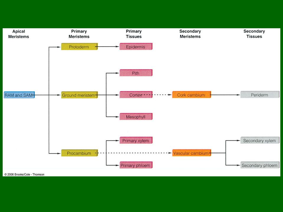
SAM ground meristem Primary meristems Region of primary growth
protoderm procambium cork cambium Secondary meristems vascular cambium procambium ground meristem Primary meristems Region of primary growth protoderm Figure 4.22: Diagram of a tomato plant showing the relative positions of the root apical meristem (RAM) and shoot apical meristem (SAM), the primary meristems (protoderm, ground meristem, and procambium), and the secondary meristems (vascular cambium and cork cambium) in both the shoot and root systems. RAM Root system Fig. 4-22, p. 66
 and shoot apical meristem (SAM), the primary meristems (protoderm, ground meristem, and procambium), and the secondary meristems (vascular cambium and cork cambium) in both the shoot and root systems. RAM. Root system. Fig. 4-22, p. 66.")
197
Root and Apical Meristems
RAM – root apical meristem SAM – shoot apical meristem New cells produced by cell division Theoretically could divide forever Does not occur Scarcity of nutrients Branch of plant can only carry so much weight Genetic regulation of growth

198
Primary Meristems Functions Form primary tissues
Elongate root and shoot

199
Primary Meristems Types of primary meristems Protoderm Procambium
Cells differentiate into epidermis Procambium Cells differentiate into primary xylem and primary phloem Ground meristem Differentiates into cells of pith and cortex of stems and roots Differentiates into mesophyll of leaves

200
young leaf SAM protoderm ground meristem procambium Fig. 4-23b, p. 67
Figure 4.23: Shoot and root tips. (b) Median longitudinal section through the SAM of a Coleus blumei plant. x49. procambium Fig. 4-23b, p. 67
 Median longitudinal section through the SAM of a Coleus blumei plant. x49. procambium. Fig. 4-23b, p. 67.")
201
Figure 4. 23: Shoot and root tips. (c) Photograph of a living root tip
Figure 4.23: Shoot and root tips. (c) Photograph of a living root tip. The root apical meristem (RAM) is located at the very tip of the root, just inside the root cap. Fig. 4-23c, p. 67
 Photograph of a living root tip. The root apical meristem (RAM) is located at the very tip of the root, just inside the root cap. Fig. 4-23c, p. 67.")
202
ground meristem procambium protoderm RAM root cap Fig. 4-23d, p. 67
Figure 4.23: Shoot and root tips. (d) Median longitudinal section through SAM of an Arabidopsis thaliana root tip showing the root cap and RAM. x426. root cap Fig. 4-23d, p. 67
 Median longitudinal section through SAM of an Arabidopsis thaliana root tip showing the root cap and RAM. x426. root cap. Fig. 4-23d, p. 67.")
203
Secondary Meristems Functions Cell division
Initiation of cell differentiation Lateral growth Increases thickness and circumference of stems and roots

204
Secondary Meristems Not found in all plants
Lacking in plants that grow only one season Leaves usually lack secondary growth Types of secondary meristems Vascular cambium Differentiates into secondary xylem and secondary phloem Cork cambium Differentiates into periderm

205
Additional Meristems Intercalary meristems Leaf specific meristems
In stems Regulates stem elongation Leaf specific meristems Regulates leaf shapes Repair of wounds Formation of buds and roots in unusual places

Similar presentations


 w There are two types of cells Prokaryotes and Eukaryotes w Prokaryotes cells that lack membrane-bound organelles. Bacteria.>")


 The McGraw-Hill Companies, Inc.>")


