Download presentation
Presentation is loading. Please wait.

1
Hemoglobin Structure & Function
HMIM224

2
Objectives of the Lecture
1- Understanding the main structural & functional details of hemoglobin as one of the hemoproteins. 2- Identify types & relative concentrations of normal adult hemoglobin with reference to HBA1c with its clinical application. 3- Recognize some of the main genetic & biochemical aspects of methemoglobinopathies with some implications on clinical features (with focusing on thalassemias).
.")
3
Hemoglobin is a globular hemoprotein
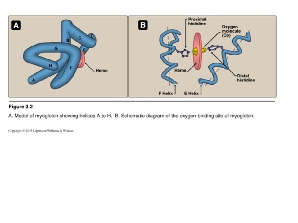
Hemeproteins are a group of specialized proteins that contain heme as a tightly bound prosthetic group. Heme is a complex of protoporphyrin IX and ferrous iron (Fe2+) . The iron is held in the center of the heme molecule by bonds to the four nitrogens of the porphyrin ring. The heme Fe2+ can form two additional bonds, one on each side of the planar porphyrin ring. In myoglobin and hemoglobin, one of these positions is coordinated to the side chain of a histidine residue of the globin molecule, whereas the other position is available to bind oxygen
 . The iron is held in the center of the heme molecule by bonds to the four nitrogens of the porphyrin ring. The heme Fe2+ can form two additional bonds, one on each side of the planar porphyrin ring. In myoglobin and hemoglobin, one of these positions is coordinated to the side chain of a histidine residue of the globin molecule, whereas the other position is available to bind oxygen.")
4
Globin of hemoglobin is a globular protein with a quaternary structure

5
Structure of heme Heme is a complex of protoporphyrin IX and ferrous iron (Fe2+). The iron is held in the center of the heme molecule by bonds of the four nitrogens of the protoporphrin ring. Heme F2+ can form two additional bonds, one on each side of the porphyrin ring. One of these positions is coordinated to the Side chain of histidine residue of the globin molecule, whereas the other position is available to bind oxygen.

7
Structure & function of hemoglobin
Hemoglobin is found exclusively in RBCs. Its main function is to transport oxygen from lungs to the tissues & carbon dioxide & hydrogen protons from tissues to lungs. Hemoglobin A is the major hemoglobin in adults, is composed of four polypeptide chains, 2 alpha (a) & 2 beta (b) chains, held together by noncovalent interactions Each subunit has stretches of a-helical structure & a heme binding pocket.
 & 2 beta (b) chains, held together by noncovalent interactions. Each subunit has stretches of a-helical structure & a heme binding pocket.")
8
Structure & function of hemoglobin (cont.)
")
9
Structure & function of hemoglobin (cont.)
Quaternary structure of hemoglobin: The hemoglobin tetramer can be envisioned as being composed of two identical dimers, (αβ)1 and (αβ)2, in which the numbers refer to dimers one and two. The two polypeptide chains within each dimer are tightly held together, primarily by hydrophobic interactions In contrast, the two dimers are able to move with respect to each other, being held together primarily by polar bonds. The weaker interactions between these mobile dimers result in the two dimers occupying different relative positions in deoxyhemoglobin as compared with oxyhemoglobin
1 and (αβ)2, in which the numbers refer to dimers one and two. The two polypeptide chains within each dimer are tightly held together, primarily by hydrophobic interactions. In contrast, the two dimers are able to move with respect to each other, being held together primarily by polar bonds. The weaker interactions between these mobile dimers result in the two dimers occupying different relative positions in deoxyhemoglobin as compared with oxyhemoglobin.")
10
oxygenation & deoxygenation of hemoglobin (oxyhemoglobin & deoxyhemoglobin)
Taut structure Oxyhemoglobin Relaxed structure

11
Types of adult hemoglobin
3–6 % HBA: the major hemoglobin in humans HBA2: first appears 12 weeks after birth- a minor component of normal adult HB HBF: normally synthesized only during fetal development HBA1C : has glucose residues attached to b-globin chains – increased amounts in DM

12
Hemoglobin A1c (HBA1c) Some of hemoglobin A is glycosylated
Extent of glycosylation depends on the plasma concentration of a particular hexose (as glucose). The most abundant form of glycosylated hemoglobin is HBA1c which has a glucose residues attached to b-globin chains in hemoglobin RBCs. Increased amounts of HBA1c are found in RBCs of patients with diabetes mellitus (DM). HbA1c could be used as a monitor for the control of the blood glucose level during the last 2 months for diabetic patients
. The most abundant form of glycosylated hemoglobin is HBA1c which has a glucose residues attached to b-globin chains in hemoglobin RBCs. Increased amounts of HBA1c are found in RBCs of patients with diabetes mellitus (DM). HbA1c could be used as a monitor for the control of the blood glucose level during the last 2 months for diabetic patients.")
13
Hemoglobinopathies Hemoglobinopathies are members of a family of genetic disorders caused by: 1- Production of a structurally abnormal hemoglobin molecule (Qualitative hemoglobinopathies) Or: 2- Synthesis of insufficient quantities of normal hemoglobin (Quantitative hemoglobinopathies) Or: 3- both (rare).
 Or: 2- Synthesis of insufficient quantities of normal hemoglobin. (Quantitative hemoglobinopathies) Or: 3- both (rare).")
14
Thalassemias Thalassemias are hereditary hemolytic diseases in which an imbalance occurs in the synthesis of globin chains. They are most common single gene disorders in humans. Normally, synthesis of a- and b- globin chains are coordinated, so that each a-globin chain has a b-globin chain partner. This leads to the formation of a2b2 (HbA). In thalassemias, the synthesis of either the a- or b-globin chain is defective.
. In thalassemias, the synthesis of either the a- or b-globin chain is defective.")
15
Thalassemias (cont.) Thalassemia can be caused by a variety of mutations, including: 1- Entire gene deletions (whole gene is absent) Or: 2- Substitutions or deletions of one or more nucleotides in the DNA. Each thalassemia can be classified as either: 1- A disorder in which no globin chains are produced (ao- or bo -thalassemia) Or: 2- Some b-chains are synthesized, but at a reduced rate. (a+- or b+- thalassemia).
 Or: 2- Some b-chains are synthesized, but at a reduced rate. (a+- or b+- thalassemia).")
16
Thalassemias (cont.) 1- b-thalassemias:
Synthesis of b-globin chains are decreased or absent, whereas a-globin chain synthesis is normal. a-globin chains cannot form stable tetramers, and therefore precipitate causing premature death of RBCs. Accumulation of a2g2 (HbF), g4 (Hb Bart's) & a-chain precipitate occurs. These factors end result in development of chronic anemia (hemolytic).
, g4 (Hb Bart s) & a-chain precipitate occurs. These factors end result in development of chronic anemia (hemolytic).")
17
Thalassemias (cont.) Some genetic aspects of thalassemia:
There are only two copies of the b -globin gene in each cell (one on each chromosome 11). So, individuals with b -globin gene defects have either: 1- b-thalassemia minor (b -thalassemia trait): if they have only one defective b-globin gene. 2- b- thalassemia major (Cooley anemia): if both genes are defective.
. So, individuals with b -globin gene defects have either: 1- b-thalassemia minor (b -thalassemia trait): if they have only one defective b-globin gene. 2- b- thalassemia major (Cooley anemia): if both genes are defective.")
18
Thalassemias (cont.) Mutation in both Mutation in one of
b-globin genes b-thalassemia major Mutation in one of b-globin genes b-thalassemia minor

19
Thalassemias (cont.) Some clinical aspects of thalassemias:
1- As b-globin gene is not expressed until late fetal gestation, the physical manifestations of b- thalassemias appear only after birth. 2- Individuals with b - thalassemias minor, make some b-chains, and usually require no specific treatment. 3- Infants born with b - thalassemias major seem healthy at birth, but become severely anemic during the first or second years of life. They require regular transfusions of blood. In these cases, bone marrow replacement therapy is recommended.

20
Thalassemias (cont.) 2- a-thalassemia:
Synthesis of a-globin chains is decreased or absent. Each individual's genome contains four copies of the a-globin (two on each chromosome 16), there are several levels of a-globin chain deficiencies
, there are several levels of a-globin chain. deficiencies.")
21
Thalassemias (cont.) Types: If one of the four genes is defective
If one of the four genes is defective the individual is termed a silent carrier of a- thalassemia as no physical manifestations of the disease occur. If two a-globin genes are defective, the individual is designated as having a-thalassemia trait. If three a-globin genes are defective; the individual has hemoglobin H (HbH) disease which is a mildly to moderately hemolytic anemia. Synthesis of unaffected g- and then b- globin chains continues, resulting in the accumulation of g tetramer in the newborn (g4, Hb Bart's) or b-tetramers (b4, HbH). The subunits do not show heme-heme interactions. So, they have very high oxygen affinities. Thus, they are essentially useless as oxygen carriers to tissues If four a-globin genes are defective, hydrops fetalis & fetal death (death at birth), occurs as a-globin chains are required for the synthesis of HbF
 disease which is a mildly to moderately hemolytic. anemia. Synthesis of unaffected g- and then b- globin chains continues, resulting in the accumulation of. g tetramer in the newborn (g4, Hb Bart s) or b-tetramers (b4, HbH). The subunits do not show heme-heme interactions. So, they have very high oxygen affinities. Thus, they are essentially useless as oxygen carriers to tissues. If four a-globin genes are defective, hydrops fetalis & fetal death (death at birth), occurs as a-globin chains are required for the. synthesis of HbF.")
22
Thalassemias (cont.) Types of a-thalassemias
 Types of a-thalassemias")
23
Assignments Methemoglobin
Sickle cell anemia (Genetic, Biochemical & Clinical Aspects)
")
Similar presentations


 Hb is found in RBCs its main function is to transport O2 to tissues. Structure: 2 parts : heme + globin Globin: four globin chains (2 α.>")


