Download presentation
Presentation is loading. Please wait.

1
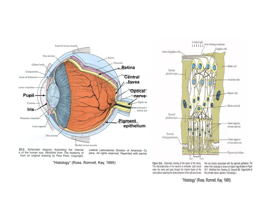
Human Eyes as an Image Sensor
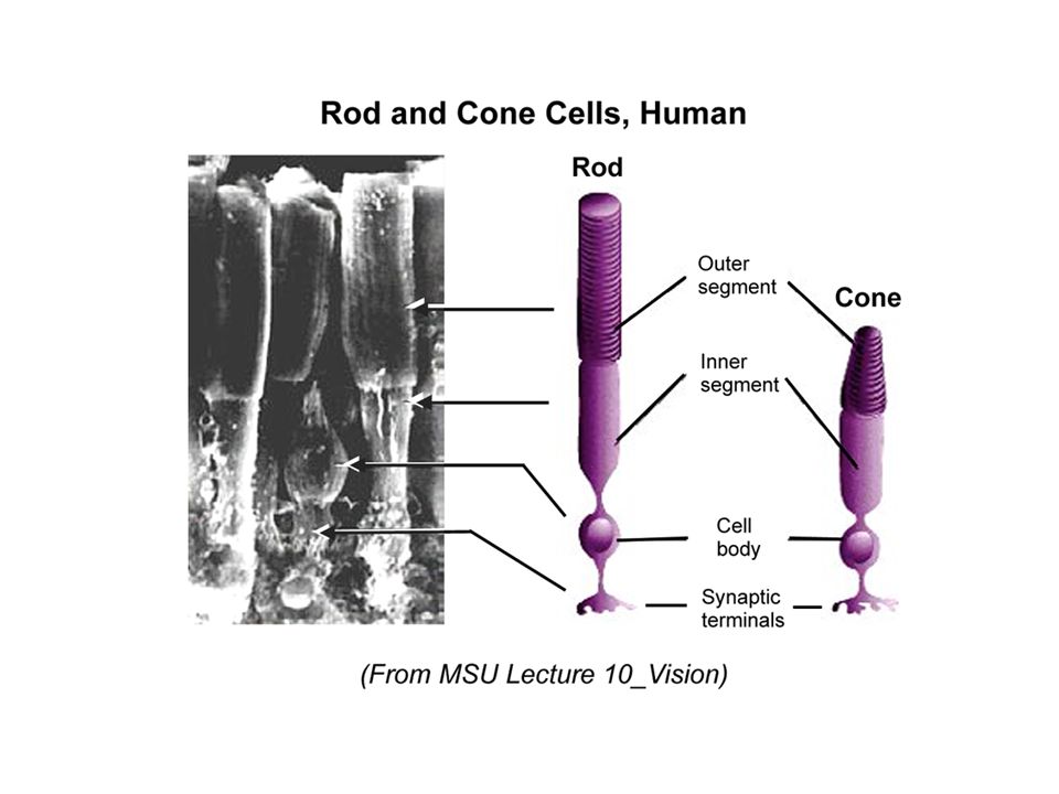
Sensors: Photochemical receptors in retina. Cone and rod cells (“photopic”/“scotopic vision”). In cone, red (570 nm), green (535 nm), blue (445 nm) pigments identified. “Rhodopsine” in rod. Spectral sensitivity: Light-adapted eyes: 400 – 700 nm (peak at 555 nm). Dark-adapted eyes: 380 – 650 nm (peak at 507 nm). Contrast sensitivity: Can distinguish about 50 discrete shades of gray, corresponding to 5- to 6-bit “gray values”. Storage time: 0.2 seconds. Signal/noise (S/N) ratio: 5. Quantum efficiency: At low luminance = 5%. At high luminance = 0.5%. Spatial resolution: For 555 nm, 4.6 mm at center of fovia. (From Inoué & Spring: “Video Microscopy”, 1997; J. Davis: UWSP.edu, 1998)
. In cone, red (570 nm), green (535 nm), blue (445 nm) pigments identified. Rhodopsine in rod. Spectral sensitivity: Light-adapted eyes: 400 – 700 nm (peak at 555 nm). Dark-adapted eyes: 380 – 650 nm (peak at 507 nm). Contrast sensitivity: Can distinguish about 50 discrete shades of gray, corresponding to 5- to 6-bit gray values . Storage time: 0.2 seconds. Signal/noise (S/N) ratio: 5. Quantum efficiency: At low luminance = 5%. At high luminance = 0.5%. Spatial resolution: For 555 nm, 4.6 mm at center of fovia. (From Inoué & Spring: Video Microscopy , 1997; J. Davis: UWSP.edu, 1998)")
4
Color Recognition of Human Eye: Product of Receptor and Neuronal Processing
(Note) Lord Reyleigh contributed significantly in study of color recognition. The standard distinguishing “color-blind” from “normal” visions was made by Lord Reyleigh. Strictly speaking, this Reyleigh standard does not work for everybody. Scientifically, we all are color-blind!
 Lord Reyleigh contributed significantly in study of color recognition. The standard. distinguishing color-blind from normal visions was made by Lord Reyleigh. Strictly speaking, this Reyleigh standard does not work for everybody. Scientifically, we all are color-blind!")
5
Color Recognition: What is Color Anyway?
1. Color is one of the modes of light recognition. 2. Color recognizes light by wavelength. 3. Color recognition results from absorption by visual pigments and neuronal processing. 4. Spatial resolution is determined by spacing of visual cells. 5. Neuronal processing is equipped with sensitivity enhancement. 6. In human, color vision reflects the presence of three pigments with different absorption spectra: red (570 nm), green (535 nm), blue (445 nm). 7. Strictly speaking, the color recognition of each individual is different. 8. Other mode of light recognition includes polarized-light vision in fish and amphibians, adapted for environment. 9. While diurnal animals depend on color recognition, nocturnal animals depend on monochromatic, low-light sensitivity.
, green (535 nm), blue (445 nm). 7. Strictly speaking, the color recognition of each individual is different. 8. Other mode of light recognition includes polarized-light vision in fish and amphibians, adapted for environment. 9. While diurnal animals depend on color recognition, nocturnal animals depend on monochromatic, low-light sensitivity.")
6
Dog’s Vision is Different from Human’s: How? and Why?
1. While human vision is trichromatic (445, 535, 570 nm), dog’s is dichromatic (429, 555 nm) (Plonsky, 1998). 2. Dog’s vision acuity is lower than human’s (12 cycles vs. 30 cycles per degree) (East, 1998). 3. Dogs are nearsighted than human’s (20/ vs. 20/20) (Davis, 1998). 4. Dog’s eye is sensitive in low light and superior for motion detection. 5. Human visual system is adapted for daylight vision and color recognition – They are diurnal and vision help them find food in day light. 6. Dogs are originally nocturnal and their visual system was adapted for night vision. 7. Each cone cell has own intermediate neuron, while a group of rod cells are connected to a single neuron, resulting in high sensitivity to low light in expense of spatial resolution.
, dog’s is dichromatic (429, 555 nm) (Plonsky, 1998). 2. Dog’s vision acuity is lower than human’s (12 cycles vs. 30 cycles per degree) (East, 1998). 3. Dogs are nearsighted than human’s (20/ vs. 20/20) (Davis, 1998). 4. Dog’s eye is sensitive in low light and superior for motion detection. 5. Human visual system is adapted for daylight vision and color recognition – They are diurnal and vision help them find food in day light. 6. Dogs are originally nocturnal and their visual system was adapted for night vision. 7. Each cone cell has own intermediate neuron, while a group of rod cells are connected to a single neuron, resulting in high sensitivity to low light in expense of spatial resolution.")
7
Dog’s Eye is Dichromatic while Human Eye is Trichmatic

8
Human Eye is Superior in Spatial Resolution than Dog
The diagram shows the difference in spatial resolution between human and dog. Because dogs cannot resolve fine B/W contrast grid as human can, the pattern just appears as a homogeneous gray to them. However, dog’s vision is superior to human’s in recognizing fast-moving object, particularly under dim light.

Similar presentations


 Related color: color.>")

 Related color: color perceived to belong to.>")


 Dept.>")

