Download presentation
Presentation is loading. Please wait.

1
Digestive System Digestive Tract

2
Digestive System Digestive system Digestive Tract: Digestive glands: oral cavity, esophagus, stomach, small and large intestines, rectum, and anus. salivary glands, liver, gallbladder and pancreas. Functions: Food intake, preparation of food for swallowing and digestion. Obtain the molecules necessary for the maintenance, growth, and energy needs for the body. Digestive tract is a protective barrier.

3
I. GENERAL STRUCTURE OF THE DIGESTIVE TRACT Mucosa Lamina propria Muscularis mucosae Epithelium Submucosa Muscularis externa Serosa or adventitia

4
Figure. Schematic structure of a portion of the digestive tract with various components Mucosa Submucosa Lamina propria Musculairs externa Adventitia Epithelium Muscularis mucosae Myenteric plexusSubmucosa nerve plexus

5
A.Mucosa 1.Epithelium 2.Lamina propria 3.Muscularis mucosae A barrier between the lumen of digestive tract and the body a.Simple columnar epithelium on most part for absorption and secretion b.Stratified squamous epithelium on both ends is capable withstanding abrasion A layer of loose connective tissue that is rich in capillaries, lymph vessels & tissue, glands. thin layer smooth muscle fibers, its contraction assist secration of minor glands. Inner circular and outer longitudinal layer Innermost and most important layer

6
B.Submucosa Loose connective tissue, many blood, lymph vessels,a submucosa (Meissner’s) nerve plexus, submucosal glands Mucosa and submucosa form projections into the lumen to increase surface area. C.Muscularis externa smooth muscle fibers (except 2 ends) arranged in inner circular and outer longitudinal layer, myenteric(Auerbach’s) nerve plexus D. Serosa or Adventitia Outer most, LCT 1.serosa- covered with simple squamous epithelium, lubricating surface and reducing friction 2. Adventitia- not covered with mesothelium
 arranged in inner circular and outer longitudinal layer, myenteric(Auerbach’s) nerve plexus D. Serosa or Adventitia Outer most, LCT 1.serosa- covered with simple squamous epithelium, lubricating surface and reducing friction 2. Adventitia- not covered with mesothelium.")
8
II. Esophagus 1.Epithelium: Stratified squamous epi A. Mucosa A. Mucosa 2.Lamina propria: Dense CT 3.Muscularis mucosae: smooth muscle longitudinally arranged B. Submucosa: Dense collagenous & elastic fibres ( Esophageal submucosal glands, BV,LV,NF) the upper one third Skeletal muscle C.Muscularis the middle third C.Muscularis the middle third intermingal the distal third smooth muscle D. adventitia: LCT
 the upper one third Skeletal muscle C.Muscularis the middle third C.Muscularis the middle third intermingal the distal third smooth muscle D. adventitia: LCT.")
10
Esophageal mucosaEsophageal submucosal glands

11
III. Stomach 1.epithelium: simple columnar epi. (VILLI) 1.epithelium: simple columnar epi. (VILLI) Mucosa 2. lamina propria: glands, lymphoid tissue 3. muscularis muscosae: smooth 3. muscularis muscosae: smooth muscle muscle Submucosa Loose, areolar CT Muscularis three layer smooth muscle Serosa
 Mucosa 2. lamina propria: glands, lymphoid tissue 3. muscularis muscosae: smooth 3. muscularis muscosae: smooth muscle muscle Submucosa Loose, areolar CT Muscularis three layer smooth muscle Serosa.")
13
Regions of the stomach and their histological structure.

14
Gastric Glands The lamina proparia contains numerous glands. Three types of glands: cardiac and pyloric glands located in the cardiac and pyloric region, respectively, and both mainly secrete mucus. Gastric or fundic glands, located over the body and fundus, consist of 5 types of cells.

15
A) Cardic Glands: simple tubular or compound glands simple tubular or compound glands cells are columnar in shape and mostly mucus-secreting cells are columnar in shape and mostly mucus-secreting B) Pyloric Glands: Branched, convoluted glands Branched, convoluted glands Secrete mucus, lysozyme and a small quantity of proteolytic enzyme. Secrete mucus, lysozyme and a small quantity of proteolytic enzyme.

16
c) Gastric (fundic) Glands Consists of 5 types of cells 1. Chief (zymogenic) cells- pepsinogen 2. Parietal (oxyntic cells)- HCL 3. Mucous neck Cells- mucus 4. Endocrine cells 5. Undifferentiated cells
 cells- pepsinogen 2. Parietal (oxyntic cells)- HCL 3. Mucous neck Cells- mucus 4. Endocrine cells 5. Undifferentiated cells.")
19
Diagram of a chief cell Apical microvilli \

20
Diagram of a parietal cell

21
parietal cell

22
APUD (amine precursor uptake and decarboxylation) system and form part of diffuse neuroendocrine system
 system and form part of diffuse neuroendocrine system")
23
IV Small Intestine Small intestine is divided into 3 segments: DuodenumJejunumIleum The mucosa and sub mucosa of the small intestine are arranged in permanent circular folds (plicae circulares)
")
24
Mucosa Mocosa lining of the small intestine is characterised by the presence of a dense mat of finger-like projection known as villi (0.5-1.5mm in length) Crypts or intestinal glands, which almost extend to the muscularis mucosa, open between the bases of villi.
 Crypts or intestinal glands, which almost extend to the muscularis mucosa, open between the bases of villi.")
25
SMALL GUT PARTS VILLI covered with simple columnar epithelium MUSCULAR smooth muscle SUBMUCOSA connective tissue suspensory MESENTERY with blood vessels covering SEROSA with mesothelium INTESTINAL GLANDS

26
Intestine glands (intestine crypts)
")
27
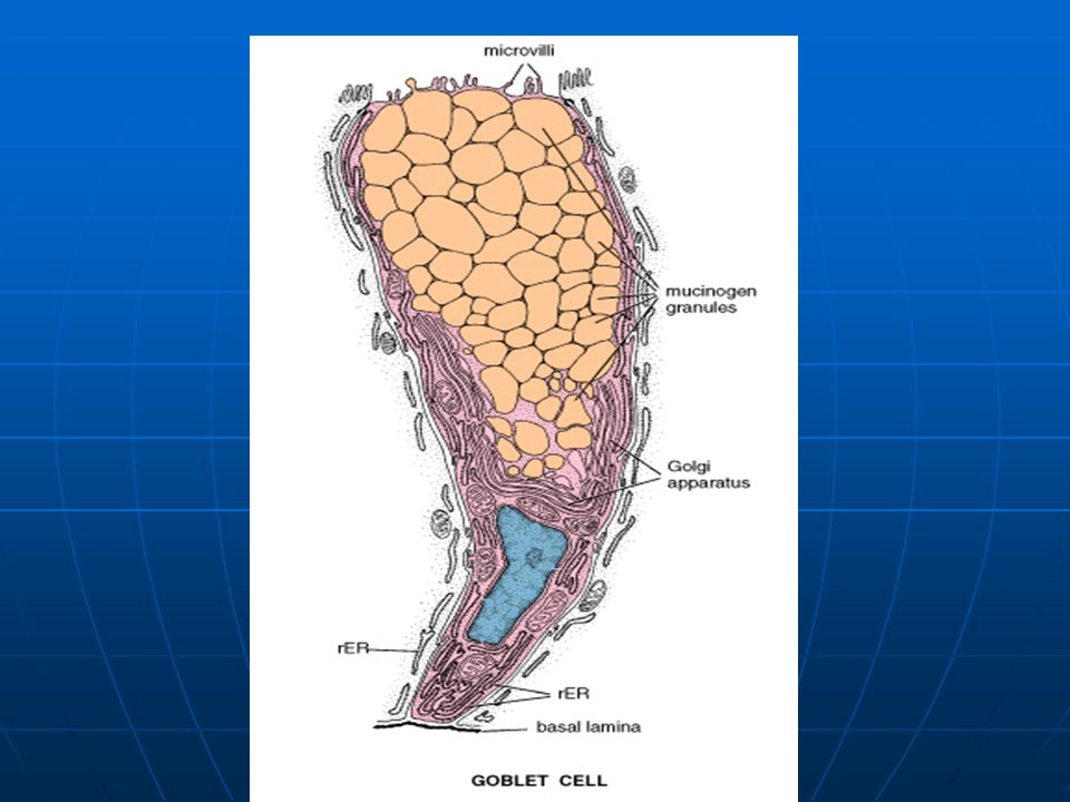
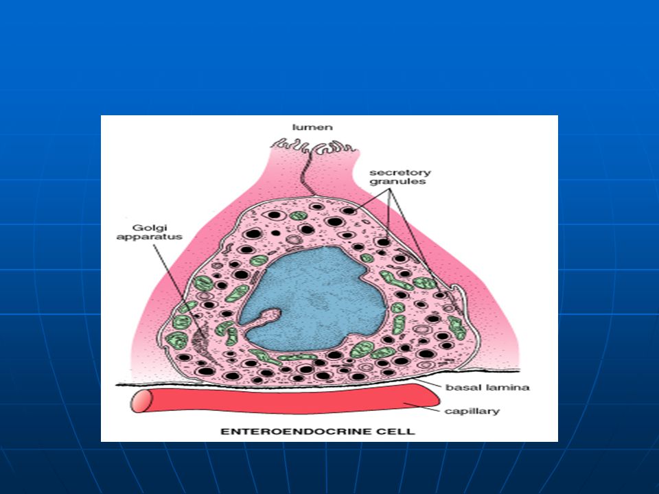
Epithelium- simple columnar epithelium 3 types of cells are found 1. Absorptive cells 2. Goblet cells 3. Enteroendocrine cells Lamina Propria – LCT with a large cellular component cellular component isolated lymphatic nodules isolated lymphatic nodules Peyer’s patches Peyer’s patches Muscularis Mucosa inner circular and outer longitudinal smooth muscle layer inner circular and outer longitudinal smooth muscle layer

31
Submucosa- DCT and contains duodenal glands in the duodenum Muscularis Externa- inner circular and outer longitudinal smooth muscle layer Serosa/adventitia- except for the duodenum, which is retroperitoneal, the rest of the small intestine has an outer layer of serosa

32
V Large Intestine The main function of the large intestine, which includes the caecum, appendix, colon, rectum and anal canal ending as the anus, is absorption of water and minerals.

33
Colon The main features of colon are: (a) An absence of plicae and villi (b) An epithelium containing large numbers of goblet cells. (c) Well-developed, long, deep and closely-packed intestinal crypts. (d) The outer longitudinal layer of the muscularis externa is organised into three thick, longitudinal bands (taeniae coli)
 Well-developed, long, deep and closely-packed intestinal crypts. (d) The outer longitudinal layer of the muscularis externa is organised into three thick, longitudinal bands (taeniae coli).")
34
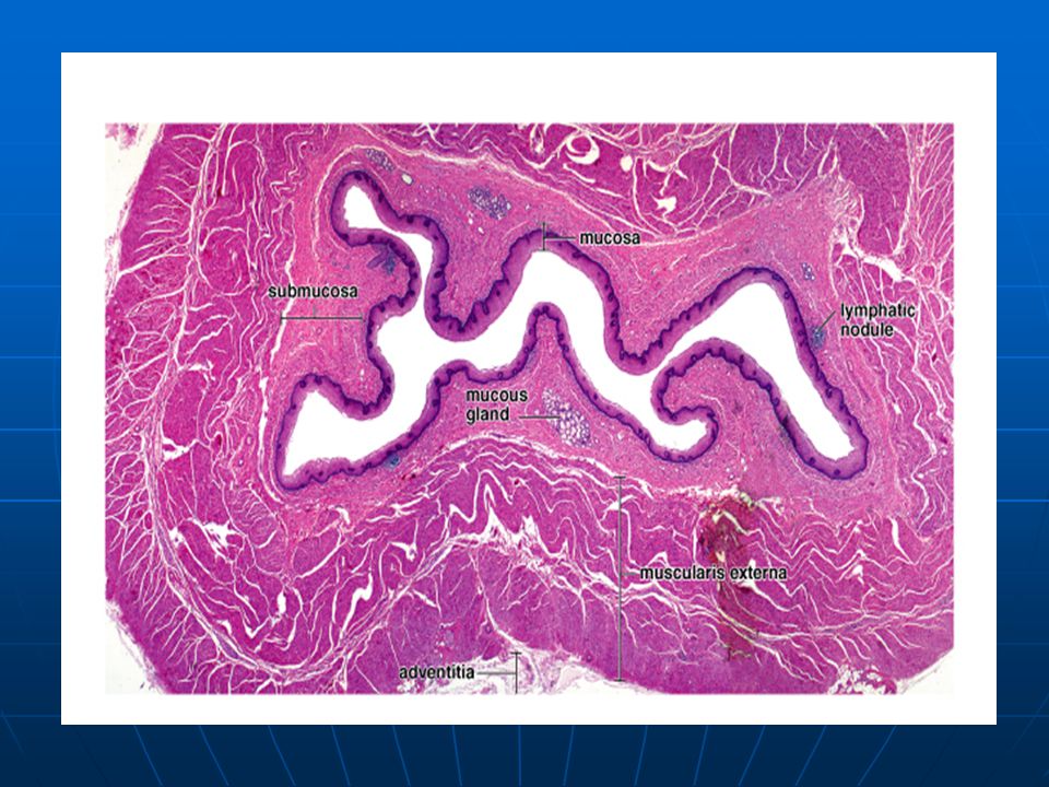
Large intestine

35
Appendix The main characteristics of the appendix are: (a) It is histologically similar to the colon but has a thinner wall, smaller lumen and fewer intestinal glands. (b) It has lymphatic tissue masses in the lamina propria and submucosa. (c) The muscularis mucosae is incomplete and very thin.
 It has lymphatic tissue masses in the lamina propria and submucosa. (c) The muscularis mucosae is incomplete and very thin..")
36
Appendix

Similar presentations