Download presentation
Presentation is loading. Please wait.

1
Large Intestine & Inferior Mesenteric Artery

2
Objectives Discuss anatomical structure of large intestine.
Enlist the characteristic features of large intestine. What are the different positions of the Appendix. Describe the blood supply of the large intestine.

3
The large bowel may vary considerably in length in different subjects;the average is approximately 5 feet (1.5 m). The large intestine is subdivided, for descriptive purposes, into: Caecum with the Appendix vermiform Colon ascending colon hepatic flexure transverse colon splenic flexure descending colon sigmoid colon Rectum & Anal canal .

4
Large Intestine

5
The general characteristics of most of the large intestine are:
Its large internal diameter compared to that of the small intestine; the appendices epiploicae (omental appendices) are fat-filled peritoneal tags The taeniae coli: three thickened bands of muscles the haustra of colon are sacculations of the colon between the taeniae
 are fat-filled peritoneal tags. The taeniae coli: three thickened bands of muscles. the haustra of colon are sacculations of the colon between the taeniae.")
6
No taeniae in the appendix or rectum.
The colon (but not the appendix, caecum or rectum), bears characteristic fat-filled peritoneal tags called appendices epiploicae scattered over its surface. These are especially numerous in the sigmoid colon. The transverse colon and sigmoid are completely peritonealized (the former being readily identified by its attachment to the greater omentum).
, bears characteristic fat-filled peritoneal tags called appendices epiploicae scattered over its surface. These are especially numerous in the sigmoid colon. The transverse colon and sigmoid are completely peritonealized (the former being readily identified by its attachment to the greater omentum).")
7
The ascending and descending colon have no mesocolon but adhere directly to the posterior abdominal wall . The caecum is usually completely peritonealized, The appendix has its own mesocolon.

8
Features of large intestine:
Taeniae Coli: Three thickened bands of muscles No taeniae in the appendix or rectum Haustra: Sacculations of the colon between the taeniae Omental Appendices: Small fatty projections of the omentum Caliber: The internal diameter is much bigger than small intestine The large intestine extends from the distal end of the ileum to the anus, a distance of approximately 1.5 m. It absorbs fluids and salts from the gut contents, thus forming feces, and consists of the cecum, appendix, colon, rectum, and anal canal

10
Cecum and Appendix Ileocecal Junction Taenia Coli Sacculations =
Haustra

11
The cecum is that part of the large intestine that lies below the level of the junction of the ileum with the large intestine . It is a blind-ended pouch that is situated in the right iliac fossa. It is about 2.5 in. (6 cm) long and is completely covered with peritoneum. The appendix is attached to the posteromedial wall of the cecum, just inferior to the end of the ileum

13
The appendix is suspended from the terminal ileum by the mesoappendix, which contains the appendicular vessels . Its point of attachment to the cecum, the base of the appendix, is consistent with the highly visible free taenia leading directly to it. But the location of the rest of the appendix varies considerably . The appendix is at the junction of the lateral and middle one-thirds of a line from the anterior superior iliac spine to the umbilicus (McBurney's point).
.")
14
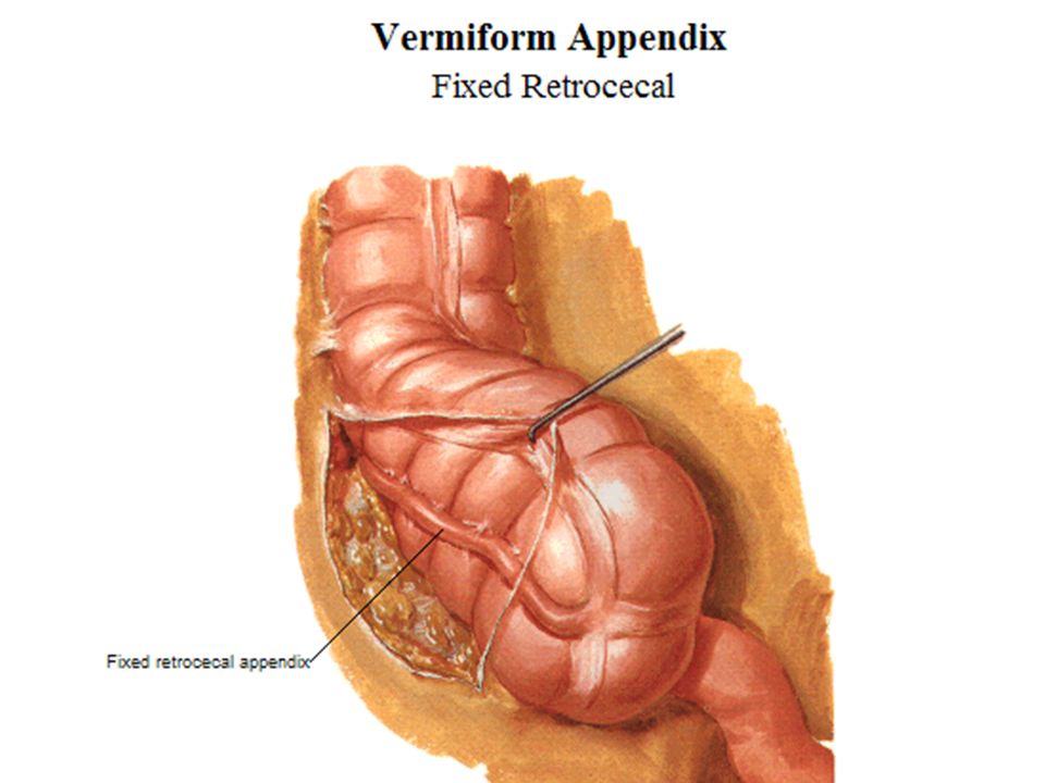
Variations in position Vermiform Appendix

18
Variations in position Vermiform Appendix
64%

22
Inferior mesenteric artery
Branches: 1. Left colic artery 2. Several sigmoid arteries 3. Superior rectal artery

23
The inferior mesenteric artery and arises anterior to the body of vertebra L3. Its branches include the left colic artery, several sigmoid arteries, superior rectal artery. The veins drain into the inferior mesenteric vein

25
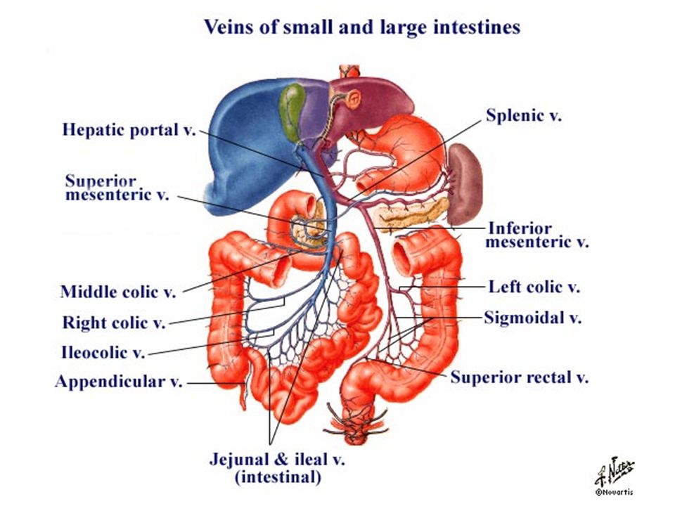
Ascending towards the liver, the portal vein passes posterior to the superior part of the duodenum and enters the right margin of the lesser omentum. As it passes through this part of the lesser omentum, it is anterior to the omental foramen and posterior to both the bile duct, which is slightly to its right, and the hepatic artery proper, which is slightly to its left (see Fig. 4.99, p. 298). Body_ID: P004360 On approaching the liver, the portal vein divides into right and left branches, which enter the liver parenchyma. Tributaries to the portal vein include: right and left gastric veins draining the lesser curvature of the stomach and abdominal esophagus; cystic veins from the gallbladder; the para-umbilical veins, which are associated with the obliterated umbilical vein and connect to veins on the anterior abdominal wall (Fig ). Body_ID: P004361 page 303 page 304 Body_ID: P0304 Splenic vein Body_ID: HC004118 The splenic vein forms from numerous smaller vessels leaving the hilum of the spleen (Fig ). It passes to the right, passing through the splenorenal ligament with the splenic artery and the tail of pancreas. Continuing to the right, the large, straight splenic vein is in contact with the body of the pancreas as it crosses the posterior abdominal wall. Posterior to the neck of pancreas, the splenic vein joins the superior mesenteric vein to form the portal vein short gastric veins from the fundus and left part of the greater curvature of the stomach; the left gastro-omental vein from the greater curvature of the stomach; pancreatic veins draining the body and tail of pancreas; usually the inferior mesenteric vein. Body_ID: P Superior mesenteric vein Body_ID: HC The superior mesenteric vein drains blood from the small intestine, cecum, ascending colon, and transverse colon (Fig ). It begins in the right iliac fossa as veins draining the terminal ileum, cecum, and appendix join, and ascends in the mesentery to the right of the superior mesenteric artery. Body_ID: P Posterior to the neck of the pancreas, the superior mesenteric vein joins the splenic vein to form the portal vein. Body_ID: P page 304 page 305 Body_ID: P0305 As a corresponding vein accompanies each branch of the superior mesenteric artery, tributaries to the superior mesenteric vein include jejunal, ileal, ileocolic, right colic, and middle colic veins. Additional tributaries include: the right gastro-omental vein, draining the right part of the greater curvature of the stomach; the anterior and posterior inferior pancreaticoduodenal veins, which pass alongside the arteries of the same name. The anterior superior pancreaticoduodenal vein usually empties into the right gastro-omental vein, and the posterior superior pancreatico duodenal vein usually empties directly into the portal vein. Body_ID: P Inferior mesenteric vein Body_ID: HC The inferior mesenteric vein drains blood from the rectum, sigmoid colon, descending colon, and splenic flexure (Fig ). It begins as the superior rectal vein and ascends, receiving tributaries from the sigmoid veins and the left colic vein. All these veins accompany arteries of the same name. Continuing to ascend, the inferior mesenteric vein passes posterior to the body of the pancreas and usually joins the splenic vein. Occasionally, it ends at the junction of the splenic and superior mesenteric veins or joins the superior mesenteric vein
. Body_ID: P page 303. page 304. Body_ID: P0304. Splenic vein. Body_ID: HC The splenic vein forms from numerous smaller vessels leaving the hilum of the spleen (Fig ). It passes to the right, passing through the splenorenal ligament with the splenic artery and the tail of pancreas. Continuing to the right, the large, straight splenic vein is in contact with the body of the pancreas as it crosses the posterior abdominal wall. Posterior to the neck of pancreas, the splenic vein joins the superior mesenteric vein to form the portal vein. short gastric veins from the fundus and left part of the greater curvature of the stomach; the left gastro-omental vein from the greater curvature of the stomach; pancreatic veins draining the body and tail of pancreas; usually the inferior mesenteric vein. Body_ID: P Superior mesenteric vein Body_ID: HC The superior mesenteric vein drains blood from the small intestine, cecum, ascending colon, and transverse colon (Fig ). It begins in the right iliac fossa as veins draining the terminal ileum, cecum, and appendix join, and ascends in the mesentery to the right of the superior mesenteric artery. Body_ID: P Posterior to the neck of the pancreas, the superior mesenteric vein joins the splenic vein to form the portal vein. Body_ID: P page 304 page 305 Body_ID: P0305 As a corresponding vein accompanies each branch of the superior mesenteric artery, tributaries to the superior mesenteric vein include jejunal, ileal, ileocolic, right colic, and middle colic veins. Additional tributaries include: the right gastro-omental vein, draining the right part of the greater curvature of the stomach; the anterior and posterior inferior pancreaticoduodenal veins, which pass alongside the arteries of the same name. The anterior superior pancreaticoduodenal vein usually empties into the right gastro-omental vein, and the posterior superior pancreatico duodenal vein usually empties directly into the portal vein. Body_ID: P Inferior mesenteric vein Body_ID: HC The inferior mesenteric vein drains blood from the rectum, sigmoid colon, descending colon, and splenic flexure (Fig ). It begins as the superior rectal vein and ascends, receiving tributaries from the sigmoid veins and the left colic vein. All these veins accompany arteries of the same name. Continuing to ascend, the inferior mesenteric vein passes posterior to the body of the pancreas and usually joins the splenic vein. Occasionally, it ends at the junction of the splenic and superior mesenteric veins or joins the superior mesenteric vein.")
31
Ascending towards the liver, the portal vein passes posterior to the superior part of the duodenum and enters the right margin of the lesser omentum. As it passes through this part of the lesser omentum, it is anterior to the omental foramen and posterior to both the bile duct, which is slightly to its right, and the hepatic artery proper, which is slightly to its left (see Fig. 4.99, p. 298). Body_ID: P004360 On approaching the liver, the portal vein divides into right and left branches, which enter the liver parenchyma. Tributaries to the portal vein include: right and left gastric veins draining the lesser curvature of the stomach and abdominal esophagus; cystic veins from the gallbladder; the para-umbilical veins, which are associated with the obliterated umbilical vein and connect to veins on the anterior abdominal wall (Fig ). Body_ID: P004361 page 303 page 304 Body_ID: P0304 Splenic vein
. Body_ID: P page 303. page 304. Body_ID: P0304. Splenic vein.")
32
Splenic vein Body_ID: HC004118 The splenic vein forms from numerous smaller vessels leaving the hilum of the spleen (Fig ). It passes to the right, passing through the splenorenal ligament with the splenic artery and the tail of pancreas. Continuing to the right, the large, straight splenic vein is in contact with the body of the pancreas as it crosses the posterior abdominal wall. Posterior to the neck of pancreas, the splenic vein joins the superior mesenteric vein to form the portal vein short gastric veins from the fundus and left part of the greater curvature of the stomach; the left gastro-omental vein from the greater curvature of the stomach; pancreatic veins draining the body and tail of pancreas; usually the inferior mesenteric vein. Body_ID: P Superior mesenteric vein Body_ID: HC The superior mesenteric vein drains blood from the small intestine, cecum, ascending colon, and transverse colon (Fig ). It begins in the right iliac fossa as veins draining the terminal ileum, cecum, and appendix join, and ascends in the mesentery to the right of the superior mesenteric artery. Body_ID: P Posterior to the neck of the pancreas, the superior mesenteric vein joins the splenic vein to form the portal vein. Body_ID: P page 304 page 305 Body_ID: P0305 As a corresponding vein accompanies each branch of the superior mesenteric artery, tributaries to the superior mesenteric vein include jejunal, ileal, ileocolic, right colic, and middle colic veins. Additional tributaries include: the right gastro-omental vein, draining the right part of the greater curvature of the stomach; the anterior and posterior inferior pancreaticoduodenal veins, which pass alongside the arteries of the same name. The anterior superior pancreaticoduodenal vein usually empties into the right gastro-omental vein, and the posterior superior pancreatico duodenal vein usually empties directly into the portal vein. Body_ID:
. It passes to the right, passing through the splenorenal ligament with the splenic artery and the tail of pancreas. Continuing to the right, the large, straight splenic vein is in contact with the body of the pancreas as it crosses the posterior abdominal wall. Posterior to the neck of pancreas, the splenic vein joins the superior mesenteric vein to form the portal vein. short gastric veins from the fundus and left part of the greater curvature of the stomach; the left gastro-omental vein from the greater curvature of the stomach; pancreatic veins draining the body and tail of pancreas; usually the inferior mesenteric vein. Body_ID: P Superior mesenteric vein Body_ID: HC The superior mesenteric vein drains blood from the small intestine, cecum, ascending colon, and transverse colon (Fig ). It begins in the right iliac fossa as veins draining the terminal ileum, cecum, and appendix join, and ascends in the mesentery to the right of the superior mesenteric artery. Body_ID: P Posterior to the neck of the pancreas, the superior mesenteric vein joins the splenic vein to form the portal vein. Body_ID: P page 304 page 305 Body_ID: P0305 As a corresponding vein accompanies each branch of the superior mesenteric artery, tributaries to the superior mesenteric vein include jejunal, ileal, ileocolic, right colic, and middle colic veins. Additional tributaries include: the right gastro-omental vein, draining the right part of the greater curvature of the stomach; the anterior and posterior inferior pancreaticoduodenal veins, which pass alongside the arteries of the same name. The anterior superior pancreaticoduodenal vein usually empties into the right gastro-omental vein, and the posterior superior pancreatico duodenal vein usually empties directly into the portal vein. Body_ID:")
33
P Inferior mesenteric vein Body_ID: HC The inferior mesenteric vein drains blood from the rectum, sigmoid colon, descending colon, and splenic flexure (Fig ). It begins as the superior rectal vein and ascends, receiving tributaries from the sigmoid veins and the left colic vein. All these veins accompany arteries of the same name. Continuing to ascend, the inferior mesenteric vein passes posterior to the body of the pancreas and usually joins the splenic vein. Occasionally, it ends at the junction of the splenic and superior mesenteric veins or joins the superior mesenteric vein
. It begins as the superior rectal vein and ascends, receiving tributaries from the sigmoid veins and the left colic vein. All these veins accompany arteries of the same name. Continuing to ascend, the inferior mesenteric vein passes posterior to the body of the pancreas and usually joins the splenic vein. Occasionally, it ends at the junction of the splenic and superior mesenteric veins or joins the superior mesenteric vein.")
Similar presentations